Defects in lipid homeostasis reflect the function of TANGO2 in phospholipid and neutral lipid metabolism
Curation statements for this article:-
Curated by eLife

eLife assessment
This important manuscript describes a series of cellular phenotypes associated with the depletion of TANGO2, a poorly characterized gene product but relevant to neurological and muscular disorders. The authors present solid data indicating that TANGO2 associates with membrane-bound organelles, mainly mitochondria, impacting lipid metabolism and the accumulation of reactive-oxygen species. A few additional experiments would help to understand the link between the lipid changes reported and the cellular phenotype.
This article has been Reviewed by the following groups
Discuss this preprint
Start a discussion What are Sciety discussions?Listed in
- Evaluated articles (eLife)
Abstract
We show that TANGO2 in mammalian cells localizes predominantly to mitochondria and partially at mitochondria sites juxtaposed to lipid droplets (LDs) and the endoplasmic reticulum. HepG2 cells and fibroblasts of patients lacking TANGO2 exhibit enlarged LDs. Quantitative lipidomics revealed a marked increase in lysophosphatidic acid (LPA) and a concomitant decrease in its biosynthetic precursor phosphatidic acid (PA). These changes were exacerbated in nutrient-starved cells. Based on our data, we suggest that TANGO2 function is linked to acyl-CoA metabolism, which is necessary for the acylation of LPA to generate PA. The defect in acyl-CoA availability impacts the metabolism of many other fatty acids, generates high levels of reactive oxygen species, and promotes lipid peroxidation. We suggest that the increased size of LDs is a combination of enrichment in peroxidized lipids and a defect in their catabolism. Our findings help explain the physiological consequence of mutations in TANGO2 that induce acute metabolic crises, including rhabdomyolysis, cardiomyopathy, and cardiac arrhythmias, often leading to fatality upon starvation and stress.
Article activity feed
-

-

Author Response
Reviewer #1 (Public Review):
The manuscript by Lujan and colleagues describes a series of cellular phenotypes associated with the depletion of TANGO2, a poorly characterized gene product but relevant to neurological and muscular disorders. The authors report that TANGO2 associates with membrane-bound organelles, mainly mitochondria, impacting in lipid metabolism and the accumulation of reactive-oxygen species. Based on these observations the authors speculate that TANGO2 function in Acyl-CoA metabolism.
The observations are generally convincing and most of the conclusions appear logical. While the function of TANGO2 remains unclear, the finding that it interferes with lipid metabolism is novel and important. This observation was not developed to a great extent and based on the data presented, the link between TANGO2 …
Author Response
Reviewer #1 (Public Review):
The manuscript by Lujan and colleagues describes a series of cellular phenotypes associated with the depletion of TANGO2, a poorly characterized gene product but relevant to neurological and muscular disorders. The authors report that TANGO2 associates with membrane-bound organelles, mainly mitochondria, impacting in lipid metabolism and the accumulation of reactive-oxygen species. Based on these observations the authors speculate that TANGO2 function in Acyl-CoA metabolism.
The observations are generally convincing and most of the conclusions appear logical. While the function of TANGO2 remains unclear, the finding that it interferes with lipid metabolism is novel and important. This observation was not developed to a great extent and based on the data presented, the link between TANGO2 and acyl-CoA, as proposed by the authors, appears rather speculative.
We thank you for your advice and now include additional data that lends support to the role of TANGO2 in lipid metabolism. We have changed the title accordingly.
- The data with overexpressed TANGO2 looks convincing but I wonder if the authors analyzed the localization of endogenous TANGO2 by immunofluorescence using the antibody described in Figure S2. The idea that TANGO2 localizes to membrane contact sites between mitochondria and the ER and LDs would also be strengthened by experiments including multiple organelle markers.
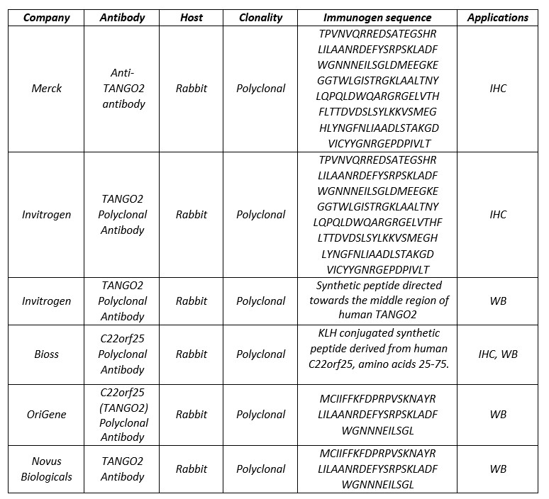
We agree that most of the data on TANGO2 localization are based on the overexpression of the protein. As suggested by the reviewer and despite the lack of commercial antibodies for immunofluorescence-based evaluation, see the following chart, we tested the commercial antibody described in Figure 2 on HepG2 and U2OS cells. Moreover, we used Förster resonance energy transfer (FRET) technology to analyze the proximity of TANGO2 and Tom20, a specific outer mitochondrial membrane protein. In addition, we visualized cells expressing tagged TANGO2 and tagged VAP-B, an integral ER protein in the mitochondria-associated membranes (doi:10.1093/hmg/ddr559) or tagged TANGO2 and tagged GPAT4-Hairpin, an integral LD protein (doi:10.1016/j.devcel.2013.01.013). These data strengthen our proposal and are presented in the revised manuscript.
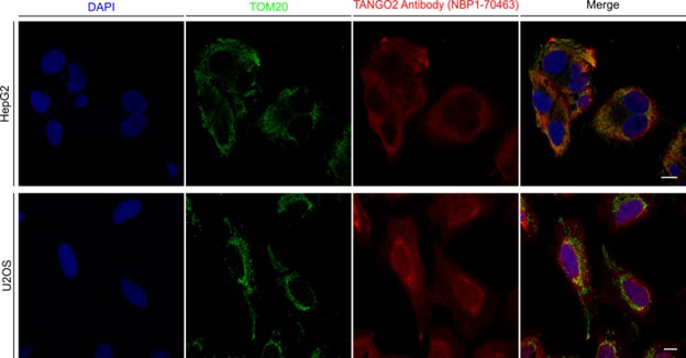
As suggested by the reviewer, we have also visualized two additional cell lines (HepG2 and U2OS) with the anti-TANGO2( from Novus Biologicals) that have been used for western blot (see chart above). As shown in the following figure, the commercial antibody shows a lot of staining in addition to mitochondria, especially in U2OS cells, where it also appears to label the nucleus.
- The changes in LD size in TANGO2-depleted cells are very interesting and consistent with the role of TANGO2 in lipid metabolism. From the lipidomics analysis, it seems that the relative levels of the main neutral lipids in TANGO2-depleted cells remain unaltered (TAG) or even decrease (CE). Therefore, it would be interesting to explore further the increase in LD size for example analyze/display the absolute levels of neutral lipids in the various conditions.
We agree with the reviewer and now present the absolute levels of lipids of interest in the various conditions of the lipidomics analyses (Figure S 3).
- Most of the lipidomics changes in TANGO2-depleted cells are observed in lipid species present in very low amounts while the relative abundance of major phospholipids (PC, PE PI) remains mostly unchanged. It would be good to also display the absolute levels of the various lipids analyzed. This is an important point to clarify as it would be unlikely that these major phospholipids are unaffected by an overall defect in Acyl-CoA metabolism, as proposed by the authors.
As stated above, we have now included the absolute levels of lipids of interest in the various conditions of the lipidomics analyses (Figure S 3).
-
eLife assessment
This important manuscript describes a series of cellular phenotypes associated with the depletion of TANGO2, a poorly characterized gene product but relevant to neurological and muscular disorders. The authors present solid data indicating that TANGO2 associates with membrane-bound organelles, mainly mitochondria, impacting lipid metabolism and the accumulation of reactive-oxygen species. A few additional experiments would help to understand the link between the lipid changes reported and the cellular phenotype.
-
Reviewer #1 (Public Review):
The manuscript by Lujan and colleagues describes a series of cellular phenotypes associated with the depletion of TANGO2, a poorly characterized gene product but relevant to neurological and muscular disorders. The authors report that TANGO2 associates with membrane-bound organelles, mainly mitochondria, impacting in lipid metabolism and the accumulation of reactive-oxygen species. Based on these observations the authors speculate that TANGO2 function in Acyl-CoA metabolism.
The observations are generally convincing and most of the conclusions appear logical. While the function of TANGO2 remains unclear, the finding that it interferes with lipid metabolism is novel and important. This observation was not developed to a great extent and based on the data presented, the link between TANGO2 and acyl-CoA, as …
Reviewer #1 (Public Review):
The manuscript by Lujan and colleagues describes a series of cellular phenotypes associated with the depletion of TANGO2, a poorly characterized gene product but relevant to neurological and muscular disorders. The authors report that TANGO2 associates with membrane-bound organelles, mainly mitochondria, impacting in lipid metabolism and the accumulation of reactive-oxygen species. Based on these observations the authors speculate that TANGO2 function in Acyl-CoA metabolism.
The observations are generally convincing and most of the conclusions appear logical. While the function of TANGO2 remains unclear, the finding that it interferes with lipid metabolism is novel and important. This observation was not developed to a great extent and based on the data presented, the link between TANGO2 and acyl-CoA, as proposed by the authors, appears rather speculative.
1. The data with overexpressed TANGO2 looks convincing but I wonder if the authors analyzed the localization of endogenous TANGO2 by immunofluorescence using the antibody described in Figure S2. The idea that TANGO2 localizes to membrane contact sites between mitochondria and the ER and LDs would also be strengthened by experiments including multiple organelle markers.
2. The changes in LD size in TANGO2-depleted cells are very interesting and consistent with the role of TANGO2 in lipid metabolism. From the lipidomics analysis, it seems that the relative levels of the main neutral lipids in TANGO2-depleted cells remain unaltered (TAG) or even decrease (CE). Therefore, it would be interesting to explore further the increase in LD size for example analyze/display the absolute levels of neutral lipids in the various conditions.
3. Most of the lipidomics changes in TANGO2-depleted cells are observed in lipid species present in very low amounts while the relative abundance of major phospholipids (PC, PE PI) remains mostly unchanged. It would be good to also display the absolute levels of the various lipids analyzed. This is an important point to clarify as it would be unlikely that these major phospholipids are unaffected by an overall defect in Acyl-CoA metabolism, as proposed by the authors.
-
Reviewer #2 (Public Review):
This is an interesting study that seeks to deorphanize Tango2, a protein linked to muscle dysfunction but with no known function. It reveals that Tango2 primarily co-localizes with mitochondria, and its loss impacts mitochondrial homeostasis. Tango2-depleted cells also accumulate LDs. Lipidomic analysis indicated a partial depletion of diacyl lipids including PA in Tango2-depleted cells, and an accumulation of lyso-lipids such as LPA. The proposed model suggests that Tango2 plays a role in lipid metabolism, potentially in acyl-CoA trafficking and or delivery to lyso-lipids to generate diacyl-lipids for mitochondrial homeostasis, which is defective in tango2-deficient diseases like rhabdomyslosis. In general, this is a well-conducted and potentially important study. The first section which deals with Tango2 …
Reviewer #2 (Public Review):
This is an interesting study that seeks to deorphanize Tango2, a protein linked to muscle dysfunction but with no known function. It reveals that Tango2 primarily co-localizes with mitochondria, and its loss impacts mitochondrial homeostasis. Tango2-depleted cells also accumulate LDs. Lipidomic analysis indicated a partial depletion of diacyl lipids including PA in Tango2-depleted cells, and an accumulation of lyso-lipids such as LPA. The proposed model suggests that Tango2 plays a role in lipid metabolism, potentially in acyl-CoA trafficking and or delivery to lyso-lipids to generate diacyl-lipids for mitochondrial homeostasis, which is defective in tango2-deficient diseases like rhabdomyslosis. In general, this is a well-conducted and potentially important study. The first section which deals with Tango2 localization and profiling of cellular changes in Tango2-depleted cells is well conducted. However, the latter half which seeks to understand how Tango2 loss impacts lipid homeostasis is more preliminary. Lyso-lipids like LPA are definitely altered with tango2 loss, but additional work is necessary to understand whether this is due to increased lyso-lipid synthesis, a block in their acylation, or some combination of factors. Delineating these possibilities will significantly enhance this study.
-
