Coupling of Slack and NaV1.6 sensitizes Slack to quinidine blockade and guides anti-seizure strategy development
Curation statements for this article:-
Curated by eLife

eLife assessment
The authors report that an interaction between the sodium-activated potassium channel Slack and Nav1.6 sensitizes Slack to inhibition by quinidine. This is an important finding because it contributes to our understanding of how the antiseizure drug quinidine affects epilepsy syndromes arising from mutations in the Slack-encoding gene KCNT1. The results are largely compelling and the work will likely spark interest in further examining the proposed channel-channel interaction in neuronal cell membranes.
This article has been Reviewed by the following groups
Discuss this preprint
Start a discussion What are Sciety discussions?Listed in
- Evaluated articles (eLife)
Abstract
Quinidine has been used as an anticonvulsant to treat patients with KCNT1-related epilepsy by targeting gain-of-function KCNT1 pathogenic mutant variants. However, the detailed mechanism underlying quinidine’s blockade against KCNT1 (Slack) remains elusive. Here, we report a functional and physical coupling of the voltage-gated sodium channel Na V 1.6 and Slack. Na V 1.6 binds to and highly sensitizes Slack to quinidine blockade. Homozygous knockout of Na V 1.6 reduces the sensitivity of native sodium-activated potassium currents to quinidine blockade. Na V 1.6-mediated sensitization requires the involvement of Na V 1.6’s N- and C-termini binding to Slack’s C-terminus and is enhanced by transient sodium influx through Na V 1.6. Moreover, disrupting the Slack-Na V 1.6 interaction by viral expression of Slack’s C-terminus can protect against Slack G269S -induced seizures in mice. These insights about a Slack-Na V 1.6 complex challenge the traditional view of ‘Slack as an isolated target’ for anti-epileptic drug discovery efforts and can guide the development of innovative therapeutic strategies for KCNT1-related epilepsy.
Article activity feed
-

-
-
-

Author Response
The following is the authors’ response to the previous reviews.
We appreciate the constructive comments made by the editor and the reviewers. We have corrected errors and provided additional experimental data and analysis to address the latest criticisms raised by the reviewers and provided point-by-point response to the reviewers as below.
Reviewer #1 (Recommendations For The Authors):
I do acknowledge the work the authors put into this manuscript and I can accept the fact that the authors decided on a minimum of additional experiments. However, I would recommend the authors to be more concise by adding more information in the method and result sections about how they performed their experiments such as which Nav and AMPAR DNA constructs they used, the age of the mice, how long time they exposed the patches to …
Author Response
The following is the authors’ response to the previous reviews.
We appreciate the constructive comments made by the editor and the reviewers. We have corrected errors and provided additional experimental data and analysis to address the latest criticisms raised by the reviewers and provided point-by-point response to the reviewers as below.
Reviewer #1 (Recommendations For The Authors):
I do acknowledge the work the authors put into this manuscript and I can accept the fact that the authors decided on a minimum of additional experiments. However, I would recommend the authors to be more concise by adding more information in the method and result sections about how they performed their experiments such as which Nav and AMPAR DNA constructs they used, the age of the mice, how long time they exposed the patches to quinidine, information on how many times they repeated their pull downs etc.
Answer: We thank the reviewer’s comments. we have incorporated the suggested modifications into our revised manuscript. Specifically, we have included detailed information on the NaV and AMPAR constructs in the Methods section. The age of the homozygous NaV1.6 knockout mice and the wild-type littermate controls is postnatal (P0-P1) (see in Results and Methods section). Prior to the application of step pulses, cells were subjected to the bath solution containing quinidine for approximately one minute (see in Methods section). Additionally, the co-immunoprecipitation assays for Slack and NaV1.6 were repeated three times (see in Methods section).
Minor detail in line 263: "...KCNT1 (Slack) have been identified to related to seizure..." I guess this should have been "...KCNT1 (Slack) have been identified and related to seizure..."?
Answer: We thank the reviewer for raising this point. We have corrected it in the revised manuscript.
Also, and again minor detail, I had a comment about the color coding in Fig 4 and by mistake, I added 4B, but I meant the use of colors in the entire figure, and mainly the use of colors in 4C, G and I.
Answer: We apologize for the confusion. We have changed the color coding of Figure 4 in the revised manuscript.
Reviewer #2 (Recommendations For The Authors):
While the paper is improved, several concerns do not seem to have been addressed. Some may have been missed because there is no response at all, but others may have been unclear because the response does not address the concern, but a related issue. Details are below.
Answer: We thank the reviewer for the criticisms. We have made changes of our manuscript to address the concerns.
Original issue:
- Remove the term in vivo.
Answer: We thank the reviewer for raising this point. In our experiments, although we did not conduct experiments directly in living organisms, our results demonstrated the coimmunoprecipitation of NaV1.6 with Slack in homogenates from mouse cortical and hippocampal tissues (Fig. 3C). This result may support that the interaction between Slack and NaV1.6 occurs in vivo.
New comment from reviewer:
The argument to use the term in vivo is not well supported by what the authors have said. Just because tissues are used from an animal does not mean experiments were conducted in vivo. As the authors say, they did not conduct experiments in living organisms. Therefore the term in vivo should be avoided. This is a minor point.
Answer: We thank the reviewer for pointing this out. We have removed the term “in vivo” in the revised manuscript.
Original:
- Figure 1C Why does Nav1.2 have a small inward current before the large inward current in the inset?
Answer: We apologize for the confusion. We would like to clarify that the small inward current can be attributed to the current of membrane capacitance (slow capacitance or C-slow). The larger inward current is mediated by NaV1.2.
New comment:
This is not well argued. Please note why the authors know the current is due to capacitance. Also, how do they know the larger current is due to NaV1.2? Please add that to the paper so readers know too.
Answer: We thank the reviewer’s comment. To provide a clearer representation of NaV1.2mediated currents in Fig. 1C, we have replaced the original example trace with a new one in which only one inward current is observed.
Original:
The slope of the rising phase of the larger sodium current seems greater than Nav1.6 or Nav1.5. Was this examined?
Answer: Additionally, we did not compare the slope of the rising phase of NaV subtypes sodium currents but primarily focused on the current amplitudes.
New comment:
This is not a strong answer. There seems to be an effect that the authors do not mention and evidently did not quantify that argues against their conclusion, which weakens the presentation.
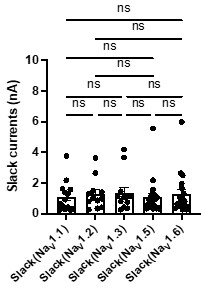
Answer: We thank the reviewer’s comment. To assess the slope of the rising phase of NaV subtype currents, we compared the activation time constants of NaV1.2, NaV1.5, and NaV1.6 peak currents in HEK293 cells co-expressing NaV channel subtypes with Slack. The results have shown no significant differences (Author response image 1). We have included this analysis (see Fig. S9A) and the corresponding fitting equation (see in Methods section) in the revised manuscript.
Author response image 1.
The activation time constants of peak sodium currents in HEK293 cells co-expressing NaV1.2 (n=6), NaV1.5 (n=5), and NaV1.6 (n=5) with Slack, respectively. ns, p > 0.05, one-way ANOVA followed by Bonferroni’s post hoc test.

Original:
2D-E For Nav1.5 the sodium current is very large compared to Nav1.6. Is it possible the greater effect of quinidine for Nav1.6 is due to the lesser sodium current of Nav1.6?
Answer: We thank the reviewer for raising this point. We would like to clarify that our results indicate that transient sodium currents contribute to the sensitization of Slack to quinidine blockade (Fig. 2C,E). Therefore, it is unlikely that the greater effect observed for NaV1.6 in sensitizing Slack is due to its lower sodium currents.
New comment:
I am not sure the question I was asking was clear. How can the authors discount the possibility that quinidine is more effective on NaV1.6 because the NaV1.6 current is relatively weak?
Answer: We thank the reviewer for raising this point. We have examined the sodium current amplitudes of NaV1.5, NaV1.5/1.6 chimeras, and NaV1.6 upon co-expression of NaV with Slack. Our analysis revealed that there are no significant differences between NaV1.5 and NaV1.5/6N, with both exhibiting much larger current amplitudes compared to NaV1.6 (Author response image 2), but only NaV1.5/6N replicates the effect of NaV1.6 in sensitizing Slack to quinidine blockade (Fig. 4H-I), suggesting the observed differences between NaV1.5 and NaV1.6 in sensitizing Slack are unlikely to be attributed to NaV1.6's lower sodium currents but may instead involve NaV1.6's Nterminus-induced physical interaction. We have included this analysis in the revised manuscript (see Fig. S9B).
Author response image 2.
Comparison of peak sodium current amplitudes of NaV1.5 (n=9), NaV1.5/6NC (n=13), NaV1.5/6N (n=10), and NaV1.6 (n=8) upon co-expressed with Slack in HEK293 cells. ns, p > 0.05, * p < 0.05, ** p < 0.01, *** p < 0.001, **** p < 0.0001; one-way ANOVA followed by Bonferroni’s post hoc test.
Original:
The differences between WT and KO in G -H are hard to appreciate. Could quantification be shown? The text uses words like "block" but this is not clear from the figure. It seems that the replacement of Na+ with Li+ did not block the outward current or effect of quinidine.
Answer: We apologize for the confusion. We would like to clarify the methods used in this experiment. The lithium ion (Li+) is a much weaker activator of sodium-activated potassium channel Slack than sodium ion (Na+)1,2.
Zhang Z, Rosenhouse-Dantsker A, Tang QY, Noskov S, Logothetis DE. The RCK2 domain uses a coordination site present in Kir channels to confer sodium sensitivity to Slo2.2 channels. J Neurosci. Jun 2 2010;30(22):7554-62. doi:10.1523/JNEUROSCI.0525-10.2010
Kaczmarek LK. Slack, Slick and Sodium-Activated Potassium Channels. ISRN Neurosci. Apr 18 2013;2013(2013)doi:10.1155/2013/354262 Therefore, we replaced Na+ with Li+ in the bath solution to measure the current amplitudes of sodium-activated potassium currents (IKNa)3.
Budelli G, Hage TA, Wei A, et al. Na+-activated K+ channels express a large delayed outward current in neurons during normal physiology. Nat Neurosci. Jun 2009;12(6):745-50. doi:10.1038/nn.2313
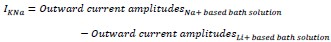
The following equation was used for quantification:
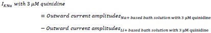
Furthermore, the remaining IKNa after application of 3 μM quinidine in the bath solution was measured as the following:
The quantification results were presented in Fig. 1K. The term "block" used in the text referred to the inhibitory effect of quinidine on IKNa.
New comment:
The fact remains that the term "block" is too strong for an effect that is incomplete. Also, the authors should add to the paper that Li+ is a weaker activator, so the reader knows some of the caveats to the approach.
Answer: We thank the reviewer for raising this point. We have added related citations and replaced the term “block” with “inhibit” in the revised manuscript.
Original:
- In K, for the WT, why is the effect of quinidine only striking for the largest currents?
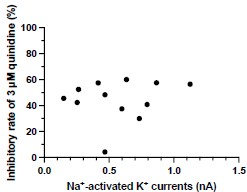
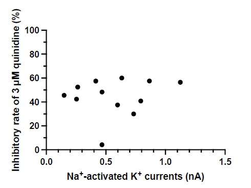
Answer: We thank the reviewer for raising this point. After conducting an analysis, we found no correlation between the inhibitory effect of quinidine and the amplitudes of baseline IKNa in WT neurons (p = 0.6294) (Author response image 3). Therefore, the effect of quinidine is not solely limited to targeting the larger currents.
Author response image 3.
The correlation between the inhibitory effect of quinidine and the amplitudes of baseline IKNa in WT neurons (data from manuscript Fig. 1K). r = 0.1555, p=0.6294, Pearson correlation analysis.
New comment:
Please add this to the paper and the figure as Supplemental.
Answer: We thank the reviewer for raising this point. We have added this figure as Fig.S3B in the revised manuscript.
Original:
- Figure 2 A. The argument could be better made if the same concentration of quinidine were used for Slack and Slack + Nav1.6. It is recognized a greater sensitivity to quinidine is to be shown but as presented the figure is a bit confusing."
Answer: We apologize for the confusion. We would like to clarify that the presented concentrations of quinidine were chosen to be near the IC50 values for Slack and Slack+NaV1.6.
New comment:
Please add this to the paper.
Answer: We thank the reviewer for raising this point. We have added the clarification about the presented concentrations in the revised manuscript.
Original:
2C. Can the authors add the effect of quinidine to the condition where the prepulse potential was 90?"
Answer: We apologize for the confusion. We would like to clarify that the condition of prepulse potential at -90 mV is the same as the condition in Fig. 1. We only changed one experiment condition where the prepulse potential was changed to -40 mV from -90 mV.
New comment:
There was no confusion. The authors should consider adding the condition where the prepulse potential was -90.
Answer: We thank the reviewer for raising this point. We have added the clarification about the voltage condition in the revised manuscript (see in Fig. 2A caption).
Original:
2A. Clarify these 6 panels."
Answer: We thank the reviewer for raising this point. We have clarified the captions of Fig. 3A in the revised manuscript.
New comment: Clarification is needed. What is the blue? DAPI? What area of hippocamps? Please label cell layers. What area of cortex? Please label layers.
Answer: We thank the reviewer for raising this point. We have included the clarification in the Figure caption.
Original:
Figure 7. The images need more clarity. They are very hard to see. Text is also hard to see."
Answer: We apologize for the lack of clarity in the images and text. we would like to provide a concise summary of the key findings shown in this figure.
Figure 7 illustrates an innovative intervention for treating SlackG269S-induced seizures in mice by disrupting the Slack-NaV1.6 interaction. Our results showed that blocking NaV1.6-mediated sodium influx significantly reduced Slack current amplitudes (Fig. 2D,G), suggesting that the Slack-NaV1.6 interaction contributes to the current amplitudes of epilepsy-related Slack mutant variants, aggravating the gain-of-function phenotype. Additionally, Slack’s C-terminus is involved in the Slack-NaV1.6 interaction (Fig. 5D). We assumed that overexpressing Slack’s C-terminus can disrupt the Slack-NaV1.6 interaction (compete with Slack) and thereby encounter the current amplitudes of epilepsy-related Slack mutant variants.
In HEK293 cells, overexpression of Slack’s C-terminus indeed significantly reduced the current amplitudes of epilepsy-related SlackG288S and SlackR398Q upon co-expression with NaV1.5/6NC (Fig. 7A,B). Subsequently, we evaluated this intervention in an in vivo epilepsy model by introducing the Slack G269S variant into C57BL/6N mice using AAV injection, mimicking the human Slack mutation G288S that we previously identified (Fig. 7C-G).
New comment:
The images do not appear to have changed. Consider moving labels above the images so they can be distinguished better. Please label cell layers. Consider adding arrows to the point in the figure the authors want the reader to notice. The study design and timeline are unclear. What is (1) + (3), (2), etc.?
Answer: We thank the reviewer for pointing this out. We have modified Figure 7 in the revised manuscript and included the cell layer information in the Figure caption.
Original:
It is not clear how data were obtained because injection of kainic acid does not lead to a convulsive seizure every 10 min for several hours, which is what appears to be shown. Individual seizures are just at the beginning and then they merge at the start of status epilepticus. After the onset of status epilepticus the animals twitch, have varied movements, sometime rear and fall, but there is not a return to normal behavior. Therefore one can not call them individual seizures. In some strains of mice, however, individual convulsive seizures do occur (even if the EEG shows status epilepticus is occurring) but there are rarely more than 5 over several hours and the graph has many more. Please explain."
Answer: We apologize for the confusion. Regarding the data acquisition in relation to kainic acid injection, we initiated the timing following intraperitoneal injection of kainic acid and recorded the seizure scores of per mouse at ten-minute intervals, following the methodology described in previous studies4.
- Huang Z, Walker MC, Shah MM. Loss of dendritic HCN1 subunits enhances cortical excitability and epileptogenesis. J Neurosci. Sep 2 2009;29(35):10979-88. doi:10.1523/JNEUROSCI.1531-09.2009
The seizure scores were determined using a modified Racine, Pinal, and Rovner scale5,6: (1) Facial movements; (2) head nodding; (3) forelimb clonus; (4) dorsal extension (rearing); (5) Loss of balance and falling; (6) Repeated rearing and failing; (7) Violent jumping and running; (8) Stage 7 with periods of tonus; (9) Dead.
Pinel JP, Rovner LI. Electrode placement and kindling-induced experimental epilepsy. Exp Neurol. Jan 15 1978;58(2):335-46. doi:10.1016/0014-4886(78)90145-0
Racine RJ. Modification of seizure activity by electrical stimulation. II. Motor seizure. Electroencephalogr Clin Neurophysiol. Mar 1972;32(3):281-94. doi:10.1016/00134694(72)90177-0
New comment:
This was clear. Perhaps my question was not clear. The question is how one can count individual seizures if animals have continuous seizures. It seems like the authors did not consider or observe status epilepticus but individual seizures. If that is true the data are hard to believe because too many seizures were counted. Animals do not have nearly this many seizures after kainic acid.
Answer: We appreciate the reviewer’s clarification. Our methodology involved assessing the maximum seizure scale during 10-minute intervals per mouse as previously described7, rather than counting individual seizures. For instance, a mouse exhibited the loss of balance and falling multiple times within 30-40 minute interval, we recorded the seizure scale as 5 for that time interval.
- Kim EC, Zhang J, Tang AY, et al. Spontaneous seizure and memory loss in mice expressing an epileptic encephalopathy variant in the calmodulin-binding domain of Kv7.2. Proc Natl Acad Sci U S A. Dec 21 2021;118(51)doi:10.1073/pnas.2021265118
Reviewer #3 (Recommendations For The Authors):
While the authors have improved the manuscript, several outstanding issues still need to be addressed. Some may have been missed because there is no response at all, but others may have been unclear.
Answer: We thank the reviewer for the criticisms. We have added additional experimental data and analysis to address the concerns.
Original issue from Public Review:
- Immunolabeling of the hippocampus CA1 suggests sodium channels as well as Slack colocalization with AnkG (Fig 3A). Proximity ligation assay for NaV1.6 and Slack or a super-resolution microscopy approach would be needed to increase confidence in the presented colocalization results. Furthermore, coimmunoprecipitation studies on the membrane fraction would bolster the functional relevance of NaV1.6-Slack interaction on the cell surface.
Answer: We thank the reviewer for good suggestions. We acknowledge that employing proximity ligation assay and high-resolution techniques would significantly enhance our understanding of the localization of the Slack-NaV1.6 coupling.
At present, the technical capabilities available in our laboratory and institution do not support highresolution testing. However, we are enthusiastic about exploring potential collaborations to address these questions in the future. Furthermore, we fully recognize the importance of conducting coimmunoprecipitation (Co-IP) assays from membrane fractions. While we have already completed Co-IP assays for total protein and quantified the FRET efficiency values between Slack and NaV1.6 in the membrane region, the Co-IP assays on membrane fractions will be conducted in our future investigations.
New comment from reviewer: so far, the authors have not demonstrated that Nav1.6 and Slack interact on the cell surface.
Answer: We thank the reviewer for pointing this out. We acknowledgement that our data did not directly demonstrate interaction between NaV1.6 and Slack on the cell surface and we have removed related terminology in the revised manuscript. Notably, our patch-clamp experiments in Fig. 2D,G and Fig. S10B showed a Na+-mediated membrane current coupling of Slack and NaV1.6. Additionally, the FRET efficiency values between Slack and NaV1.6 were quantified in the membrane region. These findings suggest that membrane-near Slack interacts with NaV1.6.
- Although hippocampal slices from Scn8a+/- were used for studies in Fig. S8, it is not clear whether Scn8a-/- or Scn8a+/- tissue was used in other studies (Fig 1J & 1K). It will be important to clarify whether genetic manipulation of NaV1.6 expression (Fig. 1K) has an impact on sodiumactivated potassium current, level of surface Slack expression, or that of NaV1.6 near Slack.
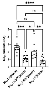
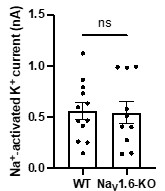
Answer: We thank the reviewer for pointing this out. In Fig. 1G,J,K, primary cortical neurons from homozygous NaV1.6 knockout (Scn8a-/-) mice were used. We will clarify this information in the revised manuscript. In terms of the effects of genetic manipulation of NaV1.6 expression on IKNa and surface Slack expression, we compared the amplitudes of IKNa measured from homozygous NaV1.6 knockout (NaV1.6-KO) neurons and wild-type (WT) neurons. The results showed that homozygous knockout of NaV1.6 does not alter the amplitudes of IKNa (Author response image 4). The level of surface Slack expression will be tested further.
Author response image 4.
The amplitudes of IKNa in WT and NaV1.6-KO neurons (data from manuscript Fig. 1K). ns, p > 0.05, unpaired two-tailed Student’s t test.
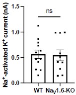
New comment from reviewer: The current version of the manuscrip>t does not contain these pertinent details and needs to be updated to include the information pertaining homozygous NaV1.6 knockouts. What age were these homozygous NaV1.6 knockout mice? These details need to be clearly stated in the manuscript.
Answer: We thank the reviewer for pointing this out. We have included this analysis in the revised manuscript (see Fig. S3A). The age of homozygous NaV1.6 knockout mice are P0-P1 and we have added this detail in the revised manuscript.
- Did the epilepsy-related Slack mutations have an impact on NaV1.6-mediated sodium current?
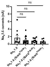
Answer: We thank the reviewer’s question. We examined the amplitudes of NaV1.6 sodium current upon expression alone or co-expression of NaV1.6 with epilepsy-related Slack mutations (K629N, R950Q, K985N). The results showed that the tested epilepsy-related Slack mutations do not alter the amplitudes of NaV1.6 sodium current (Author response image 5).
Author response image 5.
The amplitudes of NaV1.6 sodium currents upon co-expression of NaV1.6 with epilepsy-related Slack mutant variants (SlackK629N, SlackR950Q, and SlackK985N). ns, p>0.05, oneway ANOVA followed by Bonferroni’s post hoc test.
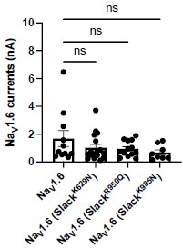
New comment from reviewer: Figure with the functional effect of co-expression of NaV1.6 with epilepsy-related Slack mutations should be included in the revised manuscript
Answer: We thank the reviewer for pointing this out. We have included this analysis in the revised manuscript (see Fig. S10A).
Original issue from Recommendations For The Authors:
- A reference to homozygous knockout is made in the abstract; however, only heterozygous mice are mentioned in the methods section. The genotype of the mice needs to be made clear in the manuscript. Furthermore, at what age were these mice used in the study. Since homozygous knockout of NaV1.6 is lethal at a very young age (<4 wks), it would be important to clarify that point as well.
Answer: We thank the reviewer for pointing this out. In the revised manuscript, we have included information about the source of the primary cortical neurons used in our study. These neurons were obtained from postnatal homozygous NaV1.6 knockout C3HeB/FeJ mice and their wild-type littermate controls.
New comment from reviewer: The answer that postnatal homozygous NaV1.6 knockout C3HeB/FeJ mice were used is insufficient. What age were these mice? This needs to be clearly stated in the manuscript.
Answer: We thank the reviewer for pointing this out. The postnatal homozygous NaV1.6 knockout C3HeB/FeJ mice and their wild-type littermate controls are in P0-P1. We have included this information in the revised manuscript.
- How long were the cells exposed to quinidine before the functional measurement were performed?
Answer: We thank the reviewer for pointing this out. The cells were exposed to the bath solution with quinidine for about one minute before applying step pulses.
New comment from reviewer: This needs to be clearly stated in the manuscript.
Answer: We thank the reviewer for pointing this out. We have included this information in the revised manuscript (see in Methods section).
- In Fig. 6B-D, it is not clear to what extent co-expression of Slack mutants and NaV1.6 increases sodium-activated potassium current.
Answer: We thank the reviewer for pointing this out. We notice that the current amplitudes of Slack mutants exhibit a considerable degree of variation, ranging from less than 1 nA to over 20 nA (n =58). To accurately measure the effects of NaV1.6 on increasing current amplitudes of Slack mutants, we plan to apply tetrodotoxin in the bath solution to block NaV1.6 sodium currents upon coexpression of Slack mutants with NaV1.6.
New comment from reviewer: Were these experiments with TTX completed? If so, they should be added to the revised manuscript.
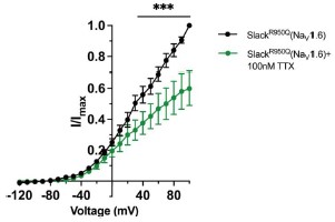
Answer: We thank the reviewer for pointing this out. We compared the current amplitudes of epilepsy-related Slack mutant (SlackR950Q) before and after bath-application of 100 nM TTX upon co-expression with NaV1.6 in HEK293 cells. The results showed that bath-application of TTX significantly reduced the current amplitudes of SlackR950Q at +100 mV by nearly 40% (Author response image 6), suggesting NaV1.6 contributes to the current amplitudes of SlackR950Q. We have included this data in the revised manuscript (see Fig. S10B).
Author response image 6.
The current amplitudes of SlackR950Q before and after bath-application of 100 nM TTX upon co-expression with NaV1.6 in HEK293 cells (n=5). ***p < 0.001, Two-way repeated measures ANOVA followed by Bonferroni’s post hoc test.
Additionally, we have corrected some errors in the methods and figure captions section:
Line 513, bath solution “5 glucose” should be “10 glucose.”
Figure 3A caption, the description “hippocampus CA1 (left) and neocortex (right)” was flipped and we have corrected it.
References
Zhang Z, Rosenhouse-Dantsker A, Tang QY, Noskov S, Logothetis DE. The RCK2 domain uses a coordination site present in Kir channels to confer sodium sensitivity to Slo2.2 channels. J Neurosci. Jun 2 2010;30(22):7554-62. doi:10.1523/JNEUROSCI.0525-10.2010
Kaczmarek LK. Slack, Slick and Sodium-Activated Potassium Channels. ISRN Neurosci. Apr 18 2013;2013(2013)doi:10.1155/2013/354262
Budelli G, Hage TA, Wei A, et al. Na+-activated K+ channels express a large delayed outward current in neurons during normal physiology. Nat Neurosci. Jun 2009;12(6):745-50. doi:10.1038/nn.2313
Huang Z, Walker MC, Shah MM. Loss of dendritic HCN1 subunits enhances cortical excitability and epileptogenesis. J Neurosci. Sep 2 2009;29(35):10979-88. doi:10.1523/JNEUROSCI.1531-09.2009
Pinel JP, Rovner LI. Electrode placement and kindling-induced experimental epilepsy. Exp Neurol. Jan 15 1978;58(2):335-46. doi:10.1016/0014-4886(78)90145-0
Racine RJ. Modification of seizure activity by electrical stimulation. II. Motor seizure. Electroencephalogr Clin Neurophysiol. Mar 1972;32(3):281-94. doi:10.1016/0013-4694(72)90177-0
Kim EC, Zhang J, Tang AY, et al. Spontaneous seizure and memory loss in mice expressing an epileptic encephalopathy variant in the calmodulin-binding domain of Kv7.2. Proc Natl Acad Sci U S A. Dec 21 2021;118(51)doi:10.1073/pnas.2021265118
-
eLife assessment
The authors report that an interaction between the sodium-activated potassium channel Slack and Nav1.6 sensitizes Slack to inhibition by quinidine. This is an important finding because it contributes to our understanding of how the antiseizure drug quinidine affects epilepsy syndromes arising from mutations in the Slack-encoding gene KCNT1. The results are largely compelling and the work will likely spark interest in further examining the proposed channel-channel interaction in neuronal cell membranes.
-
Reviewer #1 (Public Review):
Despite numerous studies on quinidine therapies for epilepsies associated with GOF mutant variants of Slack, there is no consensus on its utility due to contradictory results. In this study Yuan et al. investigated the role of different sodium selective ion channels on the sensitization of Slack to quinidine block. The study employed electrophysiological approaches, FRET studies, genetically modified proteins and biochemistry to demonstrate that Nav1.6 N- and C-tail interacts with Slack's C-terminus and significantly increases Slack sensitivity to quinidine blockade in vitro and in vivo. This finding inspired the authors to investigate whether they could rescue Slack GOF mutant variants by simply disrupting the interaction between Slack and Nav1.6. They find that the isolated C-terminus of Slack can reduce …
Reviewer #1 (Public Review):
Despite numerous studies on quinidine therapies for epilepsies associated with GOF mutant variants of Slack, there is no consensus on its utility due to contradictory results. In this study Yuan et al. investigated the role of different sodium selective ion channels on the sensitization of Slack to quinidine block. The study employed electrophysiological approaches, FRET studies, genetically modified proteins and biochemistry to demonstrate that Nav1.6 N- and C-tail interacts with Slack's C-terminus and significantly increases Slack sensitivity to quinidine blockade in vitro and in vivo. This finding inspired the authors to investigate whether they could rescue Slack GOF mutant variants by simply disrupting the interaction between Slack and Nav1.6. They find that the isolated C-terminus of Slack can reduce the current amplitude of Slack GOF mutant variants co-expressed with Nav1.6 in HEK cells and prevent Slack induced seizures in mouse models of epilepsy. This study adds to the growing list of channels that are modulated by protein-protein interactions, and is of great value for future therapeutic strategies.
-
Reviewer #2 (Public Review):
This is a very interesting paper about the coupling of Slack and Nav1.6 and the insight this brings to the effects of quinidine to treat some epilepsy syndromes.
Slack is a sodium-activated potassium channel that is important to hyperpolarization of neurons after an action potential. Slack is encoded by KNCT1 which has mutations in some epilepsy syndromes. These types of epilepsy are treated with quinidine but this is an atypical antiseizure drug, not used for other types of epilepsy. For sufficient sodium to activate Slack, Slack needs to be close to a channel that allows robust sodium entry, like Nav channels or AMPA receptors. but more mechanistic information is not available. Of particular interest to the authors is what allows quinidine to be effective in reducing Slack.
In the manuscript, the authors …
Reviewer #2 (Public Review):
This is a very interesting paper about the coupling of Slack and Nav1.6 and the insight this brings to the effects of quinidine to treat some epilepsy syndromes.
Slack is a sodium-activated potassium channel that is important to hyperpolarization of neurons after an action potential. Slack is encoded by KNCT1 which has mutations in some epilepsy syndromes. These types of epilepsy are treated with quinidine but this is an atypical antiseizure drug, not used for other types of epilepsy. For sufficient sodium to activate Slack, Slack needs to be close to a channel that allows robust sodium entry, like Nav channels or AMPA receptors. but more mechanistic information is not available. Of particular interest to the authors is what allows quinidine to be effective in reducing Slack.
In the manuscript, the authors show that Nav, not AMPA receptors, are responsible for Slack's sensitization to quinidine blockade, at least in cultured neurons (HeK293, primary cortical neurons). Most of the paper focuses on the evidence that Nav1.6 promotes Slack sensitivity to quinidine.
-
Reviewer #3 (Public Review):
Yuan et al., set out to examine the role of functional and structural interaction between Slack and NaVs on the Slack sensitivity to quinidine. Through pharmacological and genetic means they identify NaV1.6 as the privileged NaV isoform in sensitizing Slack to quinidine. Through biochemical assays, they then determine that the C-terminus of Slack physically interacts with the N- and C-termini of NaV1.6. Using the information gleaned from the in vitro experiments the authors then show that virally-mediated transduction of Slack's C-terminus lessens the extent of SlackG269S-induced seizures. These data uncover a previously unrecognized interaction between a sodium and a potassium channel, which contributes to the latter's sensitivity to quinidine.
-
-
Author Response
The following is the authors’ response to the original reviews.
We would like to thank the editor and all the reviewers for their time and thoughtful consideration of our manuscript. We appreciate the valuable comments. Our provisional response to the “public review” has been published and now we have corrected factual errors and enhanced the clarity of writings based on the “recommendations for the authors.” We believe these corrections will improve the quality and accuracy of our manuscript.
Specific responses to the reviewers' recommendations for the authors are as follows:
Reviewer #1 (Recommendations For The Authors):
- Is the Slack current amplitude dependent on the Nav subtype? Differences in Slack current amplitude might explain the sensitization of Slack to quinidine.
We appreciate the reviewer for raising …
Author Response
The following is the authors’ response to the original reviews.
We would like to thank the editor and all the reviewers for their time and thoughtful consideration of our manuscript. We appreciate the valuable comments. Our provisional response to the “public review” has been published and now we have corrected factual errors and enhanced the clarity of writings based on the “recommendations for the authors.” We believe these corrections will improve the quality and accuracy of our manuscript.
Specific responses to the reviewers' recommendations for the authors are as follows:
Reviewer #1 (Recommendations For The Authors):
- Is the Slack current amplitude dependent on the Nav subtype? Differences in Slack current amplitude might explain the sensitization of Slack to quinidine.
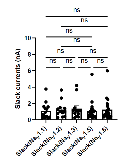
We appreciate the reviewer for raising this point. We examined Slack current amplitudes upon co-expression of Slack with specific NaV subtypes in HEK293 cells. The results have shown that there are no significant differences in Slack current amplitudes upon co-expression of Slack with different NaV channel subtypes (Author response image 1), suggesting whole-cell Slack current amplitudes cannot explain the varied ability of NaV subtypes to sensitize Slack to quinidine blockade.
Author response image 1.
The amplitudes of Slack currents upon co-expression of Slack with specific NaV subtypes in HEK293 cells. ns, p > 0.05, one-way ANOVA followed by Bonferroni’s post hoc test.
- Is the open probability changed by the presence of Nav1.6 and/or by the other Nav subtypes? Changes in open probability might explain the Nav1.6 induced sensitization of Slack to quinidine block.
We appreciate the reviewer for raising this point. To investigate the effect of different NaV channel subtypes on Slack open probability, we will perform the single-channel recordings in future studies.
- Could the authors elaborate more on the coupling between INaT mediated sensitization of Slack to block by quinidine and the Nav1.6 N-and C-tail induced sensitization?
We appreciate the reviewer for raising this point. We fully agree the importance of investigating the detailed mechanism underlying the sensitization of Slack to quinidine blockade. To address the questions, we plan to employ structural biological methods, such as cryo-electron microscopy (cryo-EM).
- Line 85: The authors use an outdated nomenclature of AMPAR subtypes. I would suggest changing to GluA1, GluA2, GluA3 and GluA4.
We appreciate the reviewer’s suggestion. We have changed the term “GluR” to “GluA” in the revised manuscript.
The authors do not explain the rationale by using the different homomeric AMPAR subtypes. Most often the AMPARs express as heteromeric receptors decorated by auxiliary subunits. Also, is the GluA2 the edited version?
We thank the reviewer for raising this point. While AMPARs are often expressed as heteromeric receptors with auxiliary subunits, we focused on the homomeric AMPAR subtypes for initial screening. Through our investigation, we found no significant effects on sensitizing Slack to quinidine blockade. Additionally, the GluA2 used in our study is unedited.
- Line 144: I expect a reduction in current amplitude caused by blocking INaT and INaP is tested at +100mV?
We thank the reviewer for raising this point. The reduction in current amplitude was indeed tested at +100 mV and we have included this information in the revised manuscript.
- Line 157 and line 162: Reference to Supplementary table S3 should be Table S2.
We thank the reviewer for pointing this out. The reference to "Table S3" has been corrected to "Table S2" in the revised manuscript.
- How many times did the authors repeat the co-immunoprecipitation? Some of the bands are very weak, and repeats are necessary for all blots.
We thank the reviewer for raising this concern. We performed the co-immunoprecipitation experiments three times independently.
- Line 288: The authors are showing the chimeric construct in Figures 7A and B but are referring to the full length Nav1.6 in the main text line 288.
We apologize for the confusion. We have clarified in the revised manuscript that we used NaV1.5/6NC in our study.
- Figure 1 line 23: 1 uM quinidine must be 30 uM quinidine?
We thank the reviewer for catching this error. We have corrected the concentration value in the caption of Figure 1 from "1 μΜ" to "30 μΜ" in the revised manuscript.
- Figure 2 line 53: I expect IC50 is measured at +100mV? Same question for line 60 in same figure text.
We thank the reviewer for pointing this out. We have now included this information in the revised manuscript.
- Figure 4B color coding is confusing.
We apologize for the confusion. We would like to clarify that Fig. 4B illustrates the domain architecture of the human NaV channel pore-forming α subunit, and we have changed the color from dark blue to black in the revised figure.
- Figure S6: Text for figure S6E and S6F has been swapped (line 96 to 106).
We thank the reviewer for raising this point. We have rectified the swapped captions for Fig. S6E and Fig. S6F in the revised manuscript.
- Methods section line 652: Kainite acid should be changed to kainic acid
We thank the reviewer for catching this typo. The term “kainite acid” has been corrected to “kainic acid” in the revised manuscript.
Reviewer #2 (Recommendations For The Authors):
- Discuss limitations about the use of non-neuronal cells or cultured primary neurons rather than a more intact system.
We thank the reviewer for raising this point. We have discussed the limitations about the use of non-neuronal cells or cultured primary neurons rather than a more intact system (line 344 to line 348).
- Riluzole is not a selective drug, so the limitations of this drug should be discussed.
We thank the reviewer for raising this point. We have discussed the limitations of riluzole in the revised manuscript (line 360 to line 364).
- Remove the term in vivo.
We thank the reviewer for raising this point. In our experiments, although we did not conduct experiments directly in living organisms, our results demonstrated the coimmunoprecipitation of NaV1.6 with Slack in homogenates from mouse cortical and hippocampal tissues (Fig. 3C). This result may support that the interaction between Slack and NaV1.6 occurs in vivo.
- Figure 1
①C Why does Nav1.2 have a small inward current before the large inward current in the inset? The slope of the rising phase of the larger sodium current seems greater than Nav1.6 or Nav1.5. Was this examined?
We apologize for the confusion. We would like to clarify that the small inward current can be attributed to the current of membrane capacitance (slow capacitance or C-slow). The larger inward current is mediated by NaV1.2. Additionally, we did not compare the slope of the rising phase of NaV subtypes sodium currents but primarily focused on the current amplitudes.
②D-E
For Nav1.5 the sodium current is very large compared to Nav1.6. Is it possible the greater effect of quinidine for Nav1.6 is due to the lesser sodium current of Nav1.6?
We thank the reviewer for raising this point. We would like to clarify that our results indicate that transient sodium currents contribute to the sensitization of Slack to quinidine blockade (Fig. 2C,E). Therefore, it is unlikely that the greater effect observed for NaV1.6 in sensitizing Slack is due to its lower sodium currents.
③The differences between WT and KO in G -H are hard to appreciate. Could quantification be shown? The text uses words like "block" but this is not clear from the figure. It seems that the replacement of Na+ with Li+ did not block the outward current or effect of quinidine.
We apologize for the confusion. We would like to clarify the methods used in this experiment. The lithium ion (Li+) is a much weaker activator of sodium-activated potassium channel Slack than sodium ion (Na+)1,2.
Zhang Z, Rosenhouse-Dantsker A, Tang QY, Noskov S, Logothetis DE. The RCK2 domain uses a coordination site present in Kir channels to confer sodium sensitivity to Slo2.2 channels. J Neurosci. Jun 2 2010;30(22):7554-62. doi:10.1523/JNEUROSCI.0525-10.2010
Kaczmarek LK. Slack, Slick and Sodium-Activated Potassium Channels. ISRN Neurosci. Apr 18 2013;2013(2013)doi:10.1155/2013/354262
Therefore, we replaced Na+ with Li+ in the bath solution to measure the current amplitudes of sodium-activated potassium currents (IKNa)3.
- Budelli G, Hage TA, Wei A, et al. Na+-activated K+ channels express a large delayed outward current in neurons during normal physiology. Nat Neurosci. Jun 2009;12(6):745-50. doi:10.1038/nn.2313
The following equation was used for quantification:
Furthermore, the remaining IKNa after application of 3 μM quinidine in the bath solution was measured as the following:
The quantification results were presented in Fig. 1K. The term "block" used in the text referred to the inhibitory effect of quinidine on IKNa.
④In K, for the WT, why is the effect of quinidine only striking for the largest currents?
We thank the reviewer for raising this point. After conducting an analysis, we found no correlation between the inhibitory effect of quinidine and the amplitudes of baseline IKNa in WT neurons (p = 0.6294) (Author response image 2). Therefore, the effect of quinidine is not solely limited to targeting the larger currents.
Author response image 2.
The correlation between the inhibitory effect of quinidine and the amplitudes of baseline IKNa in WT neurons (data from manuscript Fig. 1K). r = 0.1555, p=0.6294, Pearson correlation analysis.
- Figure 2
①A. The argument could be better made if the same concentration of quinidine were used for Slack and Slack + Nav1.6. It is recognized a greater sensitivity to quinidine is to be shown but as presented the figure is a bit confusing.
We apologize for the confusion. We would like to clarify that the presented concentrations of quinidine were chosen to be near the IC50 values for Slack and Slack+NaV1.6.
②C. Can the authors add the effect of quinidine to the condition where the prepulse potential was - 90?
We apologize for the confusion. We would like to clarify that the condition of prepulse potential at -90 mV is the same as the condition in Fig. 1. We only changed one experiment condition where the prepulse potential was changed to -40 mV from -90 mV.
- Figure 3.
①line 80 should be coronal not coronary
We thank the reviewer for catching this error. We have corrected the term “coronary” to “coronal” in the caption of Figure 3.
②A. Clarify these 6 panels.
We thank the reviewer for raising this point. We have clarified the captions of Fig. 3A in the revised manuscript.
③Please enlarge fonts in D.
We thank the reviewer’s suggestion. We’ve enlarged the fonts in Fig. 3D in the revised manuscript.
④F. The variances should be checked with a test to determine if they are significantly different because they look different - if so, data can be transformed and if transformed data have variances that are equivalent a t-test can be used on the transformed data. Otherwise, Mann-Whitney should be used.
We thank the reviewer for pointing this out. We have reanalyzed the data in Fig. 3F using Mann Whitney test after identifying the different variances in the two groups.
- Figure 7. The images need more clarity. They are very hard to see. Text is also hard to see.
We apologize for the lack of clarity in the images and text. we would like to provide a concise summary of the key findings shown in this figure.
Figure 7 illustrates an innovative intervention for treating SlackG269S-induced seizures in mice by disrupting the Slack-NaV1.6 interaction. Our results showed that blocking NaV1.6-mediated sodium influx significantly reduced Slack current amplitudes (Fig. 2D,G), suggesting that the Slack-NaV1.6 interaction contributes to the current amplitudes of epilepsy-related Slack mutant variants, aggravating the gain-of-function phenotype. Additionally, Slack’s C-terminus is involved in the Slack-NaV1.6 interaction (Fig. 5D). We assumed that overexpressing Slack’s C-terminus can disrupt the Slack-NaV1.6 interaction (compete with Slack) and thereby encounter the current amplitudes of epilepsy-related Slack mutant variants.
In HEK293 cells, overexpression of Slack’s C-terminus indeed significantly reduced the current amplitudes of epilepsy-related SlackG288S and SlackR398Q upon co-expression with NaV1.5/6NC (Fig. 7A,B). Subsequently, we evaluated this intervention in an in vivo epilepsy model by introducing the Slack G269S variant into C57BL/6N mice using AAV injection, mimicking the human Slack mutation G288S that we previously identified (Fig. 7C-G).
②It is not clear how data were obtained because injection of kainic acid does not lead to a convulsive seizure every 10 min for several hours, which is what appears to be shown. Individual seizures are just at the beginning and then they merge at the start of status epilepticus. After the onset of status epilepticus the animals twitch, have varied movements, sometime rear and fall, but there is not a return to normal behavior. Therefore one can not call them individual seizures. In some strains of mice, however, individual convulsive seizures do occur (even if the EEG shows status epilepticus is occurring) but there are rarely more than 5 over several hours and the graph has many more. Please explain.
We apologize for the confusion. Regarding the data acquisition in relation to kainic acid injection, we initiated the timing following intraperitoneal injection of kainic acid and recorded the seizure scores of per mouse at ten-minute intervals, following the methodology described in previous studies4.
- Huang Z, Walker MC, Shah MM. Loss of dendritic HCN1 subunits enhances cortical excitability and epileptogenesis. J Neurosci. Sep 2 2009;29(35):10979-88. doi:10.1523/JNEUROSCI.1531-09.2009
The seizure scores were determined using a modified Racine, Pinal, and Rovner scale5,6: (1) Facial movements; (2) head nodding; (3) forelimb clonus; (4) dorsal extension (rearing); (5) Loss of balance and falling; (6) Repeated rearing and failing; (7) Violent jumping and running; (8) Stage 7 with periods of tonus; (9) Dead.
Pinel JP, Rovner LI. Electrode placement and kindling-induced experimental epilepsy. Exp Neurol. Jan 15 1978;58(2):335-46. doi:10.1016/0014-4886(78)90145-0
Racine RJ. Modification of seizure activity by electrical stimulation. II. Motor seizure. Electroencephalogr Clin Neurophysiol. Mar 1972;32(3):281-94. doi:10.1016/0013- 4694(72)90177-0
- The graphical abstract is quite complicated and somewhat hard to follow. Please simplify and clarify. One aspect of the abstract to clarify is the direction of what is first and second and third (etc.) because arrows point to many directions.
We thank the review for raising this point. In the revised manuscript, we have included numbering of three components within the graphical abstract:
Pathological phenotype: Increased Slack currents.
Two types of interventions:
2a. Disruption of the Slack-NaV1.6 interaction.
2b. NaV1.6-mediated sensitization of Slack to quinidine blockade.
- Therapeutic effects: Reduced Slack currents.
Reviewer #3 (Recommendations For The Authors):
- A reference to homozygous knockout is made in the abstract; however, only heterozygous mice are mentioned in the methods section. The genotype of the mice needs to be made clear in the manuscript. Furthermore, at what age were these mice used in the study. Since homozygous knockout of NaV1.6 is lethal at a very young age (<4 wks), it would be important to clarify that point as well.
We thank the reviewer for pointing this out. In the revised manuscript, we have included information about the source of the primary cortical neurons used in our study. These neurons were obtained from postnatal homozygous NaV1.6 knockout C3HeB/FeJ mice and their wild-type littermate controls.
- Coimmunoprecipitation studies in Fig. 3C are not convincing. There appears to be a signal in the control lane. Furthermore, it appears that brightness levels were adjusted of that image, thereby removing completely the background.
We thank the reviewer for pointing this out. We have replaced Fig. 3C with an unadjusted version in the revised manuscript.
- In Fig. 1B, the authors indicate that 30 microM of quinidine was used, while the corresponding figure legend suggest that 1 microM. Please clarify.
We apologize for this error. We have corrected the concentration value in the caption of Figure 1 from "1 μΜ" to "30 μΜ" in the revised manuscript.
- How long were the cells exposed to quinidine before the functional measurement were performed?
We thank the reviewer for pointing this out. The cells were exposed to the bath solution with quinidine for about one minute before applying step pulses.
- In Fig. 6B-D, it is not clear to what extent co-expression of Slack mutants and NaV1.6 increases sodium-activated potassium current.
We thank the reviewer for pointing this out. We notice that the current amplitudes of Slack mutants exhibit a considerable degree of variation, ranging from less than 1 nA to over 20 nA (n = 5-8). To accurately measure the effects of NaV1.6 on increasing current amplitudes of Slack mutants, we plan to apply tetrodotoxin in the bath solution to block NaV1.6 sodium currents upon coexpression of Slack mutants with NaV1.6.
- In Fig.7A and B, it appears that some recordings had no sodium-activated potassium currents. Why were these included in analysis? How was transfection efficacy assessed?
We apologize for the confusion. We would like to clarify that all recordings included in analysis indeed exhibited outward sodium-activated potassium currents. The current density data in Fig. 7A-B are listed in Author response table 1 (in pA/pF):
Author response table 1.
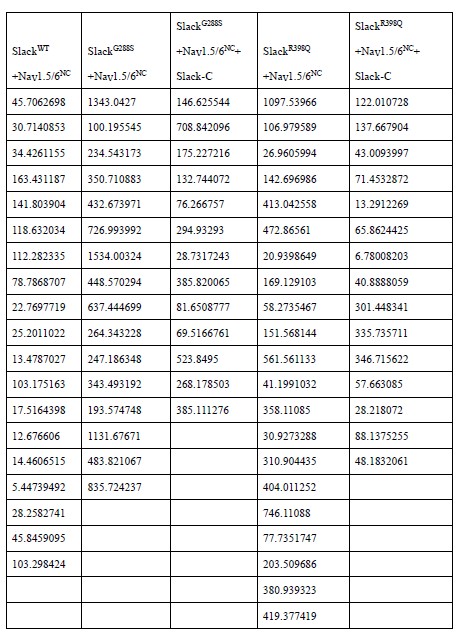
Regarding the assessment of transfection efficacy, we estimated it approximately by using fluorescence proteins as reporters, which were co-expressed with the relevant proteins via the selfcleaving 2A peptide.
- Greater detail needs to be provided for the generation of NaV1.5 and NaV1.6 chimeras. Specifically, what AA residues were changed between sodium channel isoforms?
We thank reviewer for pointing this out. In the revised manuscript, we have included the specific amino acid residues that were changed between NaV1.5 and NaV1.6 to generate the chimeric constructs.
- In line 481, the authors refer to Fig. S2d instead of Fig. S6D. This should be corrected. Furthermore, the unusual shift in sodium current kinetics that the authors observe might be due in part to junction potential. Did the authors take that into consideration?
We apologize for this error. The reference to "Fig. S2d" has been corrected to "Fig. S6D" in the revised manuscript.
Regarding the unusual shift observed in the sodium current kinetics, we agree with the reviewer's suggestion that the junction potential may contribute to this phenomenon. During patch-clamp recordings, we ensure that the junction potential was properly compensated by the amplifier. Additionally, the replacement of CsF in pipette solution may have contributed to the observed unusual shift, as CsF in pipette solution has been reported to shift the voltage dependence of activation and fast/slow inactivation of NaV channels towards more negative potentials7.
- Korngreen A. Advanced patch-clamp analysis for neuroscientists. Neuromethods. Humana Press; 2016:xii, 350 pages.
- Legends for Fig.S6E and S6F are flipped. Please correct.
We apologize for this error. We have rectified the flipped captions for figure S6E and S6F in the revised manuscript.
- Variance should be provided for the IC50 values and kinetic parameters of the sodium channels in the supplemental tables.
We thank the reviewer for raising this point. We have included the 95% confidence interval (95%CI) for the IC50 values and kinetic parameters in the revised supplementary tables.
Additionally, we have corrected some equations in the methods section:
Line 500 and line 503: We have corrected equation (1) by adding the parameter hill coefficient.
Line 514: We have revised equation (4) from
 to
to 
-
eLife assessment
The authors report that an interaction between the sodium-activated potassium channel Slack and Nav1.6 sensitizes Slack to inhibition by quinidine. This is an important finding because it contributes to our understanding of how the antiseizure drug quinidine affects epilepsy syndromes arising from mutations in the Slack-encoding gene KCNT1. The results are largely compelling, although additional data would strengthen the claims of a direct channel-channel interaction in neuronal cell membranes.
-
Reviewer #1 (Public Review):
Despite numerous studies on quinidine therapies for epilepsies associated with GOF mutant variants of Slack, there is no consensus on its utility due to contradictory results. In this study Yuan et al. investigated the role of different sodium selective ion channels on the sensitization of Slack to quinidine block. The study employed electrophysiological approaches, FRET studies, genetically modified proteins and biochemistry to demonstrate that Nav1.6 N- and C-tail interacts with Slack's C-terminus and significantly increases Slack sensitivity to quinidine blockade in vitro and in vivo. This finding inspired the authors to investigate whether they could rescue Slack GOF mutant variants by simply disrupting the interaction between Slack and Nav1.6. They find that the isolated C-terminus of Slack can reduce …
Reviewer #1 (Public Review):
Despite numerous studies on quinidine therapies for epilepsies associated with GOF mutant variants of Slack, there is no consensus on its utility due to contradictory results. In this study Yuan et al. investigated the role of different sodium selective ion channels on the sensitization of Slack to quinidine block. The study employed electrophysiological approaches, FRET studies, genetically modified proteins and biochemistry to demonstrate that Nav1.6 N- and C-tail interacts with Slack's C-terminus and significantly increases Slack sensitivity to quinidine blockade in vitro and in vivo. This finding inspired the authors to investigate whether they could rescue Slack GOF mutant variants by simply disrupting the interaction between Slack and Nav1.6. They find that the isolated C-terminus of Slack can reduce the current amplitude of Slack GOF mutant variants co-expressed with Nav1.6 in HEK cells and prevent Slack induced seizures in mouse models of epilepsy. This study adds to the growing list of channels that are modulated by protein-protein interactions, and is of great value for future therapeutic strategies.
-
Reviewer #2 (Public Review):
This is a very interesting paper about the coupling of Slack and Nav1.6 and the insight this brings to the effects of quinidine to treat some epilepsy syndromes.
Slack is a sodium-activated potassium channel that is important to hyperpolarization of neurons after an action potential. Slack is encoded by KNCT1 which has mutations in some epilepsy syndromes. These types of epilepsy are treated with quinidine but this is an atypical antiseizure drug, not used for other types of epilepsy. For sufficient sodium to activate Slack, Slack needs to be close to a channel that allows robust sodium entry, like Nav channels or AMPA receptors. but more mechanistic information is not available. Of particular interest to the authors is what allows quinidine to be effective in reducing Slack.
In the manuscript, the authors …
Reviewer #2 (Public Review):
This is a very interesting paper about the coupling of Slack and Nav1.6 and the insight this brings to the effects of quinidine to treat some epilepsy syndromes.
Slack is a sodium-activated potassium channel that is important to hyperpolarization of neurons after an action potential. Slack is encoded by KNCT1 which has mutations in some epilepsy syndromes. These types of epilepsy are treated with quinidine but this is an atypical antiseizure drug, not used for other types of epilepsy. For sufficient sodium to activate Slack, Slack needs to be close to a channel that allows robust sodium entry, like Nav channels or AMPA receptors. but more mechanistic information is not available. Of particular interest to the authors is what allows quinidine to be effective in reducing Slack.
In the manuscript, the authors show that Nav, not AMPA receptors, are responsible for Slack's sensitization to quinidine blockade, at least in cultured neurons (HeK293, primary cortical neurons). Most of the paper focuses on the evidence that Nav1.6 promotes Slack sensitivity to quinidine.
-
Reviewer #3 (Public Review):
Yuan et al., set out to examine the role of functional and structural interaction between Slack and NaVs on the Slack sensitivity to quinidine. Through pharmacological and genetic means they identify NaV1.6 as the privileged NaV isoform in sensitizing Slack to quinidine. Through biochemical assays, they then determine that the C-terminus of Slack physically interacts with the N- and C-termini of NaV1.6. Using the information gleaned from the in vitro experiments the authors then show that virally-mediated transduction of Slack's C-terminus lessens the extent of SlackG269S-induced seizures. These data uncover a previously unrecognized interaction between a sodium and a potassium channel, which contributes to the latter's sensitivity to quinidine.
-
-
Author Response:
Reviewer #1 (Public Review):
Despite numerous studies on quinidine therapies for epilepsies associated with GOF mutant variants of Slack, there is no consensus on its utility due to contradictory results. In this study Yuan et al. investigated the role of different sodium selective ion channels on the sensitization of Slack to quinidine block. The study employed electrophysiological approaches, FRET studies, genetically modified proteins and biochemistry to demonstrate that Nav1.6 N- and C-tail interacts with Slack's C-terminus and significantly increases Slack sensitivity to quinidine blockade in vitro and in vivo. This finding inspired the authors to investigate whether they could rescue Slack GOF mutant variants by simply disrupting the interaction between Slack and Nav1.6. They find that the isolated C-terminus …
Author Response:
Reviewer #1 (Public Review):
Despite numerous studies on quinidine therapies for epilepsies associated with GOF mutant variants of Slack, there is no consensus on its utility due to contradictory results. In this study Yuan et al. investigated the role of different sodium selective ion channels on the sensitization of Slack to quinidine block. The study employed electrophysiological approaches, FRET studies, genetically modified proteins and biochemistry to demonstrate that Nav1.6 N- and C-tail interacts with Slack's C-terminus and significantly increases Slack sensitivity to quinidine blockade in vitro and in vivo. This finding inspired the authors to investigate whether they could rescue Slack GOF mutant variants by simply disrupting the interaction between Slack and Nav1.6. They find that the isolated C-terminus of Slack can reduce the current amplitude of Slack GOF mutant variants co-expressed with Nav1.6 in HEK cells and prevent Slack induced seizures in mouse models of epilepsy. This study adds to the growing list of channels that are modulated by protein-protein interactions, and is of great value for future therapeutic strategies.
I have a few comments with regard to how Nav1.6 sensitize Slack to block by quinidine.
(1) It is not clear to me if the Slack induced current amplitude varies depending on the specific Nav subtype. To this end, it would be valuable to test if Slack open probability is affected by the presence of specific Nav subtypes. Nav induced differences in Slack current amplitude and open probability could explain why individual Nav subtypes show varied ability to sensitize Slack to quinidine blockade.
We appreciate the reviewer for raising this point. In order to address whether the whole-cell current amplitudes of Slack varies depending on the specific NaV subtype, we examined Slack current amplitudes upon co-expression of Slack with specific NaV subtypes in HEK293 cells. The results have shown that there are no significant differences in Slack current amplitudes upon co-expression of Slack with different NaV channel subtypes (Author response image 1), suggesting whole-cell Slack current amplitudes cannot explain the varied ability of NaV subtypes to sensitize Slack to quinidine blockade. To investigate the effect of different NaV channel subtypes on Slack open probability, we will perform the single-channel recordings in the future studies.
Author response image 1.
The amplitudes of Slack currents upon co-expression of Slack with specific NaV subtypes in HEK293 cells. ns, p > 0.05, one-way ANOVA followed by Bonferroni’s post hoc test.
(2) It has previously been shown that INaP (persistent sodium current) is important for inducing Slack currents. Here the authors show that INaT (transient sodium current) of Nav1.6 is necessary for the sensitization of Slack to quinidine block whereas INaP surprisingly has no effect. The authors then show that the N-tail together with C-tail of Nav1.6 can induce same effect on Slack as full-length Nav1.6 in presence of high intracellular concentrations of sodium. However, it is not clear to me how the isolated N- and C-tail of Nav1.6 can induce sensitization of Slack to quinidine by interacting with C-terminus of Slack, while sensitization also is dependant on INaT. The authors speculate on different slack open conformation, but one could speculate if there is a missing link, such as an un-identified additional interacting protein that causes the coupling.
We fully agree the importance of investigating the detailed mechanism underlying the sensitization of Slack to quinidine blockade mediated by the N- and C-termini of NaV1.6. Regarding the possibility of additional interacting proteins (“missing link”) that mediate the coupling between Slack and NaV1.6, our GST-pull down assays involving Slack and the N- and C-termini of NaV1.6 (Fig. S7) suggest a direct interaction between Slack and NaV1.6 channels. This finding leads us to consider the possibility of additional interacting proteins might be excluded. In order to further address these questions, we plan to employ structural biological methods, such as cryo-electron microscopy (cryo-EM).
Reviewer #2 (Public Review):
This is a very interesting paper about the coupling of Slack and Nav1.6 and the insight this brings to the effects of quinidine to treat some epilepsy syndromes.
Slack is a sodium-activated potassium channel that is important to hyperpolarization of neurons after an action potential. Slack is encoded by KNCT1 which has mutations in some epilepsy syndromes. These types of epilepsy are treated with quinidine but this is an atypical antiseizure drug, not used for other types of epilepsy. For sufficient sodium to activate Slack, Slack needs to be close to a channel that allows robust sodium entry, like Na channels or AMPA receptors. but more mechanistic information is not available. Of particular interest to the authors is what allows quinidine to be effective in reducing Slack.
In the manuscript, the authors show that Nav, not AMPA receptors are responsible for Slack activation, at least in cultured neurons (HeK293, primary cortical neurons). Most of the paper focuses on the evidence that Nav1.6 promotes Slack sensitivity to quinidine.
(1) The paper is very well written although there are reservations about the use of non-neuronal cells or cultured primary neurons rather than a more intact system.
We appreciate the reviewer's positive evaluation of our work. We acknowledge that utilizing a more intact system would provide valuable insights into the inhibitory effect of quinidine on Slack-NaV1.6. However, there are certain challenges associated with studying Slack currents in their entirety.
First, in our experiments, isolating Slack currents from Na+-activated K+ currents in an intact system is challenging as selective inhibitors for Slick are currently unavailable. To address this, we propose using Slick gene knockout mice to specifically measure Slack currents under physiological conditions in the future investigations. Second, we have observed that the interaction between Slack and NaV1.6 primarily occurs at the axon initial segment of neurons. This poses a difficulty when using brain slices for measurements, as employing the whole-cell voltage-clamp technique to assess Slack at the axon initial segment may introduce systemic errors.
We believe that testing the pharmacological effects of quinidine on Slack-NaV1.6 in primary neurons remains the optimal approach. Although non-neuronal cells or cultured primary neurons may not fully replicate the complexity of an intact system, they still provide valuable insights into the interactions between Slack and NaV1.6, and the effects of quinidine.
(2) I also have questions about the figures.
We will make the necessary modifications and clarifications based on the reviewer's comments:
(3) Finally, riluzole is not a selective drug, so the limitations of this drug should be discussed.
We thank the reviewer for raising this point. We will discuss the limitations of riluzole in our revised version of the manuscript.
(4) On a minor point, the authors use the term in vivo but there are no in vivo experiments.
We thanks the reviewer for raising this point. In our experiments, although we did not conduct experiments directly in living organisms, our results demonstrated the co-immunoprecipitation of NaV1.6 with Slack in homogenates from mouse cortical and hippocampal tissues (Fig. 3C). This result may support that the interaction between Slack and NaV1.6 occurs in vivo.
Reviewer #3 (Public Review):
Yuan et al., set out to examine the role of functional and structural interaction between Slack and NaVs on the Slack sensitivity to quinidine. Through pharmacological and genetic means they identify NaV1.6 as the privileged NaV isoform in sensitizing Slack to quinidine. Through biochemical assays, they then determine that the C-terminus of Slack physically interacts with the N- and C-termini of NaV1.6. Using the information gleaned from the in vitro experiments the authors then show that virally-mediated transduction of Slack's C-terminus lessens the extent of SlackG269S-induced seizures. These data uncover a previously unrecognized interaction between a sodium and a potassium channel, which contributes to the latter's sensitivity to quinidine.
The conclusions of this paper are mostly well supported by data, but some aspects of functional and structural studies in vivo as well as physically interaction need to be clarified and extended.
(1) Immunolabeling of the hippocampus CA1 suggests sodium channels as well as Slack colocalization with AnkG (Fig 3A). Proximity ligation assay for NaV1.6 and Slack or a super-resolution microscopy approach would be needed to increase confidence in the presented colocalization results. Furthermore, coimmunoprecipitation studies on the membrane fraction would bolster the functional relevance of NaV1.6-Slac interaction on the cell surface.
We thank the reviewer for good suggestions. We acknowledge that employing proximity ligation assay and high-resolution techniques would significantly enhance our understanding of the localization of the Slack-NaV1.6 coupling.
At present, the technical capabilities available in our laboratory and institution do not support high-resolution testing. However, we are enthusiastic about exploring potential collaborations to address these questions in the future. Furthermore, we fully recognize the importance of conducting co-immunoprecipitation (Co-IP) assays from membrane fractions. While we have already completed Co-IP assays for total protein and quantified the FRET efficiency values between Slack and NaV1.6 in the membrane region, the Co-IP assays on membrane fractions will be conducted in our future investigations.
(2) Although hippocampal slices from Scn8a+/- were used for studies in Fig. S8, it is not clear whether Scn8a-/- or Scn8a+/- tissue was used in other studies (Fig 1J & 1K). It will be important to clarify whether genetic manipulation of NaV1.6 expression (Fig. 1K) has an impact on sodium-activated potassium current, level of surface Slack expression, or that of NaV1.6 near Slack.
We thank the reviewer for pointing this out. In Fig. 1G,J,K, primary cortical neurons from homozygous NaV1.6 knockout (Scn8a-/-) mice were used. We will clarify this information in the revised manuscript. In terms of the effects of genetic manipulation of NaV1.6 expression on IKNa and surface Slack expression, we compared the amplitudes of IKNa measured from homozygous NaV1.6 knockout (NaV1.6-KO) neurons and wild-type (WT) neurons. The results showed that homozygous knockout of NaV1.6 does not alter the amplitudes of IKNa (Author response image 2). The level of surface Slack expression will be tested further.
Author response image 2.
The amplitudes of IKNa in WT and NaV1.6-KO neurons (data from manuscript Fig. 1K). ns, p > 0.05, unpaired two-tailed Student’s t test.
(3) Did the epilepsy-related Slack mutations have an impact on NaV1.6-mediated sodium current?
We thank the reviewer’s question. We examined the amplitudes of NaV1.6 sodium current upon expression alone or co-expression of NaV1.6 with epilepsy-related Slack mutations (K629N, R950Q, K985N). The results showed that the tested epilepsy-related Slack mutations do not alter the amplitudes of NaV1.6 sodium current (Author response image 3).
Author response image 3.
The amplitudes of NaV1.6 sodium currents upon co-expression of NaV1.6 with epilepsy-related Slack mutant variants (SlackK629N, SlackR950Q, and SlackK985N). ns, p>0.05, one-way ANOVA followed by Bonferroni’s post hoc test.
- Showing the impact of quinidine on persistent sodium current in neurons and on NaV1.6-expressing cells would further increase confidence in the role of persistent sodium current on sensitivity of Slack to quinidine.
We appreciate the reviewer’s question. Previous studies have shown that quinidine can inhibit persistent sodium currents at low concentrations1. In our experiments, blocking persistent sodium currents by application of riluzole in the bath solution showed no significant effects on the sensitivity of Slack to quinidine blockade upon co-expression of Slack with NaV1.6 (Fig. 2F,H). This result suggested that persistent sodium currents were not involved in the sensitization of Slack to quinidine blockade.
- Ju YK, Saint DA, Gage PW. Effects of lignocaine and quinidine on the persistent sodium current in rat ventricular myocytes. Br J Pharmacol. Oct 1992; 107(2):311-6. doi:10.1111/j.1476-5381.1992.tb12743.x
-
eLife assessment
The authors report that an interaction between the sodium-activated potassium channel Slack and Nav1.6 sensitizes Slack to inhibition by quinidine. This is an important finding because it contributes to our understanding of how the antiseizure drug quinidine affects epilepsy syndromes arising from mutations in the Slack-encoding gene KCNT1. The results are largely compelling, although additional data would strengthen the claims of a direct channel-channel interaction in vivo.
-
Reviewer #1 (Public Review):
Despite numerous studies on quinidine therapies for epilepsies associated with GOF mutant variants of Slack, there is no consensus on its utility due to contradictory results. In this study Yuan et al. investigated the role of different sodium selective ion channels on the sensitization of Slack to quinidine block. The study employed electrophysiological approaches, FRET studies, genetically modified proteins and biochemistry to demonstrate that Nav1.6 N- and C-tail interacts with Slack's C-terminus and significantly increases Slack sensitivity to quinidine blockade in vitro and in vivo. This finding inspired the authors to investigate whether they could rescue Slack GOF mutant variants by simply disrupting the interaction between Slack and Nav1.6. They find that the isolated C-terminus of Slack can reduce …
Reviewer #1 (Public Review):
Despite numerous studies on quinidine therapies for epilepsies associated with GOF mutant variants of Slack, there is no consensus on its utility due to contradictory results. In this study Yuan et al. investigated the role of different sodium selective ion channels on the sensitization of Slack to quinidine block. The study employed electrophysiological approaches, FRET studies, genetically modified proteins and biochemistry to demonstrate that Nav1.6 N- and C-tail interacts with Slack's C-terminus and significantly increases Slack sensitivity to quinidine blockade in vitro and in vivo. This finding inspired the authors to investigate whether they could rescue Slack GOF mutant variants by simply disrupting the interaction between Slack and Nav1.6. They find that the isolated C-terminus of Slack can reduce the current amplitude of Slack GOF mutant variants co-expressed with Nav1.6 in HEK cells and prevent Slack induced seizures in mouse models of epilepsy. This study adds to the growing list of channels that are modulated by protein-protein interactions, and is of great value for future therapeutic strategies.
I have a few comments with regard to how Nav1.6 sensitize Slack to block by quinidine.
It is not clear to me if the Slack induced current amplitude varies depending on the specific Nav subtype. To this end, it would be valuable to test if Slack open probability is affected by the presence of specific Nav subtypes. Nav induced differences in Slack current amplitude and open probability could explain why individual Nav subtypes show varied ability to sensitize Slack to quinidine blockade.
It has previously been shown that INaP (persistent sodium current) is important for inducing Slack currents. Here the authors show that INaT (transient sodium current) of Nav1.6 is necessary for the sensitization of Slack to quinidine block whereas INaP surprisingly has no effect. The authors then show that the N-tail together with C-tail of Nav1.6 can induce same effect on Slack as full-length Nav1.6 in presence of high intracellular concentrations of sodium. However, it is not clear to me how the isolated N- and C-tail of Nav1.6 can induce sensitization of Slack to quinidine by interacting with C-terminus of Slack, while sensitization also is dependent on INaT. The authors speculate on different slack open conformation, but one could speculate if there is a missing link, such as an un-identified additional interacting protein that causes the coupling.
-
Reviewer #2 (Public Review):
This is a very interesting paper about the coupling of Slack and Nav1.6 and the insight this brings to the effects of quinidine to treat some epilepsy syndromes.
Slack is a sodium-activated potassium channel that is important to hyperpolarization of neurons after an action potential. Slack is encoded by KNCT1 which has mutations in some epilepsy syndromes. These types of epilepsy are treated with quinidine but this is an atypical antiseizure drug, not used for other types of epilepsy. For sufficient sodium to activate Slack, Slack needs to be close to a channel that allows robust sodium entry, like Nav channels or AMPA receptors. but more mechanistic information is not available. Of particular interest to the authors is what allows quinidine to be effective in reducing Slack.
In the manuscript, the authors …
Reviewer #2 (Public Review):
This is a very interesting paper about the coupling of Slack and Nav1.6 and the insight this brings to the effects of quinidine to treat some epilepsy syndromes.
Slack is a sodium-activated potassium channel that is important to hyperpolarization of neurons after an action potential. Slack is encoded by KNCT1 which has mutations in some epilepsy syndromes. These types of epilepsy are treated with quinidine but this is an atypical antiseizure drug, not used for other types of epilepsy. For sufficient sodium to activate Slack, Slack needs to be close to a channel that allows robust sodium entry, like Nav channels or AMPA receptors. but more mechanistic information is not available. Of particular interest to the authors is what allows quinidine to be effective in reducing Slack.
In the manuscript, the authors show that Nav, not AMPA receptors, are responsible for Slack's sensitization to quinidine blockade, at least in cultured neurons (HeK293, primary cortical neurons). Most of the paper focuses on the evidence that Nav1.6 promotes Slack sensitivity to quinidine.
The paper is very well written although there are reservations about the use of non-neuronal cells or cultured primary neurons rather than a more intact system. I also have questions about the figures. Finally, riluzole is not a selective drug, so the limitations of this drug should be discussed. On a minor point, the authors use the term in vivo but there are no in vivo experiments.
-
Reviewer #3 (Public Review):
Yuan et al., set out to examine the role of functional and structural interaction between Slac and NaVs on the Slack sensitivity to quinidine. Through pharmacological and genetic means they identify NaV1.6 as the privileged NaV isoform in sensitizing Slac to quinidine. Through biochemical assays, they then determine that the C-terminus of Slack physically interacts with the N- and C-termini of NaV1.6. Using the information gleaned from the in vitro experiments the authors then show that virally-mediated transduction of Slack's C-terminus lessens the extent of SlackG269S-induced seizures. These data uncover a previously unrecognized interaction between a sodium and a potassium channel, which contributes to the latter's sensitivity to quinidine.
The conclusions of this paper are mostly well supported by data, …
Reviewer #3 (Public Review):
Yuan et al., set out to examine the role of functional and structural interaction between Slac and NaVs on the Slack sensitivity to quinidine. Through pharmacological and genetic means they identify NaV1.6 as the privileged NaV isoform in sensitizing Slac to quinidine. Through biochemical assays, they then determine that the C-terminus of Slack physically interacts with the N- and C-termini of NaV1.6. Using the information gleaned from the in vitro experiments the authors then show that virally-mediated transduction of Slack's C-terminus lessens the extent of SlackG269S-induced seizures. These data uncover a previously unrecognized interaction between a sodium and a potassium channel, which contributes to the latter's sensitivity to quinidine.
The conclusions of this paper are mostly well supported by data, but some aspects of functional and structural studies in vivo as well as physically interaction need to be clarified and extended.
Immunolabeling of the hippocampus CA1 suggests sodium channels as well as Slac colocalization with AnkG (Fig 3A). Proximity ligation assay for NaV1.6 and Slac or a super-resolution microscopy approach would be needed to increase confidence in the presented colocalization results. Furthermore, coimmunoprecipitation studies on the membrane fraction would bolster the functional relevance of NaV1.6-Slac interaction on the cell surface.
Although hippocampal slices from Scn8a+/- were used for studies in Fig. S8, it is not clear whether Scn8a-/- or Scn8a+/- tissue was used in other studies (Fig 1J & 1K). It will be important to clarify whether genetic manipulation of NaV1.6 expression (Fig. 1K) has an impact on sodium-activated potassium current, level of surface Slac expression, or that of NaV1.6 near Slac.
Did the epilepsy-related Slac mutations have an impact on NaV1.6-mediated sodium current?
Showing the impact of quinidine on persistent sodium current in neurons and on NaV1.6-expressing cells would further increase confidence in the role of persistent sodium current on sensitivity of Slac to quinidine.
-
