Two-way Dispatched function in Sonic hedgehog shedding and transfer to high-density lipoproteins
Curation statements for this article:-
Curated by eLife

eLife assessment
This useful manuscript presents an analysis of different factors that are required for release of the lipid-linked morphogen Shh from cellular membranes. The evidence is still incomplete, as experiments rely on over-expression of Shh in a single cell line and are sometimes of a correlative nature. The study, which otherwise confirms and extends previous findings, will be of interest to developmental biologists who work on Hedgehog signaling.
This article has been Reviewed by the following groups
Discuss this preprint
Start a discussion What are Sciety discussions?Listed in
- Evaluated articles (eLife)
Abstract
The Sonic hedgehog (Shh) signaling pathway controls embryonic development and tissue homeostasis after birth. This requires regulated solubilization of dual-lipidated, firmly plasma membrane-associated Shh precursors from producing cells. Although it is firmly established that the resistance-nodulation-division transporter Dispatched (Disp) drives this process, it is less clear how lipidated Shh solubilization from the plasma membrane is achieved. We have previously shown that Disp promotes proteolytic solubilization of Shh from its lipidated terminal peptide anchors. This process, termed shedding, converts tightly membrane-associated hydrophobic Shh precursors into delipidated soluble proteins. We show here that Disp-mediated Shh shedding is modulated by a serum factor that we identify as high-density lipoprotein (HDL). In addition to serving as a soluble sink for free membrane cholesterol, HDLs also accept the cholesterol-modified Shh peptide from Disp. The cholesteroylated Shh peptide is necessary and sufficient for Disp-mediated transfer because artificially cholesteroylated mCherry associates with HDL in a Disp-dependent manner, whereas an N-palmitoylated Shh variant lacking C-cholesterol does not. Disp-mediated Shh transfer to HDL is completed by proteolytic processing of the palmitoylated N-terminal membrane anchor. In contrast to dual-processed soluble Shh with moderate bioactivity, HDL-associated N-processed Shh is highly bioactive. We propose that the purpose of generating different soluble forms of Shh from the dual-lipidated precursor is to tune cellular responses in a tissue-type and time-specific manner.
Article activity feed
-

-
-

eLife assessment
This useful manuscript presents an analysis of different factors that are required for release of the lipid-linked morphogen Shh from cellular membranes. The evidence is still incomplete, as experiments rely on over-expression of Shh in a single cell line and are sometimes of a correlative nature. The study, which otherwise confirms and extends previous findings, will be of interest to developmental biologists who work on Hedgehog signaling.
-
Reviewer #1 (Public Review):
This manuscript presents a model in which combined action of the transporter-like protein DISP and the sheddases ADAM10/17 promote shedding of a mono-cholesteroylated Sonic Hedgehog (SHH) species following cleavage of palmitate from the dually lipidated precursor ligand. The authors propose that this leads to transfer of the cholesterol-modified SHH to HDL for solubilization. The minimal requirement for SHH release by this mechanism is proposed to be the covalently linked cholesterol modification because DISP could promote transfer of a cholesteroylated mCherry reporter protein to serum HDL. The authors used an in vitro system to demonstrate dependency on DISP/SCUBE2 for release of the cholesterol modified ligand. These results confirm previously published results from other groups (PMC3387659 and PMC3682496).
A strength of the work is the use of a bicistronic SHH-Hhat system to consistently generate dually-lipidated ligand to determine the quantity and lipidation status of SHH released into cell culture media.
Key shortcomings include the unusual normalization strategies used for many experiments and the lack of quantification/statistical analyses for several experiments. Due to these omissions, it is difficult to conclude that the data justify the conclusions. The significance of the data provided is overstated because many of the presented experiments confirm/support previously published work. The study provides a modest advance in understanding of the complex issue of SHH membrane extraction.
-
Reviewer #2 (Public Review):
Ehring et al. analyze contributions of Dispatched, Scube2, serum lipoproteins and Sonic Hedgehog lipid modifications to the generation of different Shh release forms. Hedgehog proteins are anchored in cellular membranes by N-terminal palmitate and C-terminal cholesterol modifications, yet spread through tissues and are released into the circulation. How Hedgehog proteins can be released, and in which form, remains controversial. The authors systematically dissect contributions of several previously identified factors, and present evidence that Disp, Scube2 and lipoproteins concertedly act to release a novel Shh variant that is cholesterol-modified but not palmitoylated. The results provide new insights into the function of Disp and Scube2 in Hedgehog release. The findings concerning the function of …
Reviewer #2 (Public Review):
Ehring et al. analyze contributions of Dispatched, Scube2, serum lipoproteins and Sonic Hedgehog lipid modifications to the generation of different Shh release forms. Hedgehog proteins are anchored in cellular membranes by N-terminal palmitate and C-terminal cholesterol modifications, yet spread through tissues and are released into the circulation. How Hedgehog proteins can be released, and in which form, remains controversial. The authors systematically dissect contributions of several previously identified factors, and present evidence that Disp, Scube2 and lipoproteins concertedly act to release a novel Shh variant that is cholesterol-modified but not palmitoylated. The results provide new insights into the function of Disp and Scube2 in Hedgehog release. The findings concerning the function of lipoproteins and cholesterol in Hedgehog release are largely confirmatory (PMID 23554573, 20685986). However, in light of the multitude of competing models for Hedgehog release, the present study is a valuable contribution that provides further insights into the relevance of lipoproteins in this process.
A novel and surprising finding of the present study is the differential removal of Shh N- or C-terminal lipid anchors depending on the presence of HDL and/or Disp. In particular, the identification of a non-palmitoylated but cholesterol-modified Shh variant that associates with lipoproteins is potentially important. The authors use RP-HPLC and defined controls to assess the properties of processed Shh forms, but their precise molecular identity remains to be defined. A caveat is the strong reliance on over-expression of Shh in a single cell line. The authors detect Shh variants that are released independently of Disp and Scube2 in secretion assays, which however are excluded from interpretation as experimental artifacts. Thus, it would be important to demonstrate key findings in cells that secrete Shh endogenously.
-
Author response:
The following is the authors’ response to the current reviews.
Reviewer #1 (Public Review):
Major shortcomings include the unusual normalization strategies used for many experiments and the lack of quantification/statistical analyses for several experiments. Because of these omissions, it is difficult to conclude that the data justify the conclusions. The significance of the data presented is overstated, as many of the experiments presented confirm/support previously published work. The study provides a modest advance in the understanding of the complex issue of SHH membrane extraction.
Major shortcomings include the unusual normalization strategies used for many experiments and the lack of quantification/statistical analysis for several experiments.
This statement is not correct for the revised manuscript: The …
Author response:
The following is the authors’ response to the current reviews.
Reviewer #1 (Public Review):
Major shortcomings include the unusual normalization strategies used for many experiments and the lack of quantification/statistical analyses for several experiments. Because of these omissions, it is difficult to conclude that the data justify the conclusions. The significance of the data presented is overstated, as many of the experiments presented confirm/support previously published work. The study provides a modest advance in the understanding of the complex issue of SHH membrane extraction.
Major shortcomings include the unusual normalization strategies used for many experiments and the lack of quantification/statistical analysis for several experiments.
This statement is not correct for the revised manuscript: The normalization strategies used are clearly described in the manuscript and are not unusual. Each experiment is now statistically analyzed.
The significance of the data presented is overstated, as many of the experiments presented confirm/support previously published work.
As reviewer 2 correctly points out, there are many competing models for Hedgehog release. Our study cannot possibly support them all - the reviewer's statement is therefore misleading. In fact, our careful biochemical analysis of the mechanistics of Dispatched- mediated Shh export supports only two of them: The model of proteolytic processing of Shh lipid anchors (shedding) and the model of lipoprotein-mediated Shh transport. In contrast, our study does not support the predominant model of Dispatched-mediated extraction of dual-lipidated Shh and delivery to Scube2, which is currently thought to act as a soluble Shh chaperone. We also do not support Dispatched function in Shh endocytic recycling and cytoneme loading, or any of the other models such as exosome-mediated or micelle Shh transport.
Reviewer #2 (Public Review):
A novel and surprising finding of the present study is the differential removal of Shh N- or C- terminal lipid anchors depending on the presence of HDL and/or Disp. In particular, the identification of a non-palmitoylated but cholesterol-modified Shh variant that associates with lipoproteins is potentially important. The authors use RP-HPLC and defined controls to assess the properties of processed forms of Shh, but their precise molecular identity remains to be defined. One caveat is the heavy reliance on overexpression of Shh in a single cell line. The authors detect Shh variants that are released independently of Disp and Scube2 in secretion assays, but these are excluded from interpretation as experimental artifacts. Therefore, it would be important to demonstrate key findings in cells that endogenously secrete Shh.
We would like to respond as follows:
The authors use RP-HPLC and defined controls to assess the properties of processed forms of Shh, but their precise molecular identity remains to be defined.
This is the original reviewers statement regarding our original manuscript submission. We believe that the biochemical and functional data presented in the VOR clearly describe the molecular identity of solubilized Shh: it is monolipidated, lipoprotein-associated, and highly biologically active in two established Shh bioassays.
One caveat is the heavy reliance on overexpression of Shh in a single cell line.
As stated by reviewer 1, the strength of our work is the use of a bicistronic SHH-Hhat system to consistently generate doubly lipidated ligand to determine the amount and lipidation status of SHH released into cell culture media. This unique system therefore eliminates the artifacts of protein overexpression. We have also added two other cell lines to our VOR that produce the same results (including Panc1 cells that endogenously produce Shh, Supplementary Figure 1).
The authors detect Shh variants that are released independently of Disp and Scube2 in secretion assays, but these are excluded from interpretation as experimental artifacts.
As the reviewer correctly points out, these variants are released independently of Disp and Scube2, both of which are known as essential release factors in vivo. These variants are therefore by definition experimental artifacts. The forms we have included in our analysis are the alternative forms that are clearly dependent on Dispatched and Scube2 for their release - as shown in the first figure in the manuscript, and in pretty much every other figure after that.
The following is the authors’ response to the previous reviews.
Reviewer #1 (Public Review):
Key shortcomings include the unusual normalization strategies used for many experiments and the lack of quantification/statistical analyses for several experiments.
In the updated version of the paper, we have addressed all of this reviewer's criticisms. Most importantly, we have performed several additional experiments to address the concern that unusual normalization strategies were used in our paper and that quantification and statistical analyses were lacking for several experiments. We have now analyzed the full set of release conditions for Shh and engineered proteins from Disp-expressing n.t. control cells and Disp-/- cells both in the presence and absence of Scube2 (Figure 1A'-D', Figure 2E added to the paper, Figure 3B'-D', Figure 5C and Figure S2F-H). Previously, we had only quantified protein release from n.t. controls and Disp-/- cells in the presence but not in the absence of Scube2 under serum-depleted conditions. Quantifications of serum-free protein release and Shh release under conditions ranging from 0.05% FCS to 10% FCS were completely missing from the earlier versions of the manuscript, but have now been added to our paper. In addition, we have reanalyzed all of the data sets in the above figures, as well as Figures 2C and S1B, to address the issue of "unusual normalization strategies": unlike previous assays in which the highest amount of protein detected in the media was set to 100% and all other proteins in that experiment were expressed relative to that value, we now directly compare the relative amounts of cellular and corresponding solubilized proteins as a method to quantify release without the need for data normalization (Figs. 1A'-D', 2C,E, 3B'-D', E, 5C, Fig. S1B, S2F-H).
We have also repeated the qPCR analyses in C3H10T1/2 cells and now show that the same Shh/C25AShh activities can be observed when using another Shh responsive cell line, NIH3T3 cells (Fig. 4B, 6B, fig. S5B).
We would like to point out that if the criticism refers to the presentation of our RP-HPLC and SEC data, the normalization of the strongest eluted protein signal to 100% for all proteins tested is necessary to put their behavior in a clearer relationship. This is because only the relative positions of protein elution, and not their amounts, are important in these experiments.
The significance of the data provided is overstated because many of the presented experiments confirm/support previously published work.
To mitigate the first reviewer's comment that the significance of the data presented is overstated, we now clearly distinguish between our novel results and the known aspect of Hh release on lipoproteins throughout our paper. We now clearly describe what is new and important in our paper: First, contrary to the general perception in the field, Disp and Scube2 are not sufficient to solubilize Shh, casting doubt on the currently accepted model that Scube2 accepts dual-lipidated Shh from Disp and transports it to the receptor Ptch. Second, lipoproteins shift dual Shh processing to N-terminal peptide processing only to generate different soluble Hh forms with different activities (as shown in Figure 4C). Third, and again contrary to popular belief, this new release mode does not inactivate Shh, as we now show in two established cellular assays for Hh biofunction (Figures 4A-C, 5B'', 6B and S5C-G). Fourth, and most importantly, we show that spatiotemporally controlled, Disp-, Scube2- and HDL-mediated Shh release absolutely requires dual lipidation of the membrane-associated Shh precursor prior to its release. This finding (as shown in Figures 1 and S2) changes the interpretation of previously published in vivo data that have long been interpreted as evidence for the requirement of dual Shh lipidation for full receptor binding and activation.
The study provides a modest advance in our understanding of the complex issue of Shh membrane extraction.
Although we agree that our results integrate our novel observations into previously established concepts of Hh release and trafficking, we also hope that our data cast well-founded doubt on the current view that the issue of Hh release and trafficking is largely resolved by the model of Disp-mediated Shh hand-over to Scube2 and then to Ptch, which requires interactions with both Shh lipids. Our data show that this is clearly not the case in the presence of lipoproteins. Thus, the significance of our data is that models of Shh lipid-regulated signaling to Ptch obtained using the dual-lipidated Shh precursor prior to its Disp- and Scube2-mediated conversion into a delipidated or monolipidated, HDL-associated soluble ligand are likely to describe a non-physiological interaction. Instead, our work describes a highly bioactive soluble ligand with only one lipid still attached, which has not been described before in the literature. The in vivo endpoint analyses presented in Fig. S8 suggest that this new protein variant is likely to play an important role during development.
Reviewer #2 (Public Review):
The precise molecular identity (of the released Shh) remains to be defined.
We would like to respond that the direct comparison of soluble proteins and their well-defined double-lipidated precursors side-by-side in the same experiment, as shown in our paper, determines all relevant molecular changes in the Shh release process. Most importantly, we show by SDS-PAGE and RP-HPLC that HDL restricts Shh processing to the N-terminus and that the absence of HDL results in double processing of Shh during its release. We also show by SEC that the C-terminus binds the protein to HDL. In addition, the fly experiments confirm the requirement for N-terminal Hh processing, but not for processing of the C-terminal peptide, and suggest that the N-terminal Cardin-Weintraub sequence replaced by the functionally blocking tag represents the physiological cleavage site.
It would be important to demonstrate key findings in cells that secrete Shh endogenously.
We now confirm the key findings of our study in Panc1 cells that endogenously produce and secrete Shh: As shown in Fig. S1D, we find that soluble proteins are processed but retain the C-cholesterol, which we now directly confirm by RP-HPLC (Fig. S4F-H). The in vivo analyses shown in Fig. S8 suggest that the key finding - that N-terminal but not C-terminal Hh shedding is required for release - can be supported, at least in the fly: here, Hh variants impaired in their ability to be processed N-terminally strongly repress the endogenous protein, whereas the same protein impaired in its ability to be processed C-terminally does not.
The authors detect Shh variants that are expressed independently of Disp and Scube2 in secretion assays, but are excluded from interpretation as experimental artifacts.
We agree with the reviewer's criticism that the amounts of Shh released independently of Disp and Scube2 in secretion assays were not quantified and analyzed statistically to justify their proposed status as not physiologically relevant. We now show that these forms are indeed secretion artifacts (Fig. 3E and Fig. S2F-H show quantification of the lower electrophoretic mobility protein fraction (i.e., the "top" band representing the double-lipidated soluble protein fraction)) because this fraction is released independently of Disp and Scube2.
-
-
Author Response
The following is the authors’ response to the original reviews.
Reviewer 1 (public):
- “It is unclear whether new in vivo experiments were conducted for this study”.
All in vivo experiments were conducted for this study by using previously published fly stocks to directly compare N- and C-terminal shedding side-by-side in two Hh-dependent developmental systems. This is now clearly stated in the revised supplement (Fig. S8). We also conducted these experiments because previous in vivo studies in flies often relied on Hh overexpression in the fat body, raising questions about their physiological relevance. Our in vivo analyses of Hh function in wing and eye discs are more physiologically relevant and can explain the previously reported presence of non-lipidated bioactive Hh in disc tissue (PMID: 23554573).
- “A critical …
Author Response
The following is the authors’ response to the original reviews.
Reviewer 1 (public):
- “It is unclear whether new in vivo experiments were conducted for this study”.
All in vivo experiments were conducted for this study by using previously published fly stocks to directly compare N- and C-terminal shedding side-by-side in two Hh-dependent developmental systems. This is now clearly stated in the revised supplement (Fig. S8). We also conducted these experiments because previous in vivo studies in flies often relied on Hh overexpression in the fat body, raising questions about their physiological relevance. Our in vivo analyses of Hh function in wing and eye discs are more physiologically relevant and can explain the previously reported presence of non-lipidated bioactive Hh in disc tissue (PMID: 23554573).
- “A critical shortcoming of the study is that experiments showing Shh secretion/export do not include a Shh(-) control condition. Without demonstration that the bands analyzed are specific for Shh(+) conditions, these experiments cannot be appropriately evaluated”.
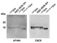
The Cell Signaling Technology C9C5 anti-Shh antibody used in our study is highly specific against Shh, and it has been used in over 60 publications. C9C5 even lacks cross-reactivity with highly similar Ihh or Dhh (https://www.cellsignal.com/products/primary-antibodies/shh-c9c5-rabbit-mab/2207?_requestid=1528451). We confirmed C9C5 specificity repeatedly (one example is shown below; another quality control that includes media of mock-transfected cells is now shown in Fig. S1) and never observed unspecific bands under any experimental condition. As shown below, C9C5 and R&D AF464 anti-Shh antibodies (the latter were previously used in our lab) detect the same bands.
Author response image 1.
Shh immunoblot. R&D 8908-SH served as a size control for full-length dual-lipidated Shh, and C25S;26-35Shh served as a size control for N-terminally truncated monolipidated Shh. Both C25SShh bands are specific: One represents the full-length protein and the bottom band represents N-truncated processed proteins. The blot was first incubated with antibody AF464 and reincubated (after stripping) with the much more sensitive antibody C9C5.
- “A stably expressing Shh/Hhat cell line would reduce condition to condition and experiment to experiment variability”.
We agree and therefore have previously aimed to establish stable Hhat-expressing cell lines. However, we found that long-term Hhat overexpression eliminated transfected cells after several passages, or cells gradually ceased to express Hhat. This prevented us from establishing stable cell lines co-expressing Shh/Hhat despite several attempts and different strategies. Instead, we established transient co-expression of Shh/Hhat from the same mRNA as the next-best strategy for reliable near-quantitative Shh palmitoylation in our assays.
- “Unusual normalization strategies are used for many experiments, and quantification/statistical analyses are missing for several experiments”.
We repeated all qPCR assays to eliminate this shortcoming. Biological activities and transcriptional responses of palmitoylated Shh and non-palmitoylated C25AShh are now directly compared and quantified (revised Fig. 4A,B, newly included Fig. 6, revised Fig. S5B). The original comparison of both proteins with dual-lipidated R&D 8908-SH is still important in order to show that both Shh and C25AShh in serum-containing media have equally high, and not equally low, activities because R&D 8908-SH is generally seen as the Shh form with the highest biological activity. These comparisons are therefore still discussed in the main manuscript text and are now shown in Fig. S5E.
- “The study provides a modest advance in the understanding of the complex issue of Shh membrane extraction”
We believe that the revised manuscript advances our understanding of Shh membrane extraction beyond the modest in three important ways. First, although Disp was indeed known as a furin-activated Hh exporter, our findings show for the first time that furin activation of Disp is strictly linked to proteolytic Shh processing as the underlying release mode, fully consistent with data obtained from the Disp-/- cells.
Second, Scube2 was known as a Shh release enhancer and several lipoproteins were previously shown to play a role in the process, but our findings are the first to show that synergistic Disp/Scube2 function depends on the presence of lipoprotein and that HDL (but no other lipoprotein) accepts free cholesterol or a novel monolipidated Shh variant from Disp. This challenges the dominant model of Scube2 chaperone function in Hh release and transport (PMID 22902404, PMID 22677548, PMID 36932157).
Third, we show that this Shh variant is fully bioactive, despite the lack of the palmitate. Therefore, N-palmitate is dispensable for Shh signaling to Ptch1 receptors, but only if the morphogen is released by, and physically linked to, HDL. In contrast, previously published studies analyzed monolipidated Shh variants in the absence of HDL, resulting in variably reduced bioactivity of these physiologically irrelevant forms. Therefore, our findings challenge the current dominating model of N-palmitate-dependent Shh signaling to Ptch1 (this model also does not postulate any role for lipoproteins, PMID 36932157) and essential roles of N-palmitate (stating that the N-palmitate is sufficient for signaling, PMID 27647915).
Reviewer 2 (public):
- “However, the results concerning the roles of lipoproteins and Shh lipid modifications are largely confirmatory of previous results, and molecular identity/physiological relevance of the newly identified Shh variant remain unclear”.
We disagree with this assessment on several points. First, our findings do not confirm, but strongly challenge, the current dogma of Disp-mediated handover of dual-lipidated Shh to Scube2 as a soluble acceptor (instead of to HDL, PMID 36932157). Second, we report three new findings: Disp, Scube2, and lipoproteins all interact to specifically increase N-terminal Shh shedding, whereas C-terminal shedding is optional; Disp function depends on the presence of HDL; and HDL modulates Shh shedding (dual Shh shedding in the absence of HDL versus N-shedding and HDL association in its presence). Our work also directly determines the molecular identity of a previously unknown Shh variant as monolipidated (by RP-HPLC), HDL associated (by SEC and density gradient centrifugation), and fully bioactive (in two cell-based reporter assays).
Third, regarding the physiological relevance of our findings: Fig. S8 demonstrates that deletion of the N-terminal sheddase target site of Hh abolishes all Hh biofunction in Drosophila eye discs and wing discs, which strongly supports physiological relevance of N-terminal Hh shedding during release. N-terminal shedding is further consistent with in vivo findings of others. These studies showed that artificial monolipidated Shh variants (C25SShh and ShhN) generate highly variable loss-of-function phenotypes in vivo, but can also generate gain-of-function phenotypes if compared with the dual-lipidated cellular protein 1, 2, 3, 4, 5. These observations are difficult to align with the dominating model of essential N-palmitate function at the level of Ptch1 (PMID 36932157), because the lack of N-palmitate is expected to always diminish signaling in all tissue contexts and developmental stages. Our finding that dual-lipidated Shh is strictly released in a Disp/Scube2-controlled manner from producing cells, while artificial monolipidated Shh variants leak uncontrolled from the cellular surface, explains these seemingly paradoxical in vivo findings much better. This is because uncontrolled Shh release can increase Shh signaling locally (when physiological release would normally be prevented at this site 6 or time), while it can also decrease it (for example, in situations requiring timed pulses of Shh release and signaling 7, 8, 9, 10, 11). This is discussed in our manuscript (Discussion, first paragraph).
- The molecular properties of the processed Shh variants are unclear – incorporation of cholesterol/palmitate and removal of peptides were not directly demonstrated…
We also disagree on this point. Our study is the only one that uses RP-HPLC and defined controls (dual-lipidated commercial R&D 9808-SH, dual-lipidated cellular proteins eluting at the same positions, non-lipidated or monolipidated controls, Fig. S1F-K) to compare the lipidation status of cellular and corresponding solubilized Shh and to determine their exact lipidation status (Figs. 1, 3, 5, Figs. S4, S6, S7). Co-expressed Hhat assures full Shh palmitoylation during biosynthesis (as shown in original Figs. 1A and S2F-K & S4A and as confirmed by R&D 9808-SH) as an essential prerequisite to reliably conduct and interpret these analyses. The removal of peptides is demonstrated by the increase in electrophoretic mobility of soluble forms, if compared with their dual-lipidated cellular precursor, because chemical delipidation results in a decrease in electrophoretic mobility in SDS-PAGE (as discussed in detail in 12 that we now cite in our work).
- This (N-terminal palmitoylation status) is particularly relevant …, as the signaling activity of non-palmitoylated Hedgehog proteins is controversial.
We agree with this comment and are aware of the published data. However, in our work, we have demonstrated strong signaling activities by using C25AShh mutants that are fully impaired in their ability to undergo N-palmitoylation (Fig. 4, Fig. S5). These are highly bioactive if associated with HDL. Therefore, we do not see any ambiguity in our findings and suggest that the reports of others resulted from different experimental conditions.
- A decrease in hydrophobicity is no proof for cleavage of palmitate, this could also be due to addition of a shorter acyl group.
As shown in the original manuscript, we have controlled for this possibility: RP-HPLC was established by using defined controls (dual-lipidated, non-lipidated, or monolipidated, Fig. S1F-K and corresponding color coding). Because the cellular Shh precursor prior to release was always dual-lipidated, whereas the soluble form was not, lipids were clearly lost during release (because a decrease in the hydrophobicity of soluble proteins is always shown relative to that in their dual-lipidated cellular precursors). The increase in electrophoretic mobility detected for the very same proteins in SDS-PAGE demonstrates delipidation during their release (please see my reply to point 2 above). Finally, the suggested possibility of palmitate exchange for shorter acyls during Shh release at the cell surface is extremely unlikely, as there is no known machinery to catalyze this exchange at the plasma membrane. Hh acylation only occurs in the ER membrane via Hhat 13.
- “It would be important to demonstrate key findings in cells that secrete Shh endogenously”.
We now show that Panc1 cells release endogenous Shh in truncated form, as our transfected cells do (Fig. S1). Moreover, the experimental data shown in Fig. S8B demonstrate that engrailed-controlled expression of sheddase-resistant Hh variants in wing disc cells completely blocks endogenous Hh produced in the same cells by stalling Disp-mediated morphogen export. Both findings strongly support our key finding that N-processing is not optional but absolutely required to finalize Hh release.
- Co-fractionation of Shh and ApoA1 is not convincing, as the two proteins peak at different molecular weights…. The authors could use an orthogonal approach, optimally a demonstration of physical interaction, or at least fractionation by a different parameter
Shifted Shh peaks upon physiologically relevant Shh transfer via Disp to HDL must be expected in SEC, because Shh association with HDL subfractions increases their size. Comparing relative peaks of Shh-loaded HDL with Shh-free reference HDL suggests 10-15 Shh molecules per HDL (adding 200kDa - 300kDa to its molecular mass). This is now stated in the revised manuscript (page 10, line 2).
Still, to further support direct Shh/HDL association, we analyzed high molecular weight Shh SEC fractions by subsequent RP-HPLC. This approach confirms direct physical interactions between cholesteroylated Shh and HDL (now shown in Fig. S6G).
We support this possibility further by density gradient centrifugation, again demonstrating that Shh and HDL interact physically (now shown in Fig. S6 E,F).
Recommendations from the reviewing editor:
- “The authors should certainly tone down statements of novelty because much of the work is confirmatory in nature”
We followed this request in our revised manuscript and now clearly point out what was known and what we add to the concept of Disp and lipoprotein-mediated Hh export. Still, as outlined in our response to reviewer 2, our findings align with only one previously published model of lipoprotein-mediated Hh transport, while they do not support the most current models of Disp-mediated handover of dual-lipidated Shh to Scube2 (PMID 36932157) and essential signaling roles of N-palmitate at the level of the receptor Ptch1. Thus, our work should not be viewed solely as confirmatory of one of the many previous models, because at the same time it also contradicts the other models of Hh solubilization and transport.
- “Inclusion of the Shh(-) control”
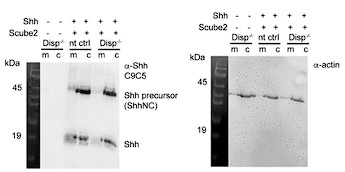
Please see our reply to reviewer 1 above. The Cell Signaling Technology C9C5 anti-Shh antibody used in our study is highly specific against Shh. We also carefully characterized the C9C5 antibody before any of the experiments shown in our work had been initiated. We never observed any unspecific C9C5 reactivity that otherwise would – of course – have prevented us from switching to this antibody from the AF464 antibodies that we had previously used. Consistent C9C5 antibody specificity is evident from the representative example shown below that was recently produced in our lab: no cellular proteins or TCA-precipitated serum-depleted media components from mock-transfected cells (left two lanes) react with C9C5.
Author response image 2.
Top left: C9C5 detects the cellular 45kDa Shh precursor and the 19 kDa signaling-active protein. No unspecific signals are detected in untransfected cells and supernatants of such cells (left two lanes). Right: Loading control on the stripped blot.
- “Clean up how the data are normalized for quantification”
Please see our reply to reviewer 1 above. Normalization has been changed for the indicated figures. We also repeated qPCR analyses and added new ones to the manuscript that include required controls. We also changed figure outlines in accordance with the request.
- “The issue of a non-specific band of this Shh antibody is critical”
Please see our replies above. In our hands, unspecific C9C5 antibody binding was never observed.
- “Regarding experimental rigor, I would add that the HPLC … should just show the real data points”
We agree and added individual data points to our revised manuscript.
Recommendations for the authors:
- I would like to see the controls in the same figure with the experimental results.
We show antibody specificity controls together with released Shh in Fig. S1.
- Figure 2 confirms previously published results. It was shown in PMC5811216 that Disp processing by furin is required for Shh release from producing cells.
Indeed, it was shown that furin processing of Disp increases Shh release (supposedly together with lipids), but we show here that furin-activated Disp specifically mediates proteolytic Shh shedding and loss of lipids – which is not the same. Indeed, we show this finding because we interpret it the other way around: Because it is known that furin activation of Disp increases Shh release by some means (PMC5811216), our observation that furin-mediated Disp activation specifically increases Shh shedding independently supports our model.
- Figure 3: it is stated that there is no increase in Shh release into the media…
We removed this statement.
- Figure S5: Scale bars are missing.
We added scale bars to the figures.
- Figure 4: A direct comparison between wt Shh and C25A conditioned media for qPCR is needed.
We agree and repeated all experiments. Results confirm our previous findings and are shown in revised Fig. 4 and in Fig. S5.
- What other components can be examined in addition to ApoA1 as a marker for HDL? Why is the Shh peak shifted to the left? What about exovesicles?
We also detected ApoE4, a mobile lipoprotein present on expanding (large) HDL (Figs. 5, 6, Figs S6, 7) 14. We also used density gradient centrifugation to support the Shh/HDL association. Regarding the leftwards Shh size shift relative to the major HDL peak in SEC, please refer to our explanation above – if loaded with Shh, a size increase of the respective HDL subfraction is expected. Finally, we did not test the role of exovesicles in our assays. However, due to their large size (60-120nm, HDL 7-12 nm), Shh associated with exovesicles should have eluted in the void volume of our gel filtration column. This we never observed.
- Why is osteoblast differentiation used?
C3H10T1/2 osteoblast differentiation is strongly driven by Ihh and Shh activity and is established as a sensitive and robust assay. Still, following this reviewer’s advice, we conducted qPCR assays on these cells and in addition on NIH3T3 cells to support our findings.
Finally, we corrected all minor mistakes regarding spelling and figure labeling. We also improved the readability of the revised manuscript, as suggested by reviewer 2.
References
Gallet A, Ruel L, Staccini-Lavenant L, Therond PP. Cholesterol modification is necessary for controlled planar long-range activity of Hedgehog in Drosophila epithelia. Development 133, 407-418 (2006).
Porter JA, et al. Hedgehog patterning activity: role of a lipophilic modification mediated by the carboxy-terminal autoprocessing domain. Cell 86, 21-34 (1996).
Lewis PM, et al. Cholesterol modification of sonic hedgehog is required for long-range signaling activity and effective modulation of signaling by Ptc1. Cell 105, 599-612 (2001).
Huang X, Litingtung Y, Chiang C. Region-specific requirement for cholesterol modification of sonic hedgehog in patterning the telencephalon and spinal cord. Development 134, 2095-2105 (2007).
Lee JD, et al. An acylatable residue of Hedgehog is differentially required in Drosophila and mouse limb development. Dev Biol 233, 122-136 (2001).
Corrales JD, Rocco GL, Blaess S, Guo Q, Joyner AL. Spatial pattern of sonic hedgehog signaling through Gli genes during cerebellum development. Development 131, 5581-5590 (2004).
Cordero D, Marcucio R, Hu D, Gaffield W, Tapadia M, Helms JA. Temporal perturbations in sonic hedgehog signaling elicit the spectrum of holoprosencephaly phenotypes. J Clin Invest 114, 485-494 (2004).
Dessaud E, et al. Interpretation of the sonic hedgehog morphogen gradient by a temporal adaptation mechanism. Nature 450, 717-720 (2007).
Garcia-Morales D, Navarro T, Iannini A, Pereira PS, Miguez DG, Casares F. Dynamic Hh signalling can generate temporal information during tissue patterning. Development 146, (2019).
Harfe BD, Scherz PJ, Nissim S, Tian H, McMahon AP, Tabin CJ. Evidence for an expansion-based temporal Shh gradient in specifying vertebrate digit identities. Cell 118, 517-528 (2004).
Nahmad M, Stathopoulos A. Dynamic interpretation of hedgehog signaling in the Drosophila wing disc. PLoS Biol 7, e1000202 (2009).
Ehring K, et al. Conserved cholesterol-related activities of Dispatched 1 drive Sonic hedgehog shedding from the cell membrane. J Cell Sci 135, (2022).
Coupland CE, et al. Structure, mechanism, and inhibition of Hedgehog acyltransferase. Mol Cell 81, 5025-5038 e5010 (2021).
Sacks FM, Jensen MK. From High-Density Lipoprotein Cholesterol to Measurements of Function: Prospects for the Development of Tests for High-Density Lipoprotein Functionality in Cardiovascular Disease. Arterioscler Thromb Vasc Biol 38, 487-499 (2018).
-
eLife assessment
The manuscript presents an analysis of different factors that are required for release of the lipid-linked morphogen Shh from cellular membranes., which will be useful in the field. The evidence is still incomplete as experiments rely on over-expression of Shh in a single cell line and are sometimes of a correlative nature. The study confirms and extends previous findings and will be of interest to developmental biologists who work on Hedgehog signaling.
-
Reviewer #1 (Public Review):
This manuscript presents a model in which combined action of the transporter-like protein DISP and the sheddases ADAM10/17 promote shedding of a mono-cholesteroylated Sonic Hedgehog (SHH) species following cleavage of palmitate from the dually lipidated precursor ligand. The authors propose that this leads to transfer of the cholesterol-modified SHH to HDL for solubilization. The minimal requirement for SHH release by this mechanism is proposed to be the covalently linked cholesterol modification because DISP could promote transfer of a cholesteroylated mCherry reporter protein to serum HDL. The authors used an in vitro system to demonstrate dependency on DISP/SCUBE2 for release of the cholesterol modified ligand. These results confirm previously published results from other groups (PMC3387659 and PMC3682496).
A strength of the work is the use of a bicistronic SHH-Hhat system to consistently generate dually-lipidated ligand to determine the quantity and lipidation status of SHH released into cell culture media.
Key shortcomings include the unusual normalization strategies used for many experiments and the lack of quantification/statistical analyses for several experiments. Due to these omissions, it is difficult to conclude that the data justify the conclusions. The significance of the data provided is overstated because many of the presented experiments confirm/support previously published work. The study provides a modest advance in understanding of the complex issue of SHH membrane extraction.
-
Reviewer #2 (Public Review):
Ehring et al. analyze contributions of Dispatched, Scube2, serum lipoproteins and Sonic Hedgehog lipid modifications to the generation of different Shh release forms. Hedgehog proteins are anchored in cellular membranes by N-terminal palmitate and C-terminal cholesterol modifications, yet spread through tissues and are released into the circulation. How Hedgehog proteins can be released, and in which form, remains controversial. The authors systematically dissect contributions of several previously identified factors, and present evidence that Disp, Scube2 and lipoproteins concertedly act to release a novel Shh variant that is cholesterol-modified but not palmitoylated. The results provide new insights into the function of Disp and Scube2 in Hedgehog release. The findings concerning the function of …
Reviewer #2 (Public Review):
Ehring et al. analyze contributions of Dispatched, Scube2, serum lipoproteins and Sonic Hedgehog lipid modifications to the generation of different Shh release forms. Hedgehog proteins are anchored in cellular membranes by N-terminal palmitate and C-terminal cholesterol modifications, yet spread through tissues and are released into the circulation. How Hedgehog proteins can be released, and in which form, remains controversial. The authors systematically dissect contributions of several previously identified factors, and present evidence that Disp, Scube2 and lipoproteins concertedly act to release a novel Shh variant that is cholesterol-modified but not palmitoylated. The results provide new insights into the function of Disp and Scube2 in Hedgehog release. The findings concerning the function of lipoproteins and cholesterol in Hedgehog release are largely confirmatory (PMID 23554573, 20685986). However, in light of the multitude of competing models for Hedgehog release, the present study is a valuable contribution that provides further insights into the relevance of lipoproteins in this process.
A novel and surprising finding of the present study is the differential removal of Shh N- or C-terminal lipid anchors depending on the presence of HDL and/or Disp. In particular, the identification of a non-palmitoylated but cholesterol-modified Shh variant that associates with lipoproteins is potentially important. The authors use RP-HPLC and defined controls to assess the properties of processed Shh forms, but their precise molecular identify remains to be defined. A caveat is the strong reliance on over-expression of Shh in a single cell line. The authors detect Shh variants that are released independently of Disp and Scube2 in secretion assays, which however are excluded from interpretation as experimental artifacts. Thus, it would be important to demonstrate key findings in cells that secrete Shh endogenously.
-
-
Author Response
Reviewer #1 (Public Review):
- “It is unclear whether new in vivo experiments were conducted for this study”.
All in vivo experiments shown were conducted independently by new researchers in the lab, using the original fly stocks. This will be more clearly stated in the revised supplement. The aim of repeating the experiments was to directly compare the consequences of impaired N- and C-terminal shedding side-by-side in two Hh-dependent developmental systems.
- “A critical shortcoming of the study is that experiments showing Shh secretion/export do not include a Shh(-) control condition. Without demonstration that the bands analyzed are specific for Shh(+) conditions, these experiments cannot be appropriately evaluated”.
C9C5 antibody reactivity and specificity is shown below, and this control will be added to the …
Author Response
Reviewer #1 (Public Review):
- “It is unclear whether new in vivo experiments were conducted for this study”.
All in vivo experiments shown were conducted independently by new researchers in the lab, using the original fly stocks. This will be more clearly stated in the revised supplement. The aim of repeating the experiments was to directly compare the consequences of impaired N- and C-terminal shedding side-by-side in two Hh-dependent developmental systems.
- “A critical shortcoming of the study is that experiments showing Shh secretion/export do not include a Shh(-) control condition. Without demonstration that the bands analyzed are specific for Shh(+) conditions, these experiments cannot be appropriately evaluated”.
C9C5 antibody reactivity and specificity is shown below, and this control will be added to the revised manuscript. We established the C9C5 immunoblotting protocol – and generated the blot shown in Author Response Image 1 - before any of the experiments in the manuscript were started. The immunoblot clearly shows Shh specificity similar to that of R&D AF464 anti-Shh antibodies that were previously used in the lab. The immunoblot also shows that both antibodies detect the same Shh signals in media, that C9C5 is more sensitive, and that AF464 and C9C5 detect 5E1-IP’d dual-lipidated and monolipidated soluble Shh equally well. Also note that, in our hands, C9C5 is highly specific: this antibody detects N-truncated C25S;Δ26-35Shh of increased electrophoretic mobility, but does not cause unspecific signals above or below, even if the blot is strongly overexposed (as shown here). Specific Shh detection by C9C5 is also discussed in our response to editor’s comments below.
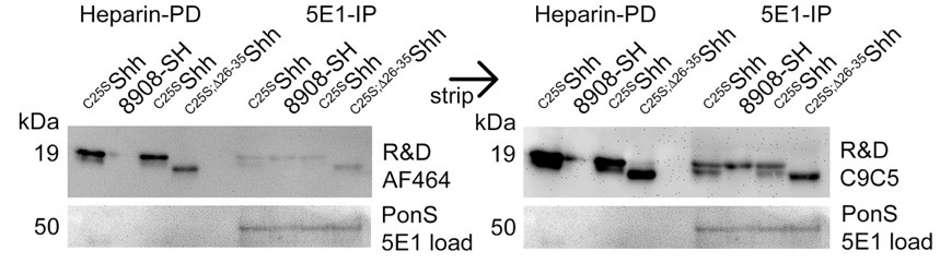
Cells were transfected with constructs encoding full-length C25SShh or truncated C25S;Δ26-35Shh, and proteins in serum-containing media were 5E1 immunoprecipitated or concentrated by heparin-sepharose pulldown. Dual-lipidated R&D 8908-SH was dissolved in the same medium and subjected to the same 5E1 immunoprecipitation or heparin pulldown. The blot was incubated with antibody AF464 and (after stripping) with antibody C9C5. Immunoblot analysis revealed high specificity of both antibodies and also revealed poor interactions of dual-lipidated 8908-SH with highly charged heparin.
- “A stably expressing Shh/Hhat cell line would reduce condition to condition and experiment to experiment variability”.
We fully agree with this reviewer and therefore aimed to establish stable Hhat expressing cell lines several years ago. However, stable Hhat expression eliminated transfected cells after several passages, or cells gradually ceased to express Hhat, preventing us to establish a stable line despite several attempts and tried strategies. For this reason, we established transient co-expression of Shh/Hhat from the same mRNA to at least eliminate variability between relative Shh/Hhat expression levels and to assure complete Shh palmitoylation in our assays.
- “Unusual normalization strategies are used for many experiments, and quantification/statistical analyses are missing for several experiments”.
This comment refers to data shown in Fig. 3 (here, no quantification of Scube2 function in Disp-/- cells had been conducted) and to qPCR data shown in Fig. 4 (here, Shh and C25AShh were compared only indirectly via dual-lipidated R&D 8908-SH, but not directly in a side-by-side experiment, and Shh variants with an N-terminal alanine or a serine were directly compared). We agree with the reviewer and therefore currently repeat qPCR assays and quantify blots to eliminate these technical shortcomings from the final manuscript.
- “The study provides a modest advance in the understanding of the complex issue of Shh membrane extraction”
Our investigation identified unexpected links between Disp as a furin-activated Hh exporter, sheddase-mediated Shh release, Scube2-mediated Shh release and lipoprotein-mediated Hh transport – established modes indeed but with no previously established direct connections – that increase their relevance. We also identified a previously unknown N-processed Shh variant attached to lipoproteins and show that Disp/Scube2 function absolutely requires lipoproteins. Therefore, although we do agree that our findings are confirmatory for the above modes, they also provide new mechanistic insight and challenge the currently dominating model of Disp-mediated hand-over of dual-lipidated Hh to Scube2 chaperones (this model does not predict a role for lipoprotein particles but for both Shh lipids in signaling, for a recent discussion, see PMID 36932157). Our findings suggest an answer to the intensely debated question of whether Disp/Ptch extract cholesterol from the outer or inner plasma membrane leaflet, and suggest that N-palmitate is dispensable for signaling of lipoprotein-associated Shh to Ptch receptors. Finally, we note that previous in vivo studies in flies often relied on Hh overexpression in the fat body, raising questions on their physiological relevance. Our in vivo analyses of Hh function in wing- and eye discs are more physiologically relevant and can explain the previously reported presence of non-lipidated bioactive Hh in disc tissue (PMID: 23554573).
Reviewer #2 (Public Review):
- “However, the results concerning the roles of lipoproteins and Shh lipid modifications are largely confirmatory of previous results, and molecular identity/physiological relevance of the newly identified Shh variant remain unclear”.
Regarding the confirmatory aspects of our work, please also refer to our response to reviewer 1. In addition, we would like to reply that our unbiased experimental approach was designed to challenge the model of Shh shedding by testing whether established Shh release regulators affect it (e.g. support it) or not. As described in our work, Disp, Scube2 and lipoproteins all contribute to increased shedding (which is new), that Disp function depends on lipoprotein presence (also new), and that lipoproteins modify the outcome of Shh shedding (dual Shh shedding versus N-shedding and lipoprotein association), which is also new.
Regarding physiological relevance, we would like to reply that our finding that artificially generated monolipidated variants (C25SShh and ShhN) solubilize in uncontrolled manner from producing cells can explain previously observed, highly variable gain-of-function or loss-of-function phenotypes upon their overexpression in vivo 1, 2, 3, 4, 5. Our data is also supported by the observed presence of variably lipidated Shh/Hh variants in vivo 6, and the in vivo observation that complete removal of Scube activity in zebrafish embryos phenocopies a complete loss of Hh function that is bypassed by increased ligand expression - and even results in wild-type-like ectopic Shh target gene expression 7. The in vivo observations are compatible with our data but are incompatible with proposed alternative models of Scube-mediated dual-lipidated Shh extraction and continued Shh/Scube association to allow for morphogen transport.
- “Thus, it would be important to demonstrate key findings in cells that secrete Shh endogenously”.
Experimental data shown in Fig. S8B demonstrates that en-controlled expression of sheddase-resistant Hh variants blocks endogenous Hh function in the same wing disc compartment. To our knowledge, this assay is the most physiologically relevant test of the mechanism of Disp-mediated Hh release. Still, we have now started to analyze Hh from Drosophila disc tissue biochemically and hope that we can include our findings in the final manuscript.
- “The authors could use an orthogonal approach, optimally a demonstration of physical interaction, or at least fractionation by a different parameter”.
We agree with this reviewer’s assessment and are currently in the process to establish co-IP and density gradient conditions to test physical HDL/Shh interactions. The results will be included in the final version of record.
-
eLife assessment
The authors present a useful analysis of different factors that are required for release of the lipid-linked morphogen Shh from cellular membranes. The evidence is currently still incomplete as experiments rely on over-expression of Shh in a single cell line and are sometimes of a correlative nature. The study confirms and extends previous findings and will be of interest to developmental biologists who work on Hedgehog signaling.
-
Reviewer #1 (Public Review):
This manuscript presents a model in which combined action of the transporter-like protein DISP and the sheddases ADAM10/17 promote shedding of a mono-cholesteroylated Sonic Hedgehog (SHH) species following cleavage of palmitate from the dually lipidated precursor ligand. The authors propose that this leads to transfer of the cholesterol-modified SHH to HDL for solubilization. The minimal requirement for SHH release by this mechanism is proposed to be the covalently linked cholesterol modification because DISP could promote transfer of a cholesteroylated mCherry reporter protein to serum HDL. The authors used an in vitro system to demonstrate dependency on DISP/SCUBE2 for release of the cholesterol modified ligand. These results confirm previously published results from other groups (PMC3387659 and …
Reviewer #1 (Public Review):
This manuscript presents a model in which combined action of the transporter-like protein DISP and the sheddases ADAM10/17 promote shedding of a mono-cholesteroylated Sonic Hedgehog (SHH) species following cleavage of palmitate from the dually lipidated precursor ligand. The authors propose that this leads to transfer of the cholesterol-modified SHH to HDL for solubilization. The minimal requirement for SHH release by this mechanism is proposed to be the covalently linked cholesterol modification because DISP could promote transfer of a cholesteroylated mCherry reporter protein to serum HDL. The authors used an in vitro system to demonstrate dependency on DISP/SCUBE2 for release of the cholesterol modified ligand. These results confirm previously published results from other groups (PMC3387659 and PMC3682496). In vivo support for these activities is provided by data from previously published studies from this group. It is unclear whether new in vivo experiments were conducted for this study.
A strength of the work is the use of a bicistronic SHH-Hhat system to consistently generate dually-lipidated ligands to determine the quantity and lipidation status of SHH released into cell culture media.
A critical shortcoming of the study is that the experiments showing SHH secretion/export by western blot of media fractions do not include a SHH(-) control condition. This is an essential control because SHH media blots can be dirty. Without demonstration that the bands being analyzed are specific for SHH(+) conditions, these experiments cannot be appropriately evaluated. Further, it appears that SHH is transiently transfected/expressed for each experimental condition. A stably expressing SHH/HHAT cell line would reduce condition to condition and experiment to experiment variability. Unusual normalization strategies are used for many experiments, and quantification/statistical analyses are missing for several experiments. Due to these shortcomings, the data do not justify the conclusions. The significance of the data provided is overstated because many of the presented experiments confirm/support previously published work. The study provides a modest advance in the understanding of the complex issue of SHH membrane extraction.
-
Reviewer #2 (Public Review):
Ehring et al. analyze contributions of Dispatched, Scube2, serum lipoproteins and Sonic Hedgehog lipid modifications to the generation of different Shh release forms. Hedgehog proteins are anchored in cellular membranes by N-terminal palmitate and C-terminal cholesterol modifications, yet spread through tissues and are released into the circulation. How Hedgehog proteins can be released, and in which form, remains unclear. The authors systematically dissect contributions of several previously identified factors, and present evidence that Disp, Scube2 and lipoproteins concertedly act to release a novel Shh variant that is cholesterol-modified but not palmitoylated. The systematic analysis of key factors that control Shh release is a commendable effort and helps to reconcile apparently disparate models. …
Reviewer #2 (Public Review):
Ehring et al. analyze contributions of Dispatched, Scube2, serum lipoproteins and Sonic Hedgehog lipid modifications to the generation of different Shh release forms. Hedgehog proteins are anchored in cellular membranes by N-terminal palmitate and C-terminal cholesterol modifications, yet spread through tissues and are released into the circulation. How Hedgehog proteins can be released, and in which form, remains unclear. The authors systematically dissect contributions of several previously identified factors, and present evidence that Disp, Scube2 and lipoproteins concertedly act to release a novel Shh variant that is cholesterol-modified but not palmitoylated. The systematic analysis of key factors that control Shh release is a commendable effort and helps to reconcile apparently disparate models. However, the results concerning the roles of lipoproteins and Shh lipid modifications are largely confirmatory of previous results, and molecular identity/physiological relevance of the newly identified Shh variant remain unclear.
The authors conclude that an important result of the study is the identification of HDL as a previously overlooked serum factor for secretion of lipid-linked Shh (p15, l24-25). This statement should be removed. A detailed analysis of Shh release on human lipoproteins was reported previously, including contributions of the major lipoprotein classes, in cells that endogenously express Shh, in human plasma and for Shh variants lacking palmitate and/or cholesterol modifications (PMID 23554573). The involvement of Disp is also not unexpected: the importance of Dips for release of cholesterol-modified Shh is well established, as is the essential function of Drosophila Disp for formation of lipoprotein-associated hemolymph Hh. A similar argument can be made for the sufficiency of sterol modification for lipoprotein association. The authors point out that GFP insertion at the C-terminus of the N-terminal Shh domain does not abrogate function. Perhaps more relevant, an mCherry-sterol that was generated using a similar strategy as in the present study associates with Drosophila lipoproteins (PMID 20685986).
A novel and surprising finding of the present study is the differential removal of Shh N- or C-terminal lipid anchors depending on the presence of HDL and/or Disp. In particular, the identification of a non-palmitoylated but cholesterol-modified Shh variant that associates with lipoproteins is potentially important. However, the significance of this result could be substantially improved in two ways: 1) The molecular properties of the processed Shh variants are unclear - incorporation of palmitate/cholesterol and removal of peptides were not directly demonstrated. This is particularly relevant for the N-terminus, as the signaling activity of non-palmitoylated Hedgehog proteins is controversial. A decrease in hydrophobicity is no proof for cleavage of palmitate, this could also be due to addition of a shorter acyl group. 2) All experiments rely on over-expression of Shh in a single cell line. The authors point out that co-overexpression of Hhat is important to ensure Shh palmitoylation, but the same argument could be made for any other protein that acts in Shh release, such as Disp or a plasma membrane sheddase. The authors detect Shh variants that are released independently of Disp and Scube2 in secretion assays, which however are excluded from interpretation as experimental artifacts. Thus, it would be important to demonstrate key findings in cells that secrete Shh endogenously.
The co-fractionation of Shh and ApoA1 in serum-containing media is not convincing (Fig. 4C), as the two proteins peak at different molecular weights. To support their conclusion, the authors could use an orthogonal approach, optimally a demonstration of physical interaction, or at least fractionation by a different parameter (density). On a technical note, all chromatography results are presented as stylized graphs. Please include individual data points.
-
