SPEx: Compartment-Resolved Proteomics via Expansion Microscopy–Guided Microdissection
Curation statements for this article:-
Curated by eLife

eLife Assessment
Franziscus et al. present an important study introducing SPEx, an elegant, broadly accessible approach to subcellular spatial proteomics that combines cell expansion, laser microdissection, and mass spectrometry to enable protein profiling within defined subcellular regions. The authors provide convincing evidence that this flexible integration of established techniques offers a robust, practical, and cost-effective strategy for compartment-resolved proteomic analysis. The method is appropriately validated across multiple subcellular compartments and complementary workflows, demonstrating its ability to recover known protein markers while identifying previously uncharacterised components. The study also provides a characterisation of the method's capabilities and limitations. Overall, SPEx is likely to be of broad interest to cell biologists seeking to map organelle composition.
This article has been Reviewed by the following groups
Listed in
- Evaluated articles (eLife)
Abstract
Cells contain different organelles and compartments that are essential for cellular function and life. These organelles and compartments need to communicate to assess cellular state in a changing environment, adapt to the new situation, and to ensure functionality and homeostasis. Moreover, organization and communication differ between cell types. However, our knowledge about these changes is still rather scarce. Subcellular spatial proteomics aims to fill this knowledge gap. While proximity labeling techniques represent a great advance, they do not provide precise spatial resolution. To overcome this limitation, we developed SPEx (Subcellular spatial Proteomics coupled to Expansion), in which we first expand cells about 10-fold, laser micro-dissect regions of interests and then perform mass spectrometry-based proteomics on these samples. We demonstrate the effectiveness of SPEx by determining the proteome of the Golgi, the nucleus and nucleoli. Importantly, we also identify novel components of these organelles. Combining inexpensive, already existing technologies makes SPEx readily usable by the wider scientific community.
Article activity feed
-

eLife Assessment
Franziscus et al. present an important study introducing SPEx, an elegant, broadly accessible approach to subcellular spatial proteomics that combines cell expansion, laser microdissection, and mass spectrometry to enable protein profiling within defined subcellular regions. The authors provide convincing evidence that this flexible integration of established techniques offers a robust, practical, and cost-effective strategy for compartment-resolved proteomic analysis. The method is appropriately validated across multiple subcellular compartments and complementary workflows, demonstrating its ability to recover known protein markers while identifying previously uncharacterised components. The study also provides a characterisation of the method's capabilities and limitations. Overall, SPEx is likely to be of broad …
eLife Assessment
Franziscus et al. present an important study introducing SPEx, an elegant, broadly accessible approach to subcellular spatial proteomics that combines cell expansion, laser microdissection, and mass spectrometry to enable protein profiling within defined subcellular regions. The authors provide convincing evidence that this flexible integration of established techniques offers a robust, practical, and cost-effective strategy for compartment-resolved proteomic analysis. The method is appropriately validated across multiple subcellular compartments and complementary workflows, demonstrating its ability to recover known protein markers while identifying previously uncharacterised components. The study also provides a characterisation of the method's capabilities and limitations. Overall, SPEx is likely to be of broad interest to cell biologists seeking to map organelle composition.
-
Reviewer #1 (Public review):
Summary:
The authors present a novel approach to subcellular spatial proteomics by combining laser microdissection with expansion microscopy and LC-MS/MS analysis (SPEx). They implement two different workflows for LMD and LC-MS/MS quantification:
(1)The standard approach in which an area of interest is cut out by LMD, subjected to proteomics analysis and compared to the rest of the cell without the dissected ROI.
(2) The subtraction approach in which ROIs are removed and the remaining cellular material is compared to samples containing both the surrounding material and the ROI.
The authors assess the technique by applying it to subcellular targets of various size, volume and protein compositions such as the nucleus, nucleoli and Golgi. They demonstrate that SPEx can identify proteins enriched or reduced in …
Reviewer #1 (Public review):
Summary:
The authors present a novel approach to subcellular spatial proteomics by combining laser microdissection with expansion microscopy and LC-MS/MS analysis (SPEx). They implement two different workflows for LMD and LC-MS/MS quantification:
(1)The standard approach in which an area of interest is cut out by LMD, subjected to proteomics analysis and compared to the rest of the cell without the dissected ROI.
(2) The subtraction approach in which ROIs are removed and the remaining cellular material is compared to samples containing both the surrounding material and the ROI.
The authors assess the technique by applying it to subcellular targets of various size, volume and protein compositions such as the nucleus, nucleoli and Golgi. They demonstrate that SPEx can identify proteins enriched or reduced in ROIs.
Strengths:
The general, relatively easy and inexpensive nature of this approach, as well as its potential applicability to many cell types and subcellular areas of interest provides an exciting alternative to subcellular fractionation, native immunoprecipitation or genetically encoded proximity labeling constructs. Moreover, by visually selecting ROIs for subsequent analysis, subcellular context or organelle morphology can be taken into account as discussed by the authors in the discussion section.
Weaknesses:
The authors addressed our concerns and technical questions in revision.
A main limitation of the current method is that it relies on isolation of structures of interest in 2D. The authors discuss some alternative approaches that might allow future access to more 3D target isolation.
There remain several unexplored minor extensions of the technical development of the method, including comparing the proteomes of organelles that are visually distinct, and applying different anchoring procedures to try to increase protein retention.
-
Reviewer #2 (Public review):
Summary:
This study introduces SPEx, an approach that combines physical expansion, imaging-guided microdissection, and mass spectrometry for compartment-resolved proteomic analysis. Using the nucleus, nucleolus, and Golgi apparatus as examples, the authors demonstrate that SPEx can identify proteins enriched or depleted in defined subcellular regions.
Strengths:
A major strength of the method is its accessibility and flexibility. It combines established technologies without requiring genetic manipulation or specialized proximity-labeling strategies, while direct imaging-based selection allows regions of interest to be defined according to their spatial and morphological context. The complementary comparative strategies used in the study provide convincing support for compartment-specific protein enrichment …
Reviewer #2 (Public review):
Summary:
This study introduces SPEx, an approach that combines physical expansion, imaging-guided microdissection, and mass spectrometry for compartment-resolved proteomic analysis. Using the nucleus, nucleolus, and Golgi apparatus as examples, the authors demonstrate that SPEx can identify proteins enriched or depleted in defined subcellular regions.
Strengths:
A major strength of the method is its accessibility and flexibility. It combines established technologies without requiring genetic manipulation or specialized proximity-labeling strategies, while direct imaging-based selection allows regions of interest to be defined according to their spatial and morphological context. The complementary comparative strategies used in the study provide convincing support for compartment-specific protein enrichment and depletion.
An important limitation is the lack of depth resolution during microdissection, which can introduce contributions from material above or below the selected region, particularly for small structures. Appropriate comparative controls are therefore important for interpreting SPEx measurements. The current study is also primarily a proof of principle in a single cultured cell type, and its ability to capture changes across biological states or perturbations remains to be established.
Overall, the study provides convincing evidence that SPEx is a useful and flexible addition to the spatial proteomics toolbox, with particular potential for applications requiring direct image-based selection of subcellular regions.
-
Reviewer #3 (Public review):
Franziscus et al. describe an elegant approach for spatially specific proteome analysis. To achieve this, they expand fixed cells and subsequently use a laser to micro-dissect a region of interest, which is then analyzed by MS.
They demonstrate the effectiveness of their approach by analyzing nucleus, nucleolus and the Golgi and benchmark their hits against previous dataset for these organelles.
The manuscript is very well written and nicely guides the reader through the applied methods. The presented data is convincing and I do not see the need for additional experimental verification of the protocol. The only minor concern is the novelty of the method and the presentation. A combination of expansion, laser microdissection and proteomics has been applied in the past (PMID: 36450705, PMID: 39477916). In the …
Reviewer #3 (Public review):
Franziscus et al. describe an elegant approach for spatially specific proteome analysis. To achieve this, they expand fixed cells and subsequently use a laser to micro-dissect a region of interest, which is then analyzed by MS.
They demonstrate the effectiveness of their approach by analyzing nucleus, nucleolus and the Golgi and benchmark their hits against previous dataset for these organelles.
The manuscript is very well written and nicely guides the reader through the applied methods. The presented data is convincing and I do not see the need for additional experimental verification of the protocol. The only minor concern is the novelty of the method and the presentation. A combination of expansion, laser microdissection and proteomics has been applied in the past (PMID: 36450705, PMID: 39477916). In the manuscript one of these studies is cited, though it does not become clear that this approach is already described. However, Franziscus et al. describe the approach better and makes it more accessible to the reader, especially since the other studies described this methodology in combination with tissue expansion and not in combination with single cell expansion as it is done here. I would ask the authors to be clearer in the introduction with what others have already done and what is there contribution here. In general, I am convinced that the community will benefit from the presented protocol to analyze organelle proteomics in detail.
Comments on revised version.
I had only minor issues with the manuscript, which have now been addressed.
-
Author response:
The following is the authors’ response to the original reviews.
Reviewer #1 (Public review):
Summary:
The authors present a novel approach to subcellular spatial proteomics by combining laser microdissection with expansion microscopy and LC-MS/MS analysis (SPEx). They implement two different workflows for LMD and LC-MS/MS quantification:
(1)The standard approach, where an area of interest is cut out by LMD, subjected to proteomics analysis, and compared to the rest of the cell without the dissected ROI.
(2) The subtraction approach, where ROIs are removed, and the remaining cellular material is compared to samples containing both the surrounding material and the ROI.
The authors assess the technique by applying it to subcellular targets of various sizes, volumes, and protein compositions such as the nucleus, …
Author response:
The following is the authors’ response to the original reviews.
Reviewer #1 (Public review):
Summary:
The authors present a novel approach to subcellular spatial proteomics by combining laser microdissection with expansion microscopy and LC-MS/MS analysis (SPEx). They implement two different workflows for LMD and LC-MS/MS quantification:
(1)The standard approach, where an area of interest is cut out by LMD, subjected to proteomics analysis, and compared to the rest of the cell without the dissected ROI.
(2) The subtraction approach, where ROIs are removed, and the remaining cellular material is compared to samples containing both the surrounding material and the ROI.
The authors assess the technique by applying it to subcellular targets of various sizes, volumes, and protein compositions such as the nucleus, nucleoli, and Golgi. They demonstrate that SPEx can identify proteins enriched or reduced in ROIs.
Strengths:
The broad, relatively easy, and inexpensive applicability of this approach to potentially many cell types and subcellular areas of interest provides an exciting alternative to subcellular fractionation, native immunoprecipitation, or genetically encoded proximity labeling constructs. Moreover, by visually selecting ROIs for subsequent analysis, subcellular context or organelle morphology can be taken into account, as discussed by the authors in the discussion section.
Weaknesses:
While strongly supporting the sharing of this approach, we have a number of comments and questions that will improve the impact of the manuscript:
We thank the reviewer for the careful evaluation of our manuscript and the generally positive assessment.
(1) General:
(a) The manuscript would benefit from restructuring and language revision. In its current form, the writing is sometimes dense and verbose (in particular, the Results section). This makes it difficult to follow the authors' arguments.
We improved readability and clarity in the revised manuscript. We added subheadings to facilitate reading.
(b) The authors mention the possibility of selecting organelles based on morphology. This is left for the discussion, but it seems like a missed opportunity - the authors could compare individual organelles in different morphological states, e.g., connected vs. fragmented mitochondria.
The authors agree with the reviewers’ assessment that investigating proteome of organelles based on morphology or cellular state is an exciting application of SPEx. While we plan experiments along this line in the future, we think that these experiments are beyond the scope of this manuscript, which is meant to describe the method and its general usefulness.
(2) Technical:
(a) Why do the authors strive and optimize for a 10x expansion factor? Is SPEx compatible with a more standard 4x expansion, as e.g., used in the classic U-ExM approach (https://www.nature.com/articles/s41592-018-0238-1)? This could be added to the discussion.
We aimed for 10x expansion solely because our ultimate goal is to cut out very small structures. 10x expansion results in a 100x increase in area, while in U-ExM the area increase would only by 16x. Thus, this makes a huge difference for small structures such as nucleoli. We did not assess the compatibility with U-ExM. Still, we performed pilots with just 4x expansion (using TREx) in the early stages of optimization. We were able to isolate single cells and obtain similar protein coverage as with 10x expansion. We further clarified our motivation to use 10x expansion in the discussion.
(b) The U-ExM approach shows improved ultrastructural preservation when using 3%FA with 0.1% glutaraldehyde fixation (GA). Is SPEx compatible with the use of low amounts of GA for fixation?
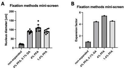
We tried different fixation methods in the early stages of this study (where expansion was not yet close to 10x). We saw a mild negative effect of GA on the expansion factor, so we avoided it in the later experiments since it also did not seem necessary to preserve the structure of our organelles of interest. However, the use of GA would generally be compatible with SPEx, potentially at the cost of a mild negative effect on expansion factor (see Author response image 1) and proteome coverage. We discussed the use of GA in the discussion section.
Author response image 1.
Fixation methods mini-screen. Cells were fixed with the indicated reagents for 10 minutes at 37°C. After TREx expansion, the diameter of the nucleus was measured (A) and the resulting expansion factor compared to the non-expanded control was determined (B).
Related to the above, was the anchoring efficiency reduced only to achieve a 10x expansion factor or does this additionally affect the proteome coverage?
We solely lowered the anchoring to increase expansion factors. In earlier pilots we performed proteomic analysis on samples that were just expanded 4x using standard TREx expansion (also using the original anchoring strategy from the TREx publication, consisting of 0.2 mg/ml AcX for overnight at RT). We presented the results of this pilot in Fig S1A. We still detected over 2,000 proteins from 10 cells, a coverage, which is highly similar to what we found in the final experiments (Figure 2F), in which the anchoring was lower for 10x expansion. Based on these data, we think that anchoring (and expansion factor!) has a negligible impact on protein coverage. We clarified this point in the manuscript.
(d) Have the authors considered using alternative anchoring approaches, such as GMA (https://journals.plos.org/plosone/article?id=10.1371/journal.pone.0291506#pone.0291506.s001), which potentially increase the amount of sample retained in the hydrogel, thus allowing for better proteome coverage? This could be added to the discussion.
We did not use alternative anchoring approaches. We modified the TREx protocol to fit our purposes and since this was sufficient, we did not explore alternatives. However, using anchoring approaches, in which higher amounts of sample could be retained in the gel might be beneficial for the proteomics coverage. We will keep this suggestion in mind for future experiments. Thank you for the suggestion!
(e) The limitation of the approach to near-2D samples should be mentioned, and alternative approaches for more 3D samples could be discussed.
The authors agree that SPEx is limited to near-2D samples at this point. We suggest that SPEx is applicable for 3D samples (e.g. in tissues) by performing cryosectioning. TREx has been shown to be compatible with sectioned tissue (Damstra et al., 2022). We discuss this point in the discussion.
(f) How are peptides that are directly anchored to the hydrogel dealt with during LC-MS/MS analysis? Are they excluded, or can they be identified during the spectral search? The latter would allow us to get a deeper structural understanding of how proteins are actually anchored into hydrogels, which so far has not been assessed.
The reviewer raises an interesting point. In general, peptides carrying the anchoring modification are analysed by LC-MS, but we did not include these specific modifications in the database search. Overall, we assumed that the labeling would be low and stochastic and hence should, if at all, only minimally affect the detection of peptides. Nevertheless, in response to the reviewers’ comment, we searched the MS data again for the crosslinking reagent linked to lysine residues. However, we could not get any confident hit for any peptide containing this modification. Since we cannot exclude that the modification precludes the identification of the corresponding peptides, we compared the number peptides generated by trypsin cleavage after arginine and lysine. As the human genome contains similar proportions of both amino acids, one would expect similar numbers for both peptide types being identified. Any modifications of lysine by the anchoring reagent, would prevent tryptic cleavage and thus reduce the number of lysine peptides. As shown in Figure S1C, the number of lysine-terminating is only slightly lower compared to arginine-terminating peptides. Notably, the proteomics results of a different fixed human tissue sample directly extracted by laser capture micro dissection without expansion showed a very similar lysine-to-arginine peptide ratio. This indicates that the large majority of lysine residues is not modified and affected by the hydrogel anchoring. We included these data into the manuscript.
An alternative approach to address this question would be to investigate if the peptide coverage of proteins detected by SPEx is enriched for peptides representing the folded core of proteins as opposed to the surface-exposed regions, which likely get more anchored into the hydrogel.
Because of the negligible amounts of modified peptides, we did not investigate this potential bias of surface-exposed versus folded-core peptides.
(g) Same question regarding peptides with NHS labeling. Can they be identified, or do they just compete for ionization and thus negatively affect coverage and dynamic range of the LC-MS/MS approach?
The reviewer raises a similar point as above for another lysine labeling used during the SPEx protocol. Again, we specifically looked for this modification by re-searching the raw MS data, but still could not identify any peptides, carrying this modification on a lysine residue. Even though we cannot exclude that this rather large modification prevents detection, considering the high number of lysine-terminating peptides in our dataset (see Figure S1 C), we would expect that also this labeling step is stochastic and affects only a minor proportion of the proteins. We discuss the potential NHS labeling in the manuscript.
(h) How are the primary and secondary antibodies affecting the proteomics analysis identified as contaminants?
We thank the reviewer for this comment. Since antibodies bind to proteins in a non-covalent manner, they will be released during the denaturing steps of the protocol. Of course, the antibodies will stay in the sample, be digested and analyzed and could, if very abundant, affect the analysis of the proteins from the samples. To check this possibility, we re-searched the MS data including the sequences of the antibodies used. To our surprise, we could not detect any peptides of these antibodies. This suggests that the concentrations of the antibodies used are much lower than those of the sample proteins and thus should not have any impact on the proteomics results. We interpret this result also as a benefit of our method compared to organellar-IP.
(i) Have the authors observed differences in proteomics coverage of only antibody vs NHS-labeling? Depending on the questions above, could pure antibody-based labeling increase proteomic coverage?
We did not perform this comparative analysis, since we always used NHS dyes. In the experiments presented in this manuscript, NHS dyes allowed easy visualization of the whole cell without the use of antibodies. This NHS staining was essential for this particular setup for sample acquisition. We cut out entire cells, cells lacking the nucleus and cells lacking the Golgi apparatus, which served as critical controls. However, other ways of detecting cell boundaries could be used to avoid NHS staining. As shown above, both, the anchor and NHS labeling are likewise sparse and stochastic. Moreover, we could not detect any impact of the antibody labeling to our results. Thus, we assume that both labeling procedures could be used.
Reviewer #1 (Recommendations for the authors):
(1) Introduce the aberration PPS-membrane.
We apologize for the omission and now explain the PPS abbreviation.
(2) Please state which cells are used already in the first section "Development of the SPEx method" as opposed to only further down in the manuscript.
We apologize for the omission, which has been fixed.
(3) In Figures 2D and E, the authors should also show the hydrated expansion factors for better comparison.
We agree that the inclusion of the hydrated expanded gels allows for better comparison. These data are now included in the manuscript.
Reviewer #2 (Public review):
Summary:
This study introduces a method that combines physical expansion of cells, imaging-guided isolation of defined regions, and protein identification to enable compartment-resolved analysis of protein composition at the subcellular scale. The authors aim to address a central limitation in existing approaches, namely the loss of spatial information during sample preparation or the indirect nature of proximity-based labeling methods. Using several cellular compartments as examples, they demonstrate that their approach can recover compartment-enriched protein sets and identify candidate proteins with previously unassigned localization.
Strengths:
A major strength of this work is the conceptual simplicity and accessibility of the approach. By combining established techniques in a modular way, the method avoids the need for genetic manipulation or specialized labeling strategies, making it broadly adaptable across experimental systems. The ability to directly select regions of interest based on imaging represents a clear advantage over indirect enrichment strategies and allows flexible targeting of both membrane-bound and non-membrane-bound compartments.
The experimental design is also a strong aspect of the study. The use of complementary comparison strategies-analyzing isolated compartments alongside matched "subtracted" controls-provides an internal framework for assessing enrichment and depletion, increasing confidence in spatial assignment. The application of the method across multiple organelles of different sizes and properties demonstrates versatility, and the reported specificity for several compartments is encouraging. In particular, the ability to profile small and biochemically challenging structures highlights a potentially important niche for the approach.
Weaknesses:
Despite these strengths, several methodological limitations constrain the interpretation of the results. The most important relates to spatial accuracy in three dimensions. While lateral resolution is improved through physical expansion, the lack of depth resolution introduces uncertainty regarding contributions from structures above and below the selected region. Although the authors argue that this does not substantially affect specificity, the current evidence is largely indirect, and a more rigorous quantification of potential contamination would strengthen this conclusion.
Quantitative interpretation also remains challenging. Because the measurements reflect total protein abundance rather than local concentration, differences in compartment size and protein density can influence enrichment values, particularly for small structures embedded within larger volumes. This issue is evident in the analysis of smaller compartments and complicates direct comparison across conditions. Additional normalization or modeling would help clarify how to interpret these measurements.
Another limitation concerns variability in the expansion process and its downstream consequences. Differences in expansion factor across samples may affect the definition of regions of interest and introduce variability in sampling, yet the impact of this variability is not fully explored. Similarly, the use of a modified chemical treatment to preserve proteins for downstream analysis is central to the workflow but is not extensively validated with respect to preservation of spatial organization.
While the identification of previously unannotated proteins is an appealing aspect of the study, validation is limited to a small number of examples, and broader support from independent datasets or literature context is lacking. In addition, the study primarily focuses on steady-state measurements in a single cell type, and therefore does not yet demonstrate the ability of the method to capture dynamic or condition-dependent changes in protein localization.
Finally, the positioning of the method relative to existing approaches could be more clearly articulated. Although qualitative comparisons are provided, a more systematic and quantitative benchmarking against alternative strategies would help readers better understand the specific advantages and trade-offs.
We thank the reviewer for the careful evaluation of the manuscript and for the constructive feedback. The reviewer raises valid points, which we addressed in the revised manuscript.
Reviewer #2 (Recommendations for the authors):
Additional experiments and analyses
(1) To strengthen confidence in the spatial specificity of the method, the authors should include experiments or analyses that directly estimate potential cross-compartment contamination. For example, using markers that are known to localize to mutually exclusive cellular regions would allow an empirical assessment of signal carryover. Alternatively, a computational or geometric model could be used to estimate how much material from adjacent regions is expected to contribute to the sampled proteome, given the loss of depth information.
We agree with the reviewer’s concern regarding spatial resolution and potential contaminations resulting from it. We had the same concerns. We have obviously no resolution in z and we detected about 700 proteins (Fig. 5F) in our all samples, likely representing proteins residing above and below of (and potentially next to) the structures we cut out. To overcome this issue, we always compare the proteome of the structure of interest with an appropriate control. We also determined that mitochondria proteins are specifically omitted from our nuclear samples. Moreover, we provide evidence that our analysis methods can be used also to determine proteins that are specifically lost from a structure. Omitting the comparative approach would clearly result in very low specificity and a high number of false positives. However, we argue that comparison to a control largely removes these unspecific contaminations and we think that this is illustrated by our excellent specificity. Nevertheless, to address these concerns further, we have determined the masses of all quantified proteins using iBAQ values. We then summed the masses of all proteins being annotated to a specific organelle and calculated the percentage to the summed masses of all quantified proteins. As expected, we found that in our excised nuclei samples, 75.5% of the protein molecules originate from the nucleus and only 24.5% of protein mass was derived from other regions. Likewise, we found 84.7% of the protein mass in the excised Golgi samples annotated with Golgi or extracellular by GO. This analysis revealed that we detect an expected amount of contaminant proteins derived from other compartments due to the lack of resolution in z, but most of protein mass excised is already organelle-specific. The additional quantification step further enhances assignment accuracy, boosting the overall specificity of our approach. In the case of the nucleoli the mass contribution from known nucleoli proteins is significantly lower (~4%), which would be expected for a structure that only covers a fraction of the cell in z-direction. In the case of such small targets comparison to a fitting control is therefore essential. We added this analysis to the revised version of the manuscript and discuss it in detail (see “SPEx is best used as a comparative approach”). We thank the reviewer for bringing this up!
(2) Given that drying results in compression along the vertical axis while largely preserving the lateral dimensions, it would be valuable to provide a quantitative analysis of how this affects spatial accuracy. This could include comparing marker distributions before and after drying or examining signal distribution across depth.
Even though we were unable to image a particular cell before and after drying (due to the physical properties of the gels and the change in size during drying), we did so for the bulk of cells. We did not observe discernible morphological alterations after drying. The investigated structures (Golgi, nucleus and nucleoli) still looked morphologically similar to expanded cells (before drying) and non-expanded cells. We cannot exclude that the drying process introduced minor distortions of the sample. We would like to point out, however, that our focus is mainly on the spatial proteomics and to a lesser extent on the perfect conservation of the ultrastructure during drying. As long as we can maintain very good spatial separation and are able to cut out individual structures, our method should work just fine.
In addition, trimming or sectioning the expanded samples along the z-axis prior to microdissection could help minimize contributions from structures above and below the region of interest, thereby improving spatial specificity.
We tried to cut expanded samples (monolayer of cells) with a microtome prior to laser dissection. This approach did unfortunately not work. Mounting a monolayer of cells for microtome cutting is non-trivial! The very thin monolayer of cells would need to be perfectly aligned with the plane where the cryo-sectioning takes place. Moreover, the different densities of the hydrogel without cells and the expanded cells in the same hydrogel appeared to be particular challenging during sectioning. Therefore, we gave up on this approach for monolayers. For a monolayer of cells, the contamination in the lateral dimension seems to be small enough so it can be eliminated by comparing the sample to appropriate controls, preventing the need for cryo-sectioning. We do think, however, that cryo-sectioning should be feasible for tissues since the combination of cryo-sectioning and TREx expansion in tissues has been described previously (Damstra et al., 2022).
(3) Because the method relies on imaging-guided spatial dissection of a three-dimensional structure, further validation of spatial precision would strengthen the manuscript. Applying z-stack trimming to reduce depth-related ambiguity and comparing results obtained with more conservative and spatially restricted cutting boundaries would help determine how sensitive the proteomic output is to segmentation strategy and provide a clearer assessment of robustness.
As pointed out above, we were unable to perform trimming in z. Therefore, the proposed analysis cannot be performed.
(4) The use of SDS-based denaturation instead of protease digestion represents a key methodological deviation from conventional expansion workflows and is central to enabling downstream proteomic analysis. The authors should provide additional validation demonstrating that this approach preserves spatial fidelity without introducing distortion or bias in protein localization.
We respectfully disagree with the reviewer. SDS denaturation was used previously in both the TREx and the U-ExM protocols (Damstra et al., 2022; Gambarotto et al., 2021). The preservation of ultrastructure has been established in both studies. Still, as pointed out by the reviewer, protease digestion is frequently used in ExM, likely because it minimizes distortion of the sample by eliminating any mechanical resistance to expansion. We omitted protease treatment to be able to perform LC-MS/MS. Protease treatment would likely reduce the amounts of peptides we would be able to detect. However, with the extended SDS denaturation, we were able to maintain excellent expansion even in the absence of protease treatment.
(5) The manuscript reports variability in expansion factors across conditions, and it would be important to more clearly establish reproducibility. The authors should quantify variability across biological replicates and discuss how differences in expansion factor influence region-of-interest selection, spatial resolution, and downstream quantitative analysis.
We were surprised that this reviewer thinks that we have variability in the expansion factors across conditions. In fact, the expansion is rather reproducible, and we only observe very minor variability in expansion factors between samples. The minor differences in expansion factor are unlikely to affect sampling because sampling happens manually and differences in the size of an organelle are accounted for by just adjusting the size of the isolated ROI. Even if we would have some discernible difference in the expansion factor between samples, this would not matter. The organelle of interest is cut out from the same cell, which will be subsequently used as control. Each sample subjected to LC-MS/MS contained at least 10 individually cut-out objects, further eliminating variability potentially introduced by minor changes in expansion levels.
(6) The authors should also address how differences in compartment size and protein density influence quantitative interpretation. Because proteomic measurements reflect total protein abundance rather than local concentration, smaller compartments may be underrepresented. Incorporating normalization strategies or simulations would help clarify how enrichment values should be interpreted across compartments.
We agree with the reviewer that comparison of structures of very different sizes might be technically difficult and that in such cases, controls should be collected in a similar size range to the actually studied compartments. We have implemented this change already in our workflow and mention this consideration in the discussion.
Regarding normalization, we employed median normalization to our proteomics data abundances which controls for different sample amounts and eventually results in a comparison of protein concentration rather than pure protein abundances. This approach eliminates any knowledge of cell or organelle size and allows a more uniform comparison. We do agree, however, with the reviewer that for differential proteomics analysis, regions of interest should be preferable of the similar size and protein amount to avoid normalization biases. We discussed this point in the revised manuscript.
(7) Validation of newly identified compartment-associated proteins should be expanded. In particular, the interpretation of proteins such as C7orf50 and SPOUT1 would benefit from deeper contextual discussion, including whether prior studies have reported alternative localizations under different cellular conditions and whether their localization may be cell-type-specific or context-dependent.
We agree that more contextual discussion of the newly localized proteins will benefit the manuscript. We added more information about the newly identified proteins to the results section.
(8) To support broader claims about the utility of the method, the authors should consider demonstrating that the approach can capture biologically meaningful, context-dependent changes in protein localization. For example, comparing different cell types or applying defined perturbations such as stress or apoptosis would help establish the ability of the method to resolve dynamic intracellular remodeling.
The authors agree with the reviewers’ assessment that studying context-dependent changes at a subcellular level is an exciting idea to explore. While we are currently planning such experiments, we think that these experiments are beyond the scope of this manuscript, which is meant to describe the method and its general usefulness.
(9) The manuscript would benefit from a more direct and quantitative comparison with existing spatial proteomics approaches. In particular, benchmarking against proximity labeling methods would help clarify relative performance in terms of specificity and false positive rates, and comparison with published datasets could provide an objective frame of reference.
We are a bit puzzled by the reviewer’s request. We already compared SPEx to a number of established spatial proteomics methods in the paper (Figure S1I-J, Figure S2F-G, Figure 5F-G) and discuss coverage and specificity. However, we expanded this comparison further to methods using Exm combined with LMD in the new manuscript (see below).
(10) In addition, the positioning of the method relative to other expansion-based spatial proteomics approaches remains insufficiently defined. The authors are encouraged to include a structured comparison, ideally in the form of a summary table, outlining key parameters such as sampling strategy, spatial resolution, proteome depth, spatial specificity, throughput, and limitations. This would help clearly delineate the unique contributions of the approach within the broader methodological landscape.
Peer reviewer’s request, we added a table (S15) on spatial proteomics methods combining expansion microscopy with manual or laser dissection and also provide information about lateral resolution, sampling strategy, proteome coverage, advantages and limitations.
Writing and presentation
(1) The presentation of expansion-related measurements would benefit from clearer organization. Currently, expansion factors from non-dried and dried samples are presented in separate figures, which may create redundancy and confusion. Reorganizing these data to directly compare pre- and post-drying conditions, or moving non-essential measurements to supplementary material, would improve clarity.
We now also show the hydrated expansion factors in Figure 2C-D for comparison.
(2) The method used for quantifying expansion should be described more explicitly. It should be clarified whether the same structures were tracked before and after expansion or whether independent samples were used, particularly for nuclei, which vary in size.
We use the nucleus because this also used by other groups in the field (Liffner et al., 2024). The tracked structures are from independent samples. Thus, we average over a number of structures and calculate the expansion factor based on the mean diameter. We made this clearer in the manuscript.
(3) Additional clarification is needed regarding the fluorescent labeling strategy. The rationale for using different dyes across experiments should be explained to distinguish whether these choices are driven by technical constraints, experimental design, or visualization preferences.
We agree with the reviewer that the manuscript will profit from a more in-depth discussion of the labeling strategy. The structure of interest was always labeled in red because in our hands more red-shifted dyes are more resistant to drying than e.g. green dyes. We added this information to the manuscript.
(4) Several figure legends and result descriptions would benefit from revision for clarity and completeness. In particular, complex panels such as intersection analyses should be more fully explained to ensure accessibility to readers.
We agree with the reviewer’s assessment. The intersection analysis and the PCA, which represent the most complex panels in the manuscript, are now more clearly described in the legends.
(5) The authors should also improve clarity in describing structural changes associated with drying. Including representative images of gels before and after drying would help illustrate the preservation of lateral dimensions and compression along the vertical axis.
We agree that this addition would improve the manuscript. We included the images as Figure S1B. The gel post-drying is outlined by black dots because the gels become very thin and near-invisible post-drying.
Minor corrections
(1) Minor issues should be addressed to improve consistency and readability. These include correcting figure cross-references, standardizing statistical notation throughout the manuscript, and resolving typographical inconsistencies.
Done.
(2) Specific figure-related issues should also be addressed, including clarifying mislabeled figure references and improving color schemes in plots where group distinctions are currently difficult to interpret.
Done.
Data and reproducibility
(1) The authors should ensure that all datasets and analysis workflows are fully accessible and sufficiently documented to enable reproducibility. This includes providing raw and processed proteomics data, as well as detailed descriptions of analysis pipelines and parameter settings.
Raw proteomics data for all experiments will be publicly available upon publication of the final version and workflows are described in a detailed manner. To improve reproducibility, we also included a step-by-step explanation of the assembly of the gelation chamber, which represents the handling-wise most challenging step of the protocol, in Figure S3D.
Reviewer #3 (Public review):
Franziscus et al. describe an elegant approach for spatially specific proteome analysis. To achieve this, they expand fixed cells and subsequently use a laser to micro-dissect a region of interest, which is then analyzed by mass spectrometry.
They demonstrate the effectiveness of their approach by analyzing the nucleus, nucleolus, and the Golgi, and benchmark their hits against previous datasets for these organelles.
The manuscript is very well written and nicely guides the reader through the applied methods. The presented data is convincing, and I do not see the need for additional experimental verification of the protocol. The only minor concern is the novelty of the method and the presentation. A combination of expansion, laser microdissection, and proteomics has been applied in the past (PMID: 36450705, PMID: 39477916). In the manuscript, one of these studies is cited, though it does not become clear that this approach is already described. However, Franziscus et al. describe the approach better and make it more accessible to the reader, especially since the other studies described this methodology in combination with tissue expansion and not in combination with single cell expansion as it is done here. I would ask the authors to be clearer in the introduction about what others have already done and what their contribution is here. In general, I am convinced that the community will benefit from the presented protocol to analyze organelle proteomics in detail.
We thank the reviewer for the careful evaluation of our manuscript and overwhelmingly positive assessment. We apologize for the omission of the mentioned citations, which are now included in the manuscript together with a direct comparison with the mentioned methods (Table S15).
References
Damstra HG, Mohar B, Eddison M, Akhmanova A, Kapitein LC, Tillberg PW. 2022. Visualizing cellular and tissue ultrastructure using Ten-fold Robust Expansion Microscopy (TREx). eLife 11:e73775. DOI: https://doi.org/10.7554/eLife.73775
Gambarotto D, Hamel V, Guichard P. 2021. Ultrastructure expansion microscopy (U-ExM). Methods in Cell Biology 161:57–81. DOI: https://doi.org/10.1016/bs.mcb.2020.05.006, PMID: 33478697
Liffner B, Silva TLA e., Vega-Rodriguez J, Absalon S. 2024. Mosquito Tissue Ultrastructure-Expansion Microscopy (MoTissU-ExM) enables ultrastructural and anatomical analysis of malaria parasites and their mosquito. BMC Methods 1:13. DOI: https://doi.org/10.1186/s44330-024-00013-4
-

-
-

-
Curdin Franziscus from @spanglab.bsky.social @biozentrum.unibas.ch Presents the SPEx method allowing to do proteomics at the subcellular scale, used to measure proteome of Golgi, nucleus and nucleoli organelles. Paper available here: elifesciences.org/reviewed-pre... #BaselSpatialOmicsSymposium
-
A preprint using: Goat anti-Rabbit IgG (RRID:AB_2534079) from @thermofishersci.bsky.social was published. SciScore made a table with this resource, see “Automated Services” module (download as csv, xml or #jats) #STMpublishing #RRID
-
-
Author Response:
Reviewer #1 (Public review):
Summary:
The authors present a novel approach to subcellular spatial proteomics by combining laser microdissection with expansion microscopy and LC-MS/MS analysis (SPEx). They implement two different workflows for LMD and LC-MS/MS quantification:
(1)The standard approach, where an area of interest is cut out by LMD, subjected to proteomics analysis, and compared to the rest of the cell without the dissected ROI.
(2) The subtraction approach, where ROIs are removed, and the remaining cellular material is compared to samples containing both the surrounding material and the ROI.
The authors assess the technique by applying it to subcellular targets of various sizes, volumes, and protein compositions such as the nucleus, nucleoli, and Golgi. They demonstrate that SPEx can identify proteins …
Author Response:
Reviewer #1 (Public review):
Summary:
The authors present a novel approach to subcellular spatial proteomics by combining laser microdissection with expansion microscopy and LC-MS/MS analysis (SPEx). They implement two different workflows for LMD and LC-MS/MS quantification:
(1)The standard approach, where an area of interest is cut out by LMD, subjected to proteomics analysis, and compared to the rest of the cell without the dissected ROI.
(2) The subtraction approach, where ROIs are removed, and the remaining cellular material is compared to samples containing both the surrounding material and the ROI.
The authors assess the technique by applying it to subcellular targets of various sizes, volumes, and protein compositions such as the nucleus, nucleoli, and Golgi. They demonstrate that SPEx can identify proteins enriched or reduced in ROIs.
Strengths:
The broad, relatively easy, and inexpensive applicability of this approach to potentially many cell types and subcellular areas of interest provides an exciting alternative to subcellular fractionation, native immunoprecipitation, or genetically encoded proximity labeling constructs. Moreover, by visually selecting ROIs for subsequent analysis, subcellular context or organelle morphology can be taken into account, as discussed by the authors in the discussion section.
Weaknesses:
While strongly supporting the sharing of this approach, we have a number of comments and questions that will improve the impact of the manuscript:
We thank the reviewer for the careful evaluation of our manuscript and the generally positive assessment. We plan on improving our manuscript based on the reviewers’ comments.
(1) General:
a) The manuscript would benefit from restructuring and language revision. In its current form, the writing is sometimes dense and verbose (in particular, the Results section). This makes it difficult to follow the authors' arguments.
We will improve readability and clarity of the results section in the revised manuscript.
b) The authors mention the possibility of selecting organelles based on morphology. This is left for the discussion, but it seems like a missed opportunity - the authors could compare individual organelles in different morphological states, e.g., connected vs. fragmented mitochondria.
The authors agree with the reviewers’ assessment that investigating proteome of organelles based on morphology or cellular state is an exciting application of SPEx. While we plan experiments along this line in the future, we think that these experiments are beyond the scope of this manuscript, which is meant to describe the method and its general usefulness.
(2) Technical:
a) Why do the authors strive and optimize for a 10x expansion factor? Is SPEx compatible with a more standard 4x expansion, as e.g., used in the classic U-ExM approach (https://www.nature.com/articles/s41592-018-0238-1)? This could be added to the discussion.
We aimed for 10x expansion solely because our ultimate goal is to cut out very small structures. Isolating structures as small as nucleoli would not be as reliable with a lower expansion factor (i.e. 4x) expansion. We did not assess the compatibility with U-ExM. We would assume that SPEx would also work with U-ExM as expansion method; omitting protease treatment, however. Still, we performed pilots with just 4x expansion (using TREx) in the early stages of optimization. We were able to isolate single cells and obtain similar protein coverage as with 10x expansion. We will further clarify our motivation to use 10x expansion in the discussion.
We would also like to point out whether to U-ExM the standard method or not is rather subjective. Even though TREx was published three years later, it is also very widely used. The original expansion microscopy method was published three years prior to U-ExM.
b) The U-ExM approach shows improved ultrastructural preservation when using 3%FA with 0.1% glutaraldehyde fixation (GA). Is SPEx compatible with the use of low amounts of GA for fixation?
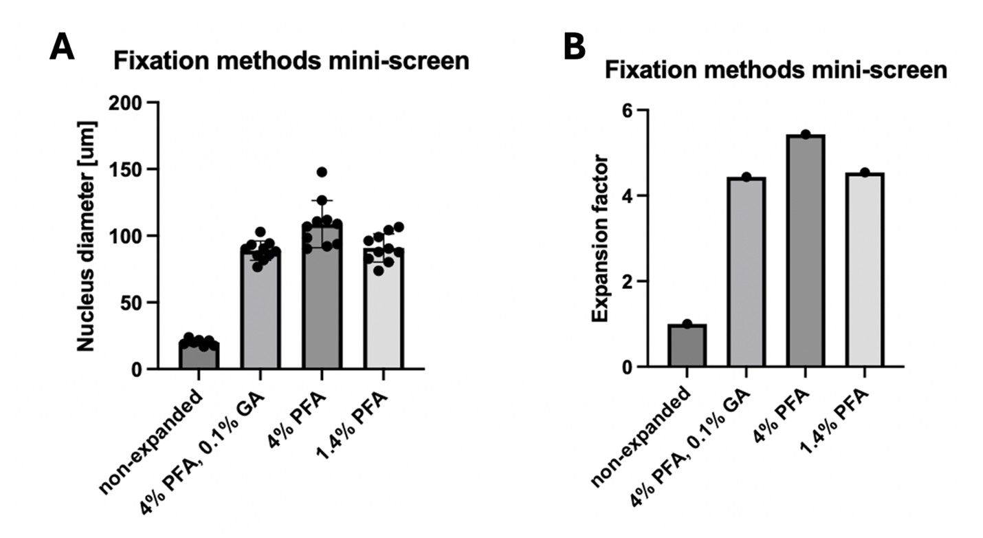
We tried different fixation methods in the early stages of this study (where expansion was not yet close to 10x). We saw a mild negative effect of GA on the expansion factor, so we avoided it in the later experiments since it also did not seem necessary to preserve the structure of our organelles of interest. However, the use of GA would generally be compatible with SPEx, potentially at the cost of a mild negative effect on expansion factor (see Author response image 1) and proteome coverage. We can add this information to the discussion.
Author response image 1.
Fixation methods mini-screen. Cells were fixed with the indicated reagents for 10 minutes at 37°C. After TREx expansion, the diameter of the nucleus was measured (A) and the resulting expansion factor compared to the non-expanded control was determined (B).
Related to the above, was the anchoring efficiency reduced only to achieve a 10x expansion factor or does this additionally affect the proteome coverage?
We solely lowered the anchoring in order to allow for higher expansion factors. In earlier pilots we performed proteomic analysis on samples that were just expanded 4x using standard TREx expansion (also using the original anchoring strategy from the TREx publication, consisting of 0.2 mg/ml AcX for overnight at RT). We presented the results of this pilot in Fig S1A. We still detected over 2,000 proteins from 10 cells, a coverage, which is highly similar to what we found in the final experiments (Figure 2F), in which the anchoring was lower yielding 10x expansion. Based on these data, we hypothesize that anchoring (and expansion factor!) has a negligible impact on protein coverage. We will clarify this in the manuscript.
d) Have the authors considered using alternative anchoring approaches, such as GMA (https://journals.plos.org/plosone/article?id=10.1371/journal.pone.0291506#pone.0291506.s001), which potentially increase the amount of sample retained in the hydrogel, thus allowing for better proteome coverage? This could be added to the discussion.
We did not use alternative anchoring approaches. We modified the TREx protocol to fit our purposes and since this was sufficient, we did not explore alternatives. However, using anchoring approaches, in which higher amounts of sample could be retained in the gel might be beneficial for the proteomics coverage. We will keep this suggestion in mind for future experiments. Thank you for the suggestion!
e) The limitation of the approach to near-2D samples should be mentioned, and alternative approaches for more 3D samples could be discussed.
The authors agree that SPEx is limited to near-2D samples at this point. We suggest that SPEx is applicable for 3D samples (e.g. in tissues) by performing cryosectioning. TREx has been shown to be compatible with sectioned tissue (Damstra et al., 2022). We will elaborate this in the discussion.
f) How are peptides that are directly anchored to the hydrogel dealt with during LC-MS/MS analysis? Are they excluded, or can they be identified during the spectral search? The latter would allow us to get a deeper structural understanding of how proteins are actually anchored into hydrogels, which so far has not been assessed.
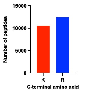
The reviewer raises an interesting point. In general, peptides carrying the anchoring modification are analysed by LC-MS, but we did not include these specific modifications in the database search. Overall, we assumed that the labeling would be low and stochastic and hence should, if at all, only minimally affect the detection of peptides. Nevertheless, in response to the reviewers’ comment, we searched the MS data again for the crosslinking reagent linked to lysine residues. However, we could not get any confident hit for any peptide containing this modification. Since we cannot exclude that the modification precludes the identification of the corresponding peptides, we compared the number peptides generated by trypsin cleavage after arginine and lysine. As the human genome contains similar proportions of both amino acids, one would expect similar numbers of both peptide types being identified. Any modifications of lysine by the anchoring reagent used, would prevent tryptic cleavage and thus reduce the number of lysine peptides. As shown in Author response image 2, the number of lysine terminating is only slightly lower compared to arginine terminating peptides. Notably, the proteomics results of a different fixed human tissue sample directly extracted by laser capture micro dissection without expansion showed a very similar lysine to arginine peptide ratio. This indicates that the large majority of lysine residues is not modified and affected by the hydrogel anchoring.
Author response image 2.
Number of peptides identified either terminating with lysine (K) or arginine (R) across all samples shown in Figure 5F.
An alternative approach to address this question would be to investigate if the peptide coverage of proteins detected by SPEx is enriched for peptides representing the folded core of proteins as opposed to the surface-exposed regions, which likely get more anchored into the hydrogel.
Because of the negligible amounts of modified peptides, we did not investigate this potential bias of surface-exposed versus folded-core peptides.
g) Same question regarding peptides with NHS labeling. Can they be identified, or do they just compete for ionization and thus negatively affect coverage and dynamic range of the LC-MS/MS approach?
The reviewer raises a similar point as above for another lysine labeling used during the SPEx protocol. Again, we specifically looked for this modification by re-searching the raw MS data, but still could not identify any peptides, carrying this modification on a lysine residue. Even though we cannot exclude that this rather large modification prevents detection, considering the high number of lysine terminating peptides in our dataset (see Figure 2), we would expect that also this labeling step is stochastic and affects only a minor proportion of the proteins.
h) How are the primary and secondary antibodies affecting the proteomics analysis identified as contaminants?
We thank the reviewer for this comment. Since antibodies bind to proteins in a non-covalent manner, they will be released during the denaturing steps of the protocol. Of course, the antibodies will stay in the sample, be digested and analyzed and could, if very abundant, affect the analysis of the proteins from the samples. To check this possibility, we re-searched the MS data including the sequences of the antibodies used. To our surprise, we could not detect any peptides of these antibodies. This suggests that the concentrations of the antibodies used are much lower than those of the sample proteins and thus should not have any impact on the proteomics results. We interpret this result also as a benefit of our method compared to organellar-IP.
i) Have the authors observed differences in proteomics coverage of only antibody vs NHS-labeling? Depending on the questions above, could pure antibody-based labeling increase proteomic coverage?
We did not perform this comparative analysis, since we always used NHS dyes. In the experiments presented in this manuscript, NHS dyes allowed easy visualization of the whole cell without the use of antibodies. This NHS staining was essential for this particular setup for sample acquisition. We cut out entire cells, cells lacking the nucleus and cells lacking the Golgi apparatus, which served as critical controls. However, other ways of detecting cell boundaries could be used to avoid NHS staining. As shown above, both, the anchor and NHS labeling are likewise sparse and stochastic. Moreover, we could not detect any impact of the antibody labeling to our results. Thus, we assume that both labeling procedures could be used.
Reviewer #2 (Public review):
Summary:
This study introduces a method that combines physical expansion of cells, imaging-guided isolation of defined regions, and protein identification to enable compartment-resolved analysis of protein composition at the subcellular scale. The authors aim to address a central limitation in existing approaches, namely the loss of spatial information during sample preparation or the indirect nature of proximity-based labeling methods. Using several cellular compartments as examples, they demonstrate that their approach can recover compartment-enriched protein sets and identify candidate proteins with previously unassigned localization.
Strengths:
A major strength of this work is the conceptual simplicity and accessibility of the approach. By combining established techniques in a modular way, the method avoids the need for genetic manipulation or specialized labeling strategies, making it broadly adaptable across experimental systems. The ability to directly select regions of interest based on imaging represents a clear advantage over indirect enrichment strategies and allows flexible targeting of both membrane-bound and non-membrane-bound compartments.
The experimental design is also a strong aspect of the study. The use of complementary comparison strategies-analyzing isolated compartments alongside matched "subtracted" controls-provides an internal framework for assessing enrichment and depletion, increasing confidence in spatial assignment. The application of the method across multiple organelles of different sizes and properties demonstrates versatility, and the reported specificity for several compartments is encouraging. In particular, the ability to profile small and biochemically challenging structures highlights a potentially important niche for the approach.
Weaknesses:
Despite these strengths, several methodological limitations constrain the interpretation of the results. The most important relates to spatial accuracy in three dimensions. While lateral resolution is improved through physical expansion, the lack of depth resolution introduces uncertainty regarding contributions from structures above and below the selected region. Although the authors argue that this does not substantially affect specificity, the current evidence is largely indirect, and a more rigorous quantification of potential contamination would strengthen this conclusion.
Quantitative interpretation also remains challenging. Because the measurements reflect total protein abundance rather than local concentration, differences in compartment size and protein density can influence enrichment values, particularly for small structures embedded within larger volumes. This issue is evident in the analysis of smaller compartments and complicates direct comparison across conditions. Additional normalization or modeling would help clarify how to interpret these measurements.
Another limitation concerns variability in the expansion process and its downstream consequences. Differences in expansion factor across samples may affect the definition of regions of interest and introduce variability in sampling, yet the impact of this variability is not fully explored. Similarly, the use of a modified chemical treatment to preserve proteins for downstream analysis is central to the workflow but is not extensively validated with respect to preservation of spatial organization.
While the identification of previously unannotated proteins is an appealing aspect of the study, validation is limited to a small number of examples, and broader support from independent datasets or literature context is lacking. In addition, the study primarily focuses on steady-state measurements in a single cell type, and therefore does not yet demonstrate the ability of the method to capture dynamic or condition-dependent changes in protein localization.
Finally, the positioning of the method relative to existing approaches could be more clearly articulated. Although qualitative comparisons are provided, a more systematic and quantitative benchmarking against alternative strategies would help readers better understand the specific advantages and trade-offs.
We thank the reviewer for the careful evaluation of the manuscript and for the constructive feedback. We think the reviewer raises valid points and will address them in the revised manuscript.
Reviewer #3 (Public review):
Franziscus et al. describe an elegant approach for spatially specific proteome analysis. To achieve this, they expand fixed cells and subsequently use a laser to micro-dissect a region of interest, which is then analyzed by mass spectrometry.
They demonstrate the effectiveness of their approach by analyzing the nucleus, nucleolus, and the Golgi, and benchmark their hits against previous datasets for these organelles.
The manuscript is very well written and nicely guides the reader through the applied methods. The presented data is convincing, and I do not see the need for additional experimental verification of the protocol. The only minor concern is the novelty of the method and the presentation. A combination of expansion, laser microdissection, and proteomics has been applied in the past (PMID: 36450705, PMID: 39477916). In the manuscript, one of these studies is cited, though it does not become clear that this approach is already described. However, Franziscus et al. describe the approach better and make it more accessible to the reader, especially since the other studies described this methodology in combination with tissue expansion and not in combination with single cell expansion as it is done here. I would ask the authors to be clearer in the introduction about what others have already done and what their contribution is here. In general, I am convinced that the community will benefit from the presented protocol to analyze organelle proteomics in detail.
We thank the reviewer for the careful evaluation of our manuscript and overwhelmingly positive assessment. We apologize for the omission of the mentioned citations, and will adjust the introduction to make it clearer what has already been done and what the advance our method provides.
References
Damstra HG, Mohar B, Eddison M, Akhmanova A, Kapitein LC, Tillberg PW. 2022. Visualizing cellular and tissue ultrastructure using Ten-fold Robust Expansion Microscopy (TREx). eLife 11:e73775. DOI: https://doi.org/10.7554/eLife.73775
Gambarotto D, Hamel V, Guichard P. 2021. Ultrastructure expansion microscopy (U-ExM). Methods in Cell Biology 161:57–81. DOI: https://doi.org/10.1016/bs.mcb.2020.05.006, PMID: 33478697
Liffner B, Silva TLA e., Vega-Rodriguez J, Absalon S. 2024. Mosquito Tissue Ultrastructure-Expansion Microscopy (MoTissU-ExM) enables ultrastructural and anatomical analysis of malaria parasites and their mosquito. BMC Methods 1:13. DOI: https://doi.org/10.1186/s44330-024-00013-4
-
eLife Assessment
This is an important study describing 'SPEx', a broadly accessible method that combines cell expansion, laser microdissection, and mass spectrometry to enable subcellular proteomic profiling. The authors provide convincing evidence that this flexible integration of established techniques provides a robust and practical approach for compartment-resolved spatial proteomics. The authors support their main claims with appropriate validation across multiple subcellular compartments and show that the method can recover known markers while also identifying previously uncharacterized components. Overall, the work is likely to be of broad interest to cell and molecular biologists, particularly those seeking scalable and cost-effective strategies for mapping organelle composition.
-
Reviewer #1 (Public review):
Summary:
The authors present a novel approach to subcellular spatial proteomics by combining laser microdissection with expansion microscopy and LC-MS/MS analysis (SPEx). They implement two different workflows for LMD and LC-MS/MS quantification:
(1)The standard approach, where an area of interest is cut out by LMD, subjected to proteomics analysis, and compared to the rest of the cell without the dissected ROI.
(2) The subtraction approach, where ROIs are removed, and the remaining cellular material is compared to samples containing both the surrounding material and the ROI.
The authors assess the technique by applying it to subcellular targets of various sizes, volumes, and protein compositions such as the nucleus, nucleoli, and Golgi. They demonstrate that SPEx can identify proteins enriched or reduced in …
Reviewer #1 (Public review):
Summary:
The authors present a novel approach to subcellular spatial proteomics by combining laser microdissection with expansion microscopy and LC-MS/MS analysis (SPEx). They implement two different workflows for LMD and LC-MS/MS quantification:
(1)The standard approach, where an area of interest is cut out by LMD, subjected to proteomics analysis, and compared to the rest of the cell without the dissected ROI.
(2) The subtraction approach, where ROIs are removed, and the remaining cellular material is compared to samples containing both the surrounding material and the ROI.
The authors assess the technique by applying it to subcellular targets of various sizes, volumes, and protein compositions such as the nucleus, nucleoli, and Golgi. They demonstrate that SPEx can identify proteins enriched or reduced in ROIs.
Strengths:
The broad, relatively easy, and inexpensive applicability of this approach to potentially many cell types and subcellular areas of interest provides an exciting alternative to subcellular fractionation, native immunoprecipitation, or genetically encoded proximity labeling constructs. Moreover, by visually selecting ROIs for subsequent analysis, subcellular context or organelle morphology can be taken into account, as discussed by the authors in the discussion section.
Weaknesses:
While strongly supporting the sharing of this approach, we have a number of comments and questions that will improve the impact of the manuscript:
(1) General:
a) The manuscript would benefit from restructuring and language revision. In its current form, the writing is sometimes dense and verbose (in particular, the Results section). This makes it difficult to follow the authors' arguments.
b) The authors mention the possibility of selecting organelles based on morphology. This is left for the discussion, but it seems like a missed opportunity - the authors could compare individual organelles in different morphological states, e.g., connected vs. fragmented mitochondria.
(2) Technical:
a) Why do the authors strive and optimize for a 10x expansion factor? Is SPEx compatible with a more standard 4x expansion, as e.g., used in the classic U-ExM approach (https://www.nature.com/articles/s41592-018-0238-1)? This could be added to the discussion.
b) The U-ExM approach shows improved ultrastructural preservation when using 3%FA with 0.1% glutaraldehyde fixation (GA). Is SPEx compatible with the use of low amounts of GA for fixation?
c) Related to the above, was the anchoring efficiency reduced only to achieve a 10x expansion factor or does this additionally affect the proteome coverage?
d) Have the authors considered using alternative anchoring approaches, such as GMA (https://journals.plos.org/plosone/article?id=10.1371/journal.pone.0291506#pone.0291506.s001), which potentially increase the amount of sample retained in the hydrogel, thus allowing for better proteome coverage? This could be added to the discussion.
e) The limitation of the approach to near-2D samples should be mentioned, and alternative approaches for more 3D samples could be discussed.
f) How are peptides that are directly anchored to the hydrogel dealt with during LC-MS/MS analysis? Are they excluded, or can they be identified during the spectral search? The latter would allow us to get a deeper structural understanding of how proteins are actually anchored into hydrogels, which so far has not been assessed.
An alternative approach to address this question would be to investigate if the peptide coverage of proteins detected by SPEx is enriched for peptides representing the folded core of proteins as opposed to the surface-exposed regions, which likely get more anchored into the hydrogel.
g) Same question regarding peptides with NHS labeling. Can they be identified, or do they just compete for ionization and thus negatively affect coverage and dynamic range of the LC-MS/MS approach?
h) How are the primary and secondary antibodies affecting the proteomics analysis identified as contaminants?
i) Have the authors observed differences in proteomics coverage of only antibody vs NHS-labeling? Depending on the questions above, could pure antibody-based labeling increase proteomic coverage?
-
Reviewer #2 (Public review):
Summary:
This study introduces a method that combines physical expansion of cells, imaging-guided isolation of defined regions, and protein identification to enable compartment-resolved analysis of protein composition at the subcellular scale. The authors aim to address a central limitation in existing approaches, namely the loss of spatial information during sample preparation or the indirect nature of proximity-based labeling methods. Using several cellular compartments as examples, they demonstrate that their approach can recover compartment-enriched protein sets and identify candidate proteins with previously unassigned localization.
Strengths:
A major strength of this work is the conceptual simplicity and accessibility of the approach. By combining established techniques in a modular way, the method …
Reviewer #2 (Public review):
Summary:
This study introduces a method that combines physical expansion of cells, imaging-guided isolation of defined regions, and protein identification to enable compartment-resolved analysis of protein composition at the subcellular scale. The authors aim to address a central limitation in existing approaches, namely the loss of spatial information during sample preparation or the indirect nature of proximity-based labeling methods. Using several cellular compartments as examples, they demonstrate that their approach can recover compartment-enriched protein sets and identify candidate proteins with previously unassigned localization.
Strengths:
A major strength of this work is the conceptual simplicity and accessibility of the approach. By combining established techniques in a modular way, the method avoids the need for genetic manipulation or specialized labeling strategies, making it broadly adaptable across experimental systems. The ability to directly select regions of interest based on imaging represents a clear advantage over indirect enrichment strategies and allows flexible targeting of both membrane-bound and non-membrane-bound compartments.
The experimental design is also a strong aspect of the study. The use of complementary comparison strategies-analyzing isolated compartments alongside matched "subtracted" controls-provides an internal framework for assessing enrichment and depletion, increasing confidence in spatial assignment. The application of the method across multiple organelles of different sizes and properties demonstrates versatility, and the reported specificity for several compartments is encouraging. In particular, the ability to profile small and biochemically challenging structures highlights a potentially important niche for the approach.
Weaknesses:
Despite these strengths, several methodological limitations constrain the interpretation of the results. The most important relates to spatial accuracy in three dimensions. While lateral resolution is improved through physical expansion, the lack of depth resolution introduces uncertainty regarding contributions from structures above and below the selected region. Although the authors argue that this does not substantially affect specificity, the current evidence is largely indirect, and a more rigorous quantification of potential contamination would strengthen this conclusion.
Quantitative interpretation also remains challenging. Because the measurements reflect total protein abundance rather than local concentration, differences in compartment size and protein density can influence enrichment values, particularly for small structures embedded within larger volumes. This issue is evident in the analysis of smaller compartments and complicates direct comparison across conditions. Additional normalization or modeling would help clarify how to interpret these measurements.Another limitation concerns variability in the expansion process and its downstream consequences. Differences in expansion factor across samples may affect the definition of regions of interest and introduce variability in sampling, yet the impact of this variability is not fully explored. Similarly, the use of a modified chemical treatment to preserve proteins for downstream analysis is central to the workflow but is not extensively validated with respect to preservation of spatial organization.
While the identification of previously unannotated proteins is an appealing aspect of the study, validation is limited to a small number of examples, and broader support from independent datasets or literature context is lacking. In addition, the study primarily focuses on steady-state measurements in a single cell type, and therefore does not yet demonstrate the ability of the method to capture dynamic or condition-dependent changes in protein localization.
Finally, the positioning of the method relative to existing approaches could be more clearly articulated. Although qualitative comparisons are provided, a more systematic and quantitative benchmarking against alternative strategies would help readers better understand the specific advantages and trade-offs.
-
Reviewer #3 (Public review):
Franziscus et al. describe an elegant approach for spatially specific proteome analysis. To achieve this, they expand fixed cells and subsequently use a laser to micro-dissect a region of interest, which is then analyzed by mass spectrometry.
They demonstrate the effectiveness of their approach by analyzing the nucleus, nucleolus, and the Golgi, and benchmark their hits against previous datasets for these organelles.
The manuscript is very well written and nicely guides the reader through the applied methods. The presented data is convincing, and I do not see the need for additional experimental verification of the protocol. The only minor concern is the novelty of the method and the presentation. A combination of expansion, laser microdissection, and proteomics has been applied in the past (PMID: 36450705, …
Reviewer #3 (Public review):
Franziscus et al. describe an elegant approach for spatially specific proteome analysis. To achieve this, they expand fixed cells and subsequently use a laser to micro-dissect a region of interest, which is then analyzed by mass spectrometry.
They demonstrate the effectiveness of their approach by analyzing the nucleus, nucleolus, and the Golgi, and benchmark their hits against previous datasets for these organelles.
The manuscript is very well written and nicely guides the reader through the applied methods. The presented data is convincing, and I do not see the need for additional experimental verification of the protocol. The only minor concern is the novelty of the method and the presentation. A combination of expansion, laser microdissection, and proteomics has been applied in the past (PMID: 36450705, PMID: 39477916). In the manuscript, one of these studies is cited, though it does not become clear that this approach is already described. However, Franziscus et al. describe the approach better and make it more accessible to the reader, especially since the other studies described this methodology in combination with tissue expansion and not in combination with single cell expansion as it is done here. I would ask the authors to be clearer in the introduction about what others have already done and what their contribution is here. In general, I am convinced that the community will benefit from the presented protocol to analyze organelle proteomics in detail.
-
SPEx: Compartment-Resolved Proteomics via Expansion Microscopy–Guided Microdissection www.biorxiv.org/content/10.6...
-
What if you could cut out sub cellular structures and measure the proteome? SPEx: Compartment-Resolved Proteomics via Expansion Microscopy–Guided Microdissection www.biorxiv.org/content/10.6... awesome work by Curdin Franziscus, @imcf-lex.bsky.social, @biehli.bsky.social, Alexander Schmidt!
-
SPEx: Compartment-Resolved Proteomics via Expansion Microscopy-Guided Microdissection https://www.biorxiv.org/content/10.64898/2026.03.28.714993v1
-
SPEx: Compartment-Resolved Proteomics via Expansion Microscopy-Guided Microdissection https://www.biorxiv.org/content/10.64898/2026.03.28.714993v1
-