Human mitochondrial DNA variants influence telomere length: evidence from a transmitochondrial cybrid model
Curation statements for this article:-
Curated by eLife

eLife Assessment
This creative and timely study uses an innovative cybrid approach to provide convincing evidence that mitochondrial variation influences telomere maintenance. Although the proposed molecular mechanism requires further validation and the evidence for telomere-length inheritance in humans remains preliminary, the work represents an important advance at the intersection of mitochondrial and telomere biology, with broad implications for aging and genome stability.
This article has been Reviewed by the following groups
Listed in
- Evaluated articles (eLife)
Abstract
Telomere shortening is a hallmark of aging, yet telomere length (TL) varies considerably among individuals and is strongly influenced by inheritance. In mice, efficient mitochondrial function-characterized by low reactive oxygen species (ROS) production-is critical for telomere elongation during early embryogenesis. Since mitochondrial DNA (mtDNA) encodes several subunits of the electron transport chain, it may influence TL at birth by regulating mitochondrial function in utero. To explore the relationship between mtDNA and TL in human, we used a transmitochondrial cybrid approach, introducing mitochondria from donor platelets with varying telomere lengths into mtDNA-depleted cells. This revealed an inverse correlation between donor blood TL and mitochondrial ROS levels measured in the resulting cybrids, suggesting that specific mtDNA variants may contribute to the maintenance of long telomeres in humans by enhancing mitochondrial fitness. During in vitro cybrid formation, a transient phase of oxidative stress precedes cellular adaptation. In this specific window, mtDNA variants associated with reduced complex I (CI) activity induced rapid telomere shortening—an effect rescued by antioxidant and NAD⁺ precursor supplementation. While these variants occur naturally in certain individuals with long telomeres, our data suggest that, at least under in vitro conditions of acute oxidative stress, CI is critical to support PARP1 activity by maintaining the NAD⁺/NADH balance, thereby preserving telomere integrity. Collectively, these findings solidify the link between mtDNA variants and human TL regulation, highlighting potential therapeutic opportunities for mitochondrial replacement strategies.
Article activity feed
-

-
-

eLife Assessment
This creative and timely study uses an innovative cybrid approach to provide convincing evidence that mitochondrial variation influences telomere maintenance. Although the proposed molecular mechanism requires further validation and the evidence for telomere-length inheritance in humans remains preliminary, the work represents an important advance at the intersection of mitochondrial and telomere biology, with broad implications for aging and genome stability.
-
Reviewer #1 (Public review):
Summary:
This is an interesting study that addresses whether mitochondrial DNA (mitoDNA) variants impact telomere length (TL), which may be relevant to potential maternal inheritance of TL in offspring. The study addresses this question using an innovative cybrid model approach in which mitochondria from donor platelets from 7 individuals that vary in TL and differ in mitoDNA variants are fused with 143B cells that lack mitochondria. MitoDNA variants that exhibited reduced complex I activity showed telomere shortening in cybrids, and increased telomere dysfunction. Interestingly, these phenotypes could be reduced with NAC antioxidant and NAD+ supplementation, suggesting that ROS and oxidative DNA damage at telomeres contributed to the telomere shortening. They further showed that cybrids with lower levels of …
Reviewer #1 (Public review):
Summary:
This is an interesting study that addresses whether mitochondrial DNA (mitoDNA) variants impact telomere length (TL), which may be relevant to potential maternal inheritance of TL in offspring. The study addresses this question using an innovative cybrid model approach in which mitochondria from donor platelets from 7 individuals that vary in TL and differ in mitoDNA variants are fused with 143B cells that lack mitochondria. MitoDNA variants that exhibited reduced complex I activity showed telomere shortening in cybrids, and increased telomere dysfunction. Interestingly, these phenotypes could be reduced with NAC antioxidant and NAD+ supplementation, suggesting that ROS and oxidative DNA damage at telomeres contributed to the telomere shortening. They further showed that cybrids with lower levels of ROS correlated with longer TL in the lymphocytes of the mitochondrial donors.
This study has many strengths, including the use of cybrid technology, which allows for analysis of how mitochondria and mtDNA variants from various human donors influence telomere length when introduced into the same genetic background of 143B mitochondria deficient cells. Other strengths include the use of human donors that vary in leukocyte telomere length, and analysis of multiple endpoints of telomere integrity and mitochondrial function in the resulting cybrids. This study provides convincing evidence that mtDNA variants influence TL, through a mechanism involving mitochondrial derived ROS, potentially causing telomeric oxidative damage. These results may help explain prior work demonstrating a link between mitochondrial function and telomere length regulation during embryogenesis and development. The data are robust, and the manuscript is well written. Overall, the results support the conclusions and will likely have an impact on the field of mitochondrial function and telomere length regulation, with important implications for development, aging, and aging-related diseases.
Given the heterogeneity in TL and mtDNA variants in the human population, the conclusion could be further strengthened by increasing the number of donors and cybrids analyzed. Other minor weaknesses stem from the limited number of samples. For example, the study presented limited telomere FISH metaphase analysis of chromosome end fusions, telomere damage, and telomere attrition rates due to low sample numbers. Furthermore, the proposal that acute oxidative stress was induced by a metabolic shift from glycolysis to OXPHOS in vitro is based primarily on assumptions from prior work, rather than direct demonstration in the current study. However, results with the NAC antioxidant combined with NR strongly support this assumption. Overall, the findings from this study are convincing and provide a solid foundation for expanding this analysis in the future to include a greater number of samples from donors for cybrid analysis.
-
Reviewer #2 (Public review):
Summary:
The authors aim to determine whether mitochondrial genotype influence telomere length. By generating cybrids harboring different mitochondrial backgrounds, the authors seek to establish a mechanistic link between mitochondrial status and telomere biology.
Strengths:
A major strength of the study is the use of cybrid technology, which provides a great approach to investigate the role of mitochondrial DNA independently of the nuclear genome. The authors also employ multiple complementary assays to assess telomere-related phenotypes associated with mitochondrial dysfunction. Together, these experiments generate an interesting dataset that will be of value to researchers interested in the intersection between mitochondrial biology, genome stability, aging and development. These results also build on …
Reviewer #2 (Public review):
Summary:
The authors aim to determine whether mitochondrial genotype influence telomere length. By generating cybrids harboring different mitochondrial backgrounds, the authors seek to establish a mechanistic link between mitochondrial status and telomere biology.
Strengths:
A major strength of the study is the use of cybrid technology, which provides a great approach to investigate the role of mitochondrial DNA independently of the nuclear genome. The authors also employ multiple complementary assays to assess telomere-related phenotypes associated with mitochondrial dysfunction. Together, these experiments generate an interesting dataset that will be of value to researchers interested in the intersection between mitochondrial biology, genome stability, aging and development. These results also build on previous work supporting roles for ROS/mitochondria in driving telomere shortening.
Overall, this study provides interesting evidence linking mitochondrial background to telomere biology. The cybrid models represent a useful resource for the field and the work raises important questions regarding mitochondria-telomere communication.
Comments on revised version.
In my view, authors have answered satisfactorily my concerns.
-
Reviewer #3 (Public review):
Strengths:
Mahieu and colleagues ask whether mitochondrial genetic variation helps explain differences in human telomere length. They selected seven donors from a Flow-FISH reference cohort of 491 individuals and compared their mitochondrial backgrounds in cybrids sharing the same nuclear background.
The combination of telomere and mitochondrial assays is a strength. The association between low complex I activity and telomere shortening, together with the NAC/NR rescue in Cyb2, provides useful evidence linking mitochondrial function to telomere maintenance. The correlation between donor telomere length and cybrid ROS is also interesting, although the cybrid results are more persuasive than the claims about inheritance.
Weaknesses:
(1) The authors have clarified that the seven donors include intermediate …
Reviewer #3 (Public review):
Strengths:
Mahieu and colleagues ask whether mitochondrial genetic variation helps explain differences in human telomere length. They selected seven donors from a Flow-FISH reference cohort of 491 individuals and compared their mitochondrial backgrounds in cybrids sharing the same nuclear background.
The combination of telomere and mitochondrial assays is a strength. The association between low complex I activity and telomere shortening, together with the NAC/NR rescue in Cyb2, provides useful evidence linking mitochondrial function to telomere maintenance. The correlation between donor telomere length and cybrid ROS is also interesting, although the cybrid results are more persuasive than the claims about inheritance.
Weaknesses:
(1) The authors have clarified that the seven donors include intermediate telomere lengths. Their analyses excluding donors #1, #3 and #6 are reassuring, since the correlation persists in each case. However, these results appear only in the response, and the full leave-one-out analysis is not reported. The small, selected sample still limits how broadly the correlation can be interpreted.
(2) The revised manuscript makes the distinction between the cybrid and donor findings clearer. However, CI activity shows no significant association with donor telomere length. The proposed PARP1 mechanism also remains untested: the authors acknowledge that they did not establish whether PARP1 is required for the NAC/NR rescue, but still describe its role too strongly.
(3) The authors appropriately acknowledge that the ATP6 A177T association is inconclusive (p = 0.153). Some inheritance claims remain stronger than this warrants. The family observations are too limited to separate mitochondrial from nuclear or shared environmental effects, and the cybrid experiments do not isolate ATP6 A177T from the other variants transferred with it.
-
Author response:
The following is the authors’ response to the original reviews.
Public Reviews:
Reviewer #1 (Public review):
Summary:
This is an interesting study that addresses whether mitochondrial DNA (mitoDNA) variants impact telomere length (TL), which may be relevant to potential maternal inheritance of TL in offspring. The study addresses this question using a cybrid model approach in which mitochondria from donor platelets from 7 individuals that vary in TL and differ in mitoDNA variants are introduced into 143B cells that lack mitochondria. MitoDNA variants that exhibited reduced complex I activity showed telomere shortening in cybrids and increased telomere dysfunction. Interestingly, these phenotypes could be reduced with NAC antioxidant and NAD+ supplementation, suggesting that ROS and oxidative DNA damage at …
Author response:
The following is the authors’ response to the original reviews.
Public Reviews:
Reviewer #1 (Public review):
Summary:
This is an interesting study that addresses whether mitochondrial DNA (mitoDNA) variants impact telomere length (TL), which may be relevant to potential maternal inheritance of TL in offspring. The study addresses this question using a cybrid model approach in which mitochondria from donor platelets from 7 individuals that vary in TL and differ in mitoDNA variants are introduced into 143B cells that lack mitochondria. MitoDNA variants that exhibited reduced complex I activity showed telomere shortening in cybrids and increased telomere dysfunction. Interestingly, these phenotypes could be reduced with NAC antioxidant and NAD+ supplementation, suggesting that ROS and oxidative DNA damage at telomeres contributed to the telomere shortening. They further showed that cybrids with lower levels of ROS correlated with longer TL in the lymphocytes of the mitochondrial donors.
Strengths:
This study provides compelling evidence that mtDNA variants influence TL through a mechanism involving mitochondrial-derived ROS, potentially causing telomeric oxidative damage. The data are robust, and the manuscript is well written. However, the study could be strengthened by addressing the following questions and minor weaknesses below.
We thank the reviewer for this very positive evaluation of our work.
Weaknesses:
(1) Introduction. Line 81, the relationship between TL and the risk of lymphoid and myeloid leukemia is not straightforward. POT1 variants associated with long TL increase the risk for lymphoid and myeloproliferative neoplasms (see PMID: 41564438 for example).
We appreciate the reviewer raising the important link between pathogenic POT1 variants and lymphoid malignancies driven by elongated telomeres. We would like to clarify, however, that our introduction focused not on the pathological telomere attrition characteristic of telomere biology disorders, but rather on the natural, non-pathological variations observed in individuals with baseline telomere lengths on the shorter end of the spectrum.
Nevertheless, the two following publications by the Armanios lab have been added in the introduction to underscore the complex relationship between telomere length and tumorigenesis:
DeBoy EA et al. Familial clonal hematopoiesis in a long telomere syndrome. 2023. N Engl J Med 388, 2422-2433.
Davidson-Swinton HR et al. Lymphoid malignancy and clonality in the POT1-mediated long telomere syndrome. 2026. Blood 147, 2226-2237.
Here is the corresponding sentence in the revised manuscript:
“However, some pathogenic mutations in POT1 gene, linked to very long telomeres, were also previously associated with increased risk for lymphoid and myeloproliferative neoplasms (5,6), underscoring the complex relationship between TL and cancer predisposition.”
(2) Figure 1. Since sex also influences TL, it would be good to know the sex of the selected individuals or explain why this is not necessary.
Because this study investigated the potential maternal inheritance of TL via the mitochondrial genome, our cohort consisted predominantly of female donors (6/7). Donor #5, the husband of Donor #7, was the only male included. We have added the information in the text:
“All were of European origin, thus excluding a possible bias linked to African ethnic groups (7), and the seven donors belonged to distinct mitochondrial subhaplogroups (Fig 1D). All but one (#5) were female donors.”
Notably, our analysis revealed minimal influence of sex on TL, which cannot account for the observed differences between the extreme groups.
(3) Please include a description of the 143B cells that were used for cybrid formation in the Results section when introducing the cybrids.
We did it:
“To evaluate the impact of distinct mitochondrial genomes on TL in human cells, we used a transmitochondrial cybrid strategy in which mtDNA-depleted Rho0 (ρ0) cells, derived from 143B human osteosarcoma cancer cells, are fused with donor platelets, thereby restoring functional mitochondria carrying donor-specific mtDNA variants (21).”
(4) Lines 155-156. The authors note that cybrids from donors 1 and 2 show "pronounced" telomere damage. This result indicates an increase in 53BP1-positive telomeres, which could be indicative of telomere dysfunction or damage. Quantification of the increased chromosome end fusions for cybrids 1 and 2 would strengthen the result.
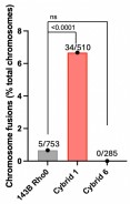
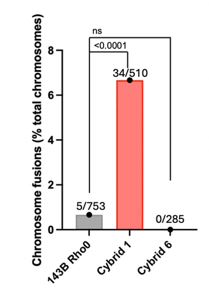
We thank the reviewer for this suggestion. Following their advice, we used our metaphase spread FISH analyses to quantify chromosome end fusions in the parental 143B Rho0, Cyb1 and Cyb6 cells. However, because Cyb2 metaphase spreads were of insufficient quality for adequate chromosome analysis, we restricted our telomere fusion comments exclusively to Cyb1 and included the quantifications in our revised manuscript (Fig 2E).
Do the increased fusions correlate with an increase in telomere signal-free ends? These should be apparent in the telomere FISH images of metaphase chromosomes.
While this is a strong argument, the telomeres within these cybrid models are critically short. Consequently, the FISH signal intensity falls below the threshold required for reliable quantification of telomere-free ends.
(5) Lines 168-169. What is the evidence that the "in vitro metabolic shift" causes acute oxidative stress?
The reviewer is correct that we have not formally demonstrated this in our experimental system. Instead, our assumption was based on the fact that the initial phase of Rho0 cell repopulation involves a temporary ROS burst, partly driven by incompletely assembled ETC supercomplexes that are known to elevate ROS levels (Maranzana et al., 2013). This is supported by our experimental observation that the NAC antioxidant, combined with NR, potently inhibits telomere shortening in cybrids with low CI activity. We added the following sentence in the revised manuscript:
“When mitochondria are replenished in ρ0 cells during the formation of transmitochondrial cybrids, a shift from glycolysis to OXPHOS occurs (Fig S4A-B) and cells need to adapt to this metabolic change. Notably, the initial phase of ρ0 cell repopulation involves a temporary ROS burst, partly driven by incompletely assembled ETC supercomplexes that are known to elevate ROS levels (22).”
(6) Why did the elevated ROS in cybrid #3 (Figure 4C) not translate to shorter telomeres in the cybrid (Figure 2A)? Perhaps there is a difference between factors that determine TL in the cybrid vs the donor's lymphocytes?
We cannot fully explain the discrepancy, but we indeed suspect in vivo oxidative stress differs significantly from cell cultures (21% O2). Additionally, early cybrid replenishment involves an unknown telomere elongation step that may offset mitochondrial ROS-induced shortening.
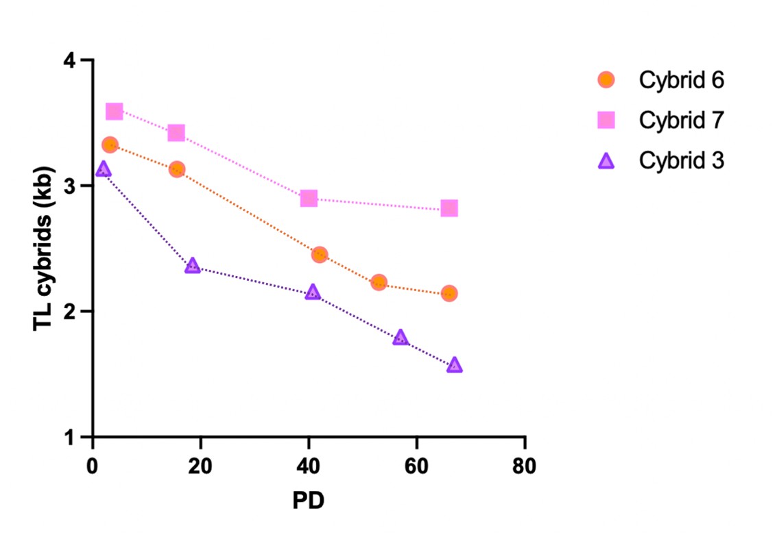
To further investigate this question, we measured telomere length across varying population doublings (PDs) and observed the following shortening after 66-67 PDs:
- Cyb3: about 1.6 kb reduction
- Cyb6: about 1.2 kb reduction
- Cyb7: about 0.8 kb reduction
These results suggest that the rate of telomere shortening in culture may be higher in Cyb3 cells, possibly due to increased ROS levels.
We included these results as Supplementary Figure 7 and have discussed these data in the revised manuscript:
“Consistently, tracking TL over sequential population doublings revealed that Cyb3 cells are more vulnerable to progressive telomere shortening than Cyb6 or Cyb7 cells (Fig S7). Together, these findings support a role for mitochondrial ROS in human TL regulation.”
In Figure 4B, it appears that the statistical comparisons for mitochondrial superoxide are all relative to Cyb3. If so, why are the comparisons not with the parental 143B rho0 cell line? Please clarify.
We excluded Rho0 cells from our superoxide measurements because these cells were grown in a different culture medium. Instead, we focused on comparing mitochondrial ROS across cybrids to accurately correlate these values with the telomere length of the corresponding donors’ lymphocytes. Consequently, Rho0 cell measurements would not have contributed to this correlation analysis.
We have discussed this in the revised manuscript:
“Mitochondrial superoxide production was measured by electron paramagnetic resonance (27) in clones #1 of all cybrids and in 143B cells (Fig 4B). We excluded 143B ρ0 cells from our superoxide measurements because these cells were grown in a different culture medium.”
(7) Given the heterogeneity in TL and mtDNA variants in the human population, the conclusions could be further strengthened by increasing the number of donors and cybrids analyzed. However, there are admittedly practical factors. Overall, these findings are compelling and provide a solid foundation for expanding this analysis in the future. This is more of a comment than a weakness.
We thank the reviewer for this constructive feedback and entirely agree that including more donors would have added depth to our findings. While we acknowledge this limitation, pursuing this further is currently impossible without acquiring new ethical approvals and establishing fresh collaborations with clinicians.
Reviewer #2 (Public review):
Summary:
The authors aim to determine whether mitochondrial genotype influences telomere length. By generating cybrids harboring different mitochondrial backgrounds, the authors seek to establish a mechanistic link between mitochondrial status and telomere biology.
Strengths:
A major strength of the study is the use of cybrid technology, which provides a great approach to investigate the role of mitochondrial DNA independently of the nuclear genome. The authors also employ multiple complementary assays to assess telomere-related phenotypes associated with mitochondrial dysfunction. Together, these experiments generate an interesting dataset that will be of value to researchers interested in the intersection between mitochondrial biology, genome stability, aging, and development. These results also build on previous work supporting roles for ROS/mitochondria in driving telomere shortening.
We thank the reviewer for this very positive evaluation of our work.
Weaknesses:
The data support the conclusion that mitochondrial background is associated with differences in telomere length and telomere-related phenotypes. However, some of the mechanistic interpretations would benefit from additional evidence. In particular, the manuscript discusses mitochondrial influences on telomere shortening, yet telomere length in some experiments is assessed at a single time point. Consequently, the current data do not directly address the rate of telomere attrition. Differences observed between cybrid lines could potentially arise from events occurring during cybrid formation, clonal selection, or subsequent cell expansion. Longitudinal analyses across multiple passages, ideally beginning immediately after cybrid generation and controlling for population doublings, would help establish whether mitochondrial function directly affects telomere shortening dynamics. Some experimental results would also benefit from additional quantification, clarification, and some biological replicates are missing.
We limited our TL measurements to the earliest viable time point after cybrid formation to avoid the confounding effects of cellular adaptation in culture. For instance, the early telomere shortening observed in Cyb1 and Cyb2 cells was later alleviated—likely due to the upregulation of the NAD+ salvage pathway genes NAMPT and NAPRT1. To accurately capture the effects specific to cybrid formation, we isolated 4 independent clones per donor, all of which showed highly consistent TL values, as shown in Figure 2A and S3D.
While we acknowledge the reviewer's point about multi-passage longitudinal analyses, we did, in fact, measure telomere length across varying population doublings (PDs) in Cyb3 (high mito ROS levels), Cyb6 and Cyb7 (moderate to low mito ROS levels) and observed the following shortening after 66-67 PDs:
- Cybrid 3: about 1.6 kb reduction
- Cybrid 6: about 1.2 kb reduction
- Cybrid 7: about 0.8 kb reduction
These results suggest that the rate of telomere shortening in culture may be higher in Cyb3 cells, possibly due to increased ROS levels.
We included these results as Supplementary Figure 7 and have discussed these data in the revised manuscript:
“Consistently, tracking TL over sequential population doublings revealed that Cyb3 cells are more vulnerable to progressive telomere shortening than Cyb6 or Cyb7 cells (Fig S7). Together, these findings support a role for mitochondrial ROS in human TL regulation.”
We have also carefully edited the manuscript so as to clarify the text and, whenever possible, have added quantifications. Among others, as suggested by Reviewer #1, we added the quantification of chromosome end fusions in the parental 143B Rho0, Cyb1 and Cyb6 cells in our revised manuscript:
Author response image 1.
Overall, this study provides interesting evidence linking mitochondrial background to telomere biology. The cybrid models represent a useful resource for the field, and the work raises important questions regarding mitochondria-telomere communication.
Reviewer #3 (Public review):
Strengths:
Mahieu and colleagues address an interesting and underexplored question: whether non-pathogenic variation in the mitochondrial genome contributes to the inter-individual variability of human telomere length (TL). Using a Belgian Flow-FISH reference cohort (n=491) to identify donors at TL extremes, they generate trans mitochondrial cybrids from platelets of seven donors of distinct mtDNA subhaplogroups and characterize the resulting cells with a broad and well-executed toolkit (TRF, TeSLA, ddTRAP, EPR-based mitoROS, Seahorse with permeabilized-cell ETC dissection, LC-MS metabolomics, telomeric PARFISH). The most compelling finding is that cybrids derived from donors with low complex I (CI) activity undergo rapid telomere shortening during the glycolysis-to-OXPHOS transition of cybrid formation, and that this is largely prevented by co-treatment with NAC and the NAD+ precursor nicotinamide riboside, supporting a model in which CI sustains the NAD+ pool required for PARP1-mediated repair of oxidative damage at telomeres. The authors further report an inverse correlation between donor lymphocyte TL and mitoROS in the corresponding cybrids, and provide preliminary evidence that the K1a-defining ATP6 A177T variant (m.G9055>A) may be enriched in long-telomere individuals.
We thank the reviewer for this very positive evaluation of our work.
Weaknesses:
(1) Statistical support and donor sampling for the central in vivo correlation (Figure 4C).
The inverse correlation between donor lymphocyte TL and cybrid mitoROS (R2=0.794, p=0.007) is the principal in vivo claim of the paper, but it is built on seven donors deliberately selected from the extremes of the Flow-FISH distribution. Sampling at the tails of the outcome variable can substantially inflate apparent correlation strength and significance. I would encourage the authors to (i) explicitly state this sampling structure where the correlation is introduced, (ii) report a leave-one-out sensitivity analysis to confirm the relationship is not driven by one or two donors (Cyb3 and Cyb6 appear to anchor the line), and (iii) where feasible, extend the analysis to additional donors with intermediate TL to test whether the relationship holds across the full distribution. Even a modest expansion (e.g., 4 to 5 additional donors at P25 to P75) would substantially strengthen this central claim.
We thank the reviewer for this insightful comment. We have conducted the requested leave-one-out analysis:
- removing Cyb3: R2=0.730; p=0.0302
- removing Cyb6 : R2=0.782 ; p=0.0194
- removing Cyb1 : R2=0.740; p=0.0278
While we agree that additional donors would enhance the study, further experiments are however currently impossible without new ethical clearances and additional clinical partnerships.
(2) Reconciling the cybrid CI / TL relationship (Fig 3B) with the absence of a CI / TL relationship in donor lymphocytes (Figure 4A).
Figure 3B shows a strong correlation between CI activity and TL in cybrids (R2=0.87), while Figure 4A shows no correlation between donor CI activity (measured in the same cybrids) and donor lymphocyte TL. The authors acknowledge this, but the manuscript subsequently builds toward a CI-centric model of in vivo TL regulation, which seems to outrun the data. The most internally consistent interpretation is that the cybrid CI phenotype reports a sensitized in vitro response to the acute oxidative stress of the metabolic shift, rather than a steady-state determinant of leukocyte TL. I would suggest reframing the abstract, significance statement, and Discussion to make this distinction clearer. The in vitro CI / NAD+ / PARP1 axis is a strong finding on its own, while the in vivo role of CI activity (as opposed to ROS more broadly) is not yet established here. Donor #1's profile (very long lymphocyte TL, low CI activity, severe shortening in cybrids, no telomere inheritance in offspring) is informative in this regard and could be discussed more directly as a case that helps delineate where the cybrid model does and does not recapitulate in vivo biology.
We acknowledge that our study does not establish the in vivo role of CI activity in TL regulation. Our abstract specifically highlights an in vitro phenomenon: “Under the specific conditions of cybrid formation, which involve a metabolic shift from glycolysis to oxidative phosphorylation, mtDNA variants associated with reduced CI activity induced rapid telomere shortening, …”.
We nevertheless extensively revised the Abstract/Significance Statement to clearly separate our in vitro results from in vivo biology as requested:
“Abstract
Telomere shortening is a hallmark of aging, yet telomere length (TL) varies considerably among individuals and is strongly influenced by inheritance. In mice, efficient mitochondrial function, characterised by low reactive oxygen species (ROS) production, is critical for telomere elongation during early embryogenesis. Since mitochondrial DNA (mtDNA) encodes several subunits of the electron transport chain, it may influence TL at birth by regulating mitochondrial function in utero. To explore the relationship between mtDNA and TL in human, we used a transmitochondrial cybrid approach, introducing mitochondria from donor platelets with varying telomere lengths into mtDNA-depleted cells. This revealed an inverse correlation between donor blood TL and mitochondrial ROS levels measured in the resulting cybrids, suggesting that specific mtDNA variants may contribute to the maintenance of long telomeres in humans by enhancing mitochondrial fitness. During in vitro cybrid formation, a transient phase of oxidative stress precedes cellular adaptation. In this specific window, mtDNA variants associated with reduced complex I (CI) activity induced rapid telomere shortening—an effect rescued by antioxidant and NAD+ precursor supplementation. While these variants occur naturally in certain individuals with long telomeres, our data suggest that, at least under in vitro conditions of acute oxidative stress, CI is critical to support PARP1 activity by maintaining the NAD+/NADH balance, thereby preserving telomere integrity. Collectively, these findings solidify the link between mtDNA variants and human TL regulation, highlighting potential therapeutic opportunities for mitochondrial replacement strategies.”
“Significance Statement
“Telomere length at birth influences aging trajectories and disease risk later in life, yet the mechanisms governing this trait remain incompletely understood. Using a trans mitochondrial cybrid approach, we show that single-nucleotide variants in the mitochondrial genome of healthy donors directly affect mitochondrial metabolism and reactive oxygen species production. In addition, mitochondrial ROS levels measured in cybrids inversely correlate with blood cell telomere length in donors. During cybrid formation, mitochondrial DNA variants associated with reduced CI activity promote telomere shortening. Attrition was reversed by antioxidant and NAD+ precursor supplementation, pointing to an essential role for robust CI function in sustaining telomere length during acute oxidative stress, at least under in vitro conditions. Together, these findings establish a direct link between mitochondrial genetics, redox homeostasis, and telomere maintenance in human cells.”
Overall, the manuscript has also been carefully edited to account for this comment.
(3) The K1a / ATP6 A177T inheritance claim.
The proposal that K1a (and specifically ATP6 A177T) contributes to maternal inheritance of long telomeres is intriguing but currently rests on three pedigrees (one of which, donor #1, does not support the hypothesis) and a chi-square test that does not reach significance (p=0.153, Figure 4F). The supporting evidence is also limited by the fact that platelet-mediated mitochondrial transfer delivers donor mitochondrial proteins, lipids, and residual mtRNA in addition to mtDNA, making it difficult to attribute the cybrid phenotype of donor #6 specifically to the ATP6 A177T variant. I would recommend either: (a) extending the genotyping screen to additional unrelated donors and, if feasible, confirming the effect of ATP6 A177T through an isogenic approach (e.g., mtDNA base editing in a clean background), or (b) softening the relevant statements to "suggestive trend warranting larger studies," and presenting the K1a observation as hypothesis-generating rather than supportive. The Ashkenazi-centenarian connection raised in the Discussion is an excellent direction for follow-up and could be framed accordingly.
We agree that the evidence for the AT6 A177T inheritance claim remains inconclusive. To clarify, we do not argue that this mitochondrial variant is solely responsible for longer telomeres; indeed, the mtDNA genome of donor #1 suggests otherwise. Furthermore, the phenotypic impact of such mtDNA variants likely depends on nuclear variants in other telomere-related genes (e.g., hTERT or hTR), meaning AT6 A177T may not consistently result in elongated telomeres. Unfortunately, our ethical protocol precludes screening additional unrelated donors. We have revised the text to soften our statements accordingly.
New references:
DeBoy EA et al. Familial clonal hematopoiesis in a long telomere syndrome. 2023. N Engl J Med 388, 2422-2433.
Davidson-Swinton HR et al. Lymphoid malignancy and clonality in the POT1-mediated long telomere syndrome. 2026. Blood 147, 2226-2237.
Maranzana E et al. Mitochondrial respiratory supercomplex association limits production of reactive oxygen species from complex I. 2013. Antioxid Redox Signal 19, 1469-1480.
Recommendations for the authors:
Reviewer #1 (Recommendations for the authors):
Figure 3C. Please clarify. Do the first two bars represent NAMPT, and bars 3-4 represent NAPRT1?
We thank the reviewer for pointing this out, and we have fixed the problem.
Reviewer #2 (Recommendations for the authors):
The authors discuss telomere shortening throughout the manuscript, but telomere length is measured only at a single time point. It would be more informative to pass the cells over several population doublings and determine the rate of telomere shortening. Longitudinal analysis across multiple passages, ideally starting immediately after cybrid generation and controlling for population doublings, would be required to determine whether mitochondria directly influence telomere attrition rates. Overall, I like the manuscript- I think it is interesting and impactful, but some controls are missing.
We thank the reviewer for this positive evaluation of our work and have inserted the TRF results of our longitudinal analysis of telomere length in Cybrids #3 (high ROS), #6 (low ROS) and #7 (very low ROS) cultured until PD 66-67 after cloning. Data are shown as Fig S7.
We observed the following shortening after 66-67 PDs:
- Cyb3: about 1.6 kb reduction
- Cyb6: about 1.2 kb reduction
- Cyb7: about 0.8 kb reduction
These results suggest that the rate of telomere shortening in culture may be higher in Cyb3 cells, possibly due to increased ROS levels.
(1) Why would rho (0) have shorter telomeres than parental cells? Or decreased telomerase activity?
We will look at this soon. Our current hypothesis, based on the recent work by the Agarwal’s lab, is that the short telomeres in 143B Rho0 cells happen because these cells lack the nuclear-encoded TK1 enzyme and cannot make pyrimidines through the salvage pathway or de novo (since dihydroorotate dehydrogenase needs working mitochondria). When mitochondria return, thymidine levels may rise and help telomerase work again, potentially through an allosteric mechanism as previosuly suggested (Mannherz and Agarwal, Nat Genet 2023).
This was already discussed in our manuscript (Discussion):
“Our study also unexpectedly revealed a reduction in telomere length in 143B cells following mitochondrial depletion. This telomere shortening was associated with decreased telomerase activity, as measured by ddTRAP, but not with reduced expression of the telomerase subunits hTERT or hTR. 143B osteosarcoma cells are deficient in thymidine kinase 1 and therefore lack an effective pyrimidine salvage pathway. In the absence of oxidative phosphorylation, the de novo thymidine synthesis pathway is also compromised, rendering 143B r0 cells dependent on uridine supplementation for survival. Although this hypothesis requires further investigation, we propose that the shorter telomeres observed in 143B r0 cells may be linked to the emerging role of thymidine nucleotide metabolism in the regulation of human telomere length, potentially through an allosteric effect of dTTP on telomerase activity (4143).”
(2) Why does 2C data show no variation? How many biological replicates?
We showed hTR and hTERT expression levels in one biological replicate of two independent cybrid clones from each donor, with technical replicates for each measurement. Measurements of hTERT and hTR expression levels over successive PDs were performed for some clones and showed good consistency.
(3) Why did the TIF analysis not compare parental and Rho (0) cells?
We have now added the data in Fig 2D.
Figure 2E is only a representative image; no quantification is provided, so no conclusions about fusion can be made.
We have now quantified chromosome end fusions in the parental 143B Rho0, Cyb1 and Cyb6 cells and have included the quantifications in our revised manuscript (Fig 2E):
“E. Left: Visualisation of telomere fusions (white arrowheads) in Cyb1 cells at PD7. 143B r0 and Cyb6 cells are shown as controls. Telomeres are detected by FISH (red) and DAPI stains DNA (blue). Scale bar: 5 µm. Right: Quantification of telomere fusions. The total number of analyzed chromosomes is shown for each cell line. Χ2 test. ns: p>0.05.”
(4) In Figure 3, I found it difficult to interpret the data shown in D (what is each lane?).
Each lane shows TL of an independent hybrid clone. We have clarified the legend accordingly.
Why is the data in E shown this way? How many biological replicates, and why no experimental validation?
Panel E quantifies the TRF analyses conducted on independent cybrid clones. These clones were isolated from new cybridizations of donor 2’s platelets with 143B Rho0 cells under the specified conditions. The specific number of independent cybrid clones (which we consider as biological replicates) for each condition is shown in the graph: NAA: 5, NAC: 7, NAC + NR: 8, NAA + NR: 8. We have clarified the legend as follows. We hope this is clearer now.
“E. Quantification of the TRF analyses conducted on independent cybrid clones isolated in the conditions detailed in D. The percentage of clones with average TL>2.5 kb was calculated. The specific number of independent cybrid clones analyzed for each condition is shown in the graph: NAA: 5, NAC: 7, NAC + NR: 8, NAA + NR: 8.”
(5) Did NAC and NR also reduce TIF or telomere fusions?
We did not test this. We expect NAC/NR treatment to reduce TIF and telomere fusions indeed.
For the Parylation experiments, how many independent experiments were performed?
Eight independent cybrid clones obtained from 4 independent cybridisations were analyzed. This has now been detailed in the legend as follows :
“F. Representative images and quantification of telomeric parylation in 8 independent Cyb2 clones obtained from 4 independent cybridisations, comparing cells that did or did not exhibit telomere shortening during the initial PDs following cybridisation (from panel D-E).”
(6) Figure 4. Why were mitochondrial superoxide levels not measured in parental cells?
We excluded Rho0 cells from our superoxide measurements because these cells were grown in a different culture medium. Instead, we focused on comparing mitochondrial ROS across cybrids to accurately correlate these values with the telomere length of the corresponding donors’ lymphocytes. Consequently, Rho0 cell measurements would not have contributed to this correlation analysis.
We have discussed this in the revised manuscript:
“Mitochondrial superoxide production was measured by electron paramagnetic resonance (24) in clones #1 of all cybrids and in 143B cells (Fig 4B). We excluded 143B r0 cells from our superoxide measurements because these cells were grown in a different culture medium.”
Reviewer #3 (Recommendations for the authors):
Additional analyses for Figure 4C. Please report a leave-one-out sensitivity analysis for the lymphocyte TL versus cybrid mitoROS correlation,
We have conducted the requested leave-one-out analysis:
- removing Cyb3: R2=0.730; p=0.0302
- removing Cyb6: R2=0.782; p=0.0194
- removing Cyb1: R2=0.740; p=0.0278
We chose not to include this analysis in the revised manuscript.
and state in the legend that donors were selected at the extremes of the Flow-FISH distribution.
This is not exactly the case, but we nevertheless added this sentence in the legend:
“TL in the donor lymphocytes was distributed as follows: >P90 (#1, #6 and #7), around P50 (#2 and #4), between P10 and P50 (#5) and at P10 (#3).”
If feasible, extend the analysis to 4 or 5 additional donors with intermediate TL (P25 to P75) to test whether the relationship holds across the full distribution.
While we agree that additional donors would enhance the study, further experiments are however currently impossible without new ethical clearances and additional clinical partnerships.
Reframing of the in vivo claims. Please calibrate the language in the abstract, significance statement, and Discussion so that the in vitro complex I / NAD+ / PARP1 mechanism and the in vivo role of the mitochondrial genome are presented at the appropriate levels of evidence. A brief paragraph in the Discussion explicitly addressing donor #1 (long lymphocyte telomeres, low complex I activity, telomere shortening in cybrids, no inheritance of long telomeres in offspring) would be helpful in delineating where the cybrid model does and does not recapitulate in vivo biology.
The text has been thoroughly revised in accordance with this suggestion. We kindly invite you to review the updated manuscript for full details.
Strengthening the K1a / ATP6 A177T claim. Two options would address the concern raised in the public review. First, extending the genotyping screen to additional unrelated donors until the chi-square test reaches the level of evidence consistent with the wording in the abstract.
Unfortunately, further experiments are currently impossible without new ethical clearances.
Second, if a collaboration is available, an isogenic introduction of the m. G9055>A variant in a clean mitochondrial background through mitochondrial base editing or mitoTALENs would directly test causality.
This would certainly be an excellent approach to test causality indeed and we fully agree that future collaborations with specialized labs would be highly relevant in this context.
If neither is feasible, please soften the statements to a suggestive trend warranting larger studies.
We chose to soften our claims instead; please see the revised manuscript for details.
Direct test of PARP1 involvement. A PARP1 knockdown or a PARP inhibitor co-treatment in Cyb2 during fusion would directly test whether the NAC + nicotinamide riboside rescue depends on PARP1 activity, as the model proposes. If this experiment is not feasible, please state so in the Discussion.
Again, redoing cybridisations is currently impossible without new ethical clearances. We nevertheless acknowledge that the suggested experiment is interesting and we discussed it in the revised manuscript as follows:
“Mitochondrial platelets with low CI activity triggered significant telomere shortening in recipient cells during early cybrid formation -a loss that was prevented by co-treatment with an antioxidant and an NAD+ precursor. Although we did not determine whether the rescue was dependent on PARP1, these results support the essential role of CI in maintaining PARP1 activity via regulation of the NAD+/NADH balance.”
Figure 1C/D legend says "eight selected donors" twice (lines 635, 636), but the text says "seven donors" (lines 132, 258), and donors are numbered #1-#7 throughout. This is a real internal inconsistency in the manuscript that the authors should fix.
We have fixed this mistake. Thanks for bringing this to our attention.
-

-
eLife Assessment
This important study addresses a timely issue at the intersection of mitochondrial and telomere biology by focusing on the relationship between naturally occurring variants in the mitochondrial genome and telomere length. This work thereby provides a conceptual and experimental framework for investigating communication between mitochondria and telomeres. Using an innovative transmitochondrial cybrid approach, the authors provide evidence that mitochondrial DNA variants influence telomere maintenance through effects on mitochondrial function, reactive oxygen species, and NAD⁺-dependent repair processes. The evidence supporting the central conclusion that mitochondrial genotype influences telomere-associated phenotypes is convincing and is strengthened by the use of complementary functional and rescue experiments. …
eLife Assessment
This important study addresses a timely issue at the intersection of mitochondrial and telomere biology by focusing on the relationship between naturally occurring variants in the mitochondrial genome and telomere length. This work thereby provides a conceptual and experimental framework for investigating communication between mitochondria and telomeres. Using an innovative transmitochondrial cybrid approach, the authors provide evidence that mitochondrial DNA variants influence telomere maintenance through effects on mitochondrial function, reactive oxygen species, and NAD⁺-dependent repair processes. The evidence supporting the central conclusion that mitochondrial genotype influences telomere-associated phenotypes is convincing and is strengthened by the use of complementary functional and rescue experiments. However, some of the mechanistic interpretations and broader conclusions regarding telomere length inheritance in humans would benefit from additional donors and longitudinal analyses following cybrid generation, or more cautious framing.
-
Reviewer #1 (Public review):
Summary:
This is an interesting study that addresses whether mitochondrial DNA (mitoDNA) variants impact telomere length (TL), which may be relevant to potential maternal inheritance of TL in offspring. The study addresses this question using a cybrid model approach in which mitochondria from donor platelets from 7 individuals that vary in TL and differ in mitoDNA variants are introduced into 143B cells that lack mitochondria. MitoDNA variants that exhibited reduced complex I activity showed telomere shortening in cybrids and increased telomere dysfunction. Interestingly, these phenotypes could be reduced with NAC antioxidant and NAD+ supplementation, suggesting that ROS and oxidative DNA damage at telomeres contributed to the telomere shortening. They further showed that cybrids with lower levels of ROS …
Reviewer #1 (Public review):
Summary:
This is an interesting study that addresses whether mitochondrial DNA (mitoDNA) variants impact telomere length (TL), which may be relevant to potential maternal inheritance of TL in offspring. The study addresses this question using a cybrid model approach in which mitochondria from donor platelets from 7 individuals that vary in TL and differ in mitoDNA variants are introduced into 143B cells that lack mitochondria. MitoDNA variants that exhibited reduced complex I activity showed telomere shortening in cybrids and increased telomere dysfunction. Interestingly, these phenotypes could be reduced with NAC antioxidant and NAD+ supplementation, suggesting that ROS and oxidative DNA damage at telomeres contributed to the telomere shortening. They further showed that cybrids with lower levels of ROS correlated with longer TL in the lymphocytes of the mitochondrial donors.
Strengths:
This study provides compelling evidence that mtDNA variants influence TL through a mechanism involving mitochondrial-derived ROS, potentially causing telomeric oxidative damage. The data are robust, and the manuscript is well written. However, the study could be strengthened by addressing the following questions and minor weaknesses below.
Weaknesses:
(1) Introduction. Line 81, the relationship between TL and the risk of lymphoid and myeloid leukemia is not straightforward. POT1 variants associated with long TL increase the risk for lymphoid and myeloproliferative neoplasms (see PMID: 41564438 for example).
(2) Figure 1. Since sex also influences TL, it would be good to know the sex of the selected individuals or explain why this is not necessary.
(3) Please include a description of the 143B cells that were used for cybrid formation in the Results section when introducing the cybrids.
(4) Lines 155-156. The authors note that cybrids from donors 1 and 2 show "pronounced" telomere damage. This result indicates an increase in 53BP1-positive telomeres, which could be indicative of telomere dysfunction or damage. Quantification of the increased chromosome end fusions for cybrids 1 and 2 would strengthen the result. Do the increased fusions correlate with an increase in telomere signal-free ends? These should be apparent in the telomere FISH images of metaphase chromosomes.
(5) Lines 168-169. What is the evidence that the "in vitro metabolic shift" causes acute oxidative stress?
(6) Why did the elevated ROS in cybrid #3 (Figure 4C) not translate to shorter telomeres in the cybrid (Figure 2A)? Perhaps there is a difference between factors that determine TL in the cybrid vs the donor's lymphocytes? In Figure 4B, it appears that the statistical comparisons for mitochondrial superoxide are all relative to Cyb3. If so, why are the comparisons not with the parental 143B rho0 cell line? Please clarify.
(7) Given the heterogeneity in TL and mtDNA variants in the human population, the conclusions could be further strengthened by increasing the number of donors and cybrids analyzed. However, there are admittedly practical factors. Overall, these findings are compelling and provide a solid foundation for expanding this analysis in the future. This is more of a comment than a weakness.
-
Reviewer #2 (Public review):
Summary:
The authors aim to determine whether mitochondrial genotype influences telomere length. By generating cybrids harboring different mitochondrial backgrounds, the authors seek to establish a mechanistic link between mitochondrial status and telomere biology.
Strengths:
A major strength of the study is the use of cybrid technology, which provides a great approach to investigate the role of mitochondrial DNA independently of the nuclear genome. The authors also employ multiple complementary assays to assess telomere-related phenotypes associated with mitochondrial dysfunction. Together, these experiments generate an interesting dataset that will be of value to researchers interested in the intersection between mitochondrial biology, genome stability, aging, and development. These results also build on …
Reviewer #2 (Public review):
Summary:
The authors aim to determine whether mitochondrial genotype influences telomere length. By generating cybrids harboring different mitochondrial backgrounds, the authors seek to establish a mechanistic link between mitochondrial status and telomere biology.
Strengths:
A major strength of the study is the use of cybrid technology, which provides a great approach to investigate the role of mitochondrial DNA independently of the nuclear genome. The authors also employ multiple complementary assays to assess telomere-related phenotypes associated with mitochondrial dysfunction. Together, these experiments generate an interesting dataset that will be of value to researchers interested in the intersection between mitochondrial biology, genome stability, aging, and development. These results also build on previous work supporting roles for ROS/mitochondria in driving telomere shortening.
Weaknesses:
The data support the conclusion that mitochondrial background is associated with differences in telomere length and telomere-related phenotypes. However, some of the mechanistic interpretations would benefit from additional evidence. In particular, the manuscript discusses mitochondrial influences on telomere shortening, yet telomere length in some experiments is assessed at a single time point. Consequently, the current data do not directly address the rate of telomere attrition. Differences observed between cybrid lines could potentially arise from events occurring during cybrid formation, clonal selection, or subsequent cell expansion. Longitudinal analyses across multiple passages, ideally beginning immediately after cybrid generation and controlling for population doublings, would help establish whether mitochondrial function directly affects telomere shortening dynamics. Some experimental results would also benefit from additional quantification, clarification, and some biological replicates are missing.
Overall, this study provides interesting evidence linking mitochondrial background to telomere biology. The cybrid models represent a useful resource for the field, and the work raises important questions regarding mitochondria-telomere communication.
-
Reviewer #3 (Public review):
Strengths:
Mahieu and colleagues address an interesting and underexplored question: whether non-pathogenic variation in the mitochondrial genome contributes to the inter-individual variability of human telomere length (TL). Using a Belgian Flow-FISH reference cohort (n=491) to identify donors at TL extremes, they generate transmitochondrial cybrids from platelets of seven donors of distinct mtDNA subhaplogroups and characterize the resulting cells with a broad and well-executed toolkit (TRF, TeSLA, ddTRAP, EPR-based mitoROS, Seahorse with permeabilized-cell ETC dissection, LC-MS metabolomics, telomeric PAR-FISH). The most compelling finding is that cybrids derived from donors with low complex I (CI) activity undergo rapid telomere shortening during the glycolysis-to-OXPHOS transition of cybrid formation, and …
Reviewer #3 (Public review):
Strengths:
Mahieu and colleagues address an interesting and underexplored question: whether non-pathogenic variation in the mitochondrial genome contributes to the inter-individual variability of human telomere length (TL). Using a Belgian Flow-FISH reference cohort (n=491) to identify donors at TL extremes, they generate transmitochondrial cybrids from platelets of seven donors of distinct mtDNA subhaplogroups and characterize the resulting cells with a broad and well-executed toolkit (TRF, TeSLA, ddTRAP, EPR-based mitoROS, Seahorse with permeabilized-cell ETC dissection, LC-MS metabolomics, telomeric PAR-FISH). The most compelling finding is that cybrids derived from donors with low complex I (CI) activity undergo rapid telomere shortening during the glycolysis-to-OXPHOS transition of cybrid formation, and that this is largely prevented by co-treatment with NAC and the NAD⁺ precursor nicotinamide riboside, supporting a model in which CI sustains the NAD⁺ pool required for PARP1-mediated repair of oxidative damage at telomeres. The authors further report an inverse correlation between donor lymphocyte TL and mitoROS in the corresponding cybrids, and provide preliminary evidence that the K1a-defining ATP6 A177T variant (m.G9055>A) may be enriched in long-telomere individuals.
Weaknesses:
(1) Statistical support and donor sampling for the central in vivo correlation (Figure 4C).
The inverse correlation between donor lymphocyte TL and cybrid mitoROS (R²=0.794, p=0.007) is the principal in vivo claim of the paper, but it is built on seven donors deliberately selected from the extremes of the Flow-FISH distribution. Sampling at the tails of the outcome variable can substantially inflate apparent correlation strength and significance. I would encourage the authors to (i) explicitly state this sampling structure where the correlation is introduced, (ii) report a leave-one-out sensitivity analysis to confirm the relationship is not driven by one or two donors (Cyb3 and Cyb6 appear to anchor the line), and (iii) where feasible, extend the analysis to additional donors with intermediate TL to test whether the relationship holds across the full distribution. Even a modest expansion (e.g., 4 to 5 additional donors at P25 to P75) would substantially strengthen this central claim.
(2) Reconciling the cybrid CI / TL relationship (Fig 3B) with the absence of a CI / TL relationship in donor lymphocytes (Figure 4A).
Figure 3B shows a strong correlation between CI activity and TL in cybrids (R²=0.87), while Figure 4A shows no correlation between donor CI activity (measured in the same cybrids) and donor lymphocyte TL. The authors acknowledge this, but the manuscript subsequently builds toward a CI-centric model of in vivo TL regulation, which seems to outrun the data. The most internally consistent interpretation is that the cybrid CI phenotype reports a sensitized in vitro response to the acute oxidative stress of the metabolic shift, rather than a steady-state determinant of leukocyte TL. I would suggest reframing the abstract, significance statement, and Discussion to make this distinction clearer. The in vitro CI / NAD⁺ / PARP1 axis is a strong finding on its own, while the in vivo role of CI activity (as opposed to ROS more broadly) is not yet established here. Donor #1's profile (very long lymphocyte TL, low CI activity, severe shortening in cybrids, no telomere inheritance in offspring) is informative in this regard and could be discussed more directly as a case that helps delineate where the cybrid model does and does not recapitulate in vivo biology.
(3) The K1a / ATP6 A177T inheritance claim.
The proposal that K1a (and specifically ATP6 A177T) contributes to maternal inheritance of long telomeres is intriguing but currently rests on three pedigrees (one of which, donor #1, does not support the hypothesis) and a chi-square test that does not reach significance (p=0.153, Figure 4F). The supporting evidence is also limited by the fact that platelet-mediated mitochondrial transfer delivers donor mitochondrial proteins, lipids, and residual mtRNA in addition to mtDNA, making it difficult to attribute the cybrid phenotype of donor #6 specifically to the ATP6 A177T variant. I would recommend either: (a) extending the genotyping screen to additional unrelated donors and, if feasible, confirming the effect of ATP6 A177T through an isogenic approach (e.g., mtDNA base editing in a clean background), or (b) softening the relevant statements to "suggestive trend warranting larger studies," and presenting the K1a observation as hypothesis-generating rather than supportive. The Ashkenazi-centenarian connection raised in the Discussion is an excellent direction for follow-up and could be framed accordingly.
-
Author response:
Public Reviews:
Reviewer #1 (Public review):
Summary:
This is an interesting study that addresses whether mitochondrial DNA (mitoDNA) variants impact telomere length (TL), which may be relevant to potential maternal inheritance of TL in offspring. The study addresses this question using a cybrid model approach in which mitochondria from donor platelets from 7 individuals that vary in TL and differ in mitoDNA variants are introduced into 143B cells that lack mitochondria. MitoDNA variants that exhibited reduced complex I activity showed telomere shortening in cybrids and increased telomere dysfunction. Interestingly, these phenotypes could be reduced with NAC antioxidant and NAD+ supplementation, suggesting that ROS and oxidative DNA damage at telomeres contributed to the telomere shortening. They further showed that …
Author response:
Public Reviews:
Reviewer #1 (Public review):
Summary:
This is an interesting study that addresses whether mitochondrial DNA (mitoDNA) variants impact telomere length (TL), which may be relevant to potential maternal inheritance of TL in offspring. The study addresses this question using a cybrid model approach in which mitochondria from donor platelets from 7 individuals that vary in TL and differ in mitoDNA variants are introduced into 143B cells that lack mitochondria. MitoDNA variants that exhibited reduced complex I activity showed telomere shortening in cybrids and increased telomere dysfunction. Interestingly, these phenotypes could be reduced with NAC antioxidant and NAD+ supplementation, suggesting that ROS and oxidative DNA damage at telomeres contributed to the telomere shortening. They further showed that cybrids with lower levels of ROS correlated with longer TL in the lymphocytes of the mitochondrial donors.
Strengths:
This study provides compelling evidence that mtDNA variants influence TL through a mechanism involving mitochondrial-derived ROS, potentially causing telomeric oxidative damage. The data are robust, and the manuscript is well written. However, the study could be strengthened by addressing the following questions and minor weaknesses below.
We thank the reviewer for this very positive evaluation of our work.
Weaknesses:
(1) Introduction. Line 81, the relationship between TL and the risk of lymphoid and myeloid leukemia is not straightforward. POT1 variants associated with long TL increase the risk for lymphoid and myeloproliferative neoplasms (see PMID: 41564438 for example).
We appreciate the reviewer raising the important link between pathogenic POT1 variants and lymphoid malignancies driven by elongated telomeres. We would like to clarify, however, that our introduction focused not on the pathological telomere attrition characteristic of telomere biology disorders, but rather on the natural, non-pathological variations observed in individuals with baseline telomere lengths on the shorter end of the spectrum.
Nevertheless, we will include this observation regarding patients with long telomeres in the introduction to underscore the complex relationship between telomere length and tumorigenesis.
The two following publications by the Armanios lab will be added:
DeBoy EA et al. Familial clonal hematopoiesis in a long telomere syndrome. 2023. N Engl J Med 388, 2422-2433.
Davidson-Swinton HR et al. Lymphoid malignancy and clonality in the POT1-mediated long telomere syndrome. 2026. Blood 147, 2226-2237.
(2) Figure 1. Since sex also influences TL, it would be good to know the sex of the selected individuals or explain why this is not necessary.
Because this study investigated the potential maternal inheritance of TL via the mitochondrial genome, our cohort consisted predominantly of female donors (6/7). Donor #5, the husband of Donor #7, was the only male included. We will update Figure 1D to include the sex of each donor and clarify this rationale in the text.
Notably, our analysis revealed minimal influence of sex on TL, which cannot account for the observed differences between the extreme groups.
(3) Please include a description of the 143B cells that were used for cybrid formation in the Results section when introducing the cybrids.
We will do so.
(4) Lines 155-156. The authors note that cybrids from donors 1 and 2 show "pronounced" telomere damage. This result indicates an increase in 53BP1-positive telomeres, which could be indicative of telomere dysfunction or damage. Quantification of the increased chromosome end fusions for cybrids 1 and 2 would strengthen the result.
We thank the reviewer for this suggestion. Following their advice, we used our metaphase spread FISH analyses to quantify chromosome end fusions in the parental 143B Rho0, Cybrid 1 and Cybrid 6 cells. However, because Cybrid 2 metaphase spreads were of insufficient quality for adequate chromosome analysis, we will restrict our telomere fusion comments exclusively to Cybrid 1 and include the quantifications in our revised manuscript.
Author response image 1.
The number of fusions and total chromosomes analyzed are indicated on each bar.
Do the increased fusions correlate with an increase in telomere signal-free ends? These should be apparent in the telomere FISH images of metaphase chromosomes.
While this is a strong argument, the telomeres within these cybrid models are critically short. Consequently, the FISH signal intensity falls below the threshold required for reliable quantification of telomere-free ends.
(5) Lines 168-169. What is the evidence that the "in vitro metabolic shift" causes acute oxidative stress?
The reviewer is correct that we have not formally demonstrated this in our experimental system. Instead, our assumption was based on the fact that the initial phase of Rho0 cell repopulation involves a temporary ROS burst, partly driven by incompletely assembled ETC supercomplexes that are known to elevate ROS levels (Maranzana et al, 2013). This is supported by our experimental observation that the NAC antioxidant, combined with NR, potently inhibits telomere shortening in cybrids with low CI activity. We propose to add this explanation in the revised manuscript.
(6) Why did the elevated ROS in cybrid #3 (Figure 4C) not translate to shorter telomeres in the cybrid (Figure 2A)? Perhaps there is a difference between factors that determine TL in the cybrid vs the donor's lymphocytes?
We cannot fully explain the discrepancy, but we indeed suspect in vivo oxidative stress differs significantly from cell cultures (21% O2). Additionally, early cybrid replenishment involves an unknown telomere elongation step that may offset mitochondrial ROS-induced shortening.
To further investigate this question, we measured telomere length across varying population doublings (PDs) and observed the following shortening after 66-67 PDs:
- Cybrid 3: about 1.6 kb reduction
- Cybrid 6: about 1.2 kb reduction
- Cybrid 7: about 0.8 kb reduction
These results suggest that the rate of telomere shortening in culture may be higher in Cybrid 3 cells, possibly due to increased ROS levels.
We propose to include (as Supplementary figure) and discuss these data in the revised manuscript.
Author response image 2.
In Figure 4B, it appears that the statistical comparisons for mitochondrial superoxide are all relative to Cyb3. If so, why are the comparisons not with the parental 143B rho0 cell line? Please clarify.
We excluded Rho0 cells from our superoxide measurements because these cells were grown in a different culture medium. Instead, we focused on comparing mitochondrial ROS across cybrids to accurately correlate these values with the telomere length of the corresponding donors’ lymphocytes. Consequently, Rho0 cell measurements would not have contributed to this correlation analysis.
We propose to discuss this in the revised manuscript.
(7) Given the heterogeneity in TL and mtDNA variants in the human population, the conclusions could be further strengthened by increasing the number of donors and cybrids analyzed. However, there are admittedly practical factors. Overall, these findings are compelling and provide a solid foundation for expanding this analysis in the future. This is more of a comment than a weakness.
We thank the reviewer for this constructive feedback and entirely agree that including more donors would have added depth to our findings. While we acknowledge this limitation, pursuing this further is currently impossible without acquiring new ethical approvals and establishing fresh collaborations with clinicians.
Reviewer #2 (Public review):
Summary:
The authors aim to determine whether mitochondrial genotype influences telomere length. By generating cybrids harboring different mitochondrial backgrounds, the authors seek to establish a mechanistic link between mitochondrial status and telomere biology.
Strengths:
A major strength of the study is the use of cybrid technology, which provides a great approach to investigate the role of mitochondrial DNA independently of the nuclear genome. The authors also employ multiple complementary assays to assess telomere-related phenotypes associated with mitochondrial dysfunction. Together, these experiments generate an interesting dataset that will be of value to researchers interested in the intersection between mitochondrial biology, genome stability, aging, and development. These results also build on previous work supporting roles for ROS/mitochondria in driving telomere shortening.
We thank the reviewer for this very positive evaluation of our work.
Weaknesses:
The data support the conclusion that mitochondrial background is associated with differences in telomere length and telomere-related phenotypes. However, some of the mechanistic interpretations would benefit from additional evidence. In particular, the manuscript discusses mitochondrial influences on telomere shortening, yet telomere length in some experiments is assessed at a single time point. Consequently, the current data do not directly address the rate of telomere attrition. Differences observed between cybrid lines could potentially arise from events occurring during cybrid formation, clonal selection, or subsequent cell expansion. Longitudinal analyses across multiple passages, ideally beginning immediately after cybrid generation and controlling for population doublings, would help establish whether mitochondrial function directly affects telomere shortening dynamics. Some experimental results would also benefit from additional quantification, clarification, and some biological replicates are missing.
We limited our TL measurements to the earliest viable time point after cybrid formation to avoid the confounding effects of cellular adaptation in culture. For instance, the early telomere shortening observed in Cybrid 1 and Cybrid 2 was later alleviated—likely due to the upregulation of the NAD+ salvage pathway genes NAMPT and NAPRT1.
To accurately capture the effects specific to cybrid formation, we isolated 4 independent clones per donor, all of which showed highly consistent TL values, as shown in Figure 2A and S3D.
While we acknowledge the reviewer's point about multi-passage longitudinal analyses, we did, in fact, measure telomere length across varying population doublings (PDs) in Cybrid 3 (high mito ROS levels), 6 and 7 (low mito ROS levels) and observed the following shortening after 66-67 PDs:
- Cybrid 3: about 1.6 kb reduction
- Cybrid 6: about 1.2 kb reduction
- Cybrid 7: about 0.8 kb reduction
These results suggest that the rate of telomere shortening in culture may be higher in Cybrid 3 cells, possibly due to increased ROS levels.
We propose to include a Supplementary figure and discuss these data in the revised manuscript.
We further propose to carefully edit the manuscript so as to clarify the text and, whenever possible, add quantifications. Among others, as suggested by Reviewer #1, we will add the quantification of chromosome end fusions in the parental 143B Rho0, Cybrid 1 and Cybrid 6 cells in our revised manuscript (See Author response image 1).
Overall, this study provides interesting evidence linking mitochondrial background to telomere biology. The cybrid models represent a useful resource for the field, and the work raises important questions regarding mitochondria-telomere communication.
Reviewer #3 (Public review):
Strengths:
Mahieu and colleagues address an interesting and underexplored question: whether non-pathogenic variation in the mitochondrial genome contributes to the inter-individual variability of human telomere length (TL). Using a Belgian Flow-FISH reference cohort (n=491) to identify donors at TL extremes, they generate transmitochondrial cybrids from platelets of seven donors of distinct mtDNA subhaplogroups and characterize the resulting cells with a broad and well-executed toolkit (TRF, TeSLA, ddTRAP, EPR-based mitoROS, Seahorse with permeabilized-cell ETC dissection, LC-MS metabolomics, telomeric PAR-FISH). The most compelling finding is that cybrids derived from donors with low complex I (CI) activity undergo rapid telomere shortening during the glycolysis-to-OXPHOS transition of cybrid formation, and that this is largely prevented by co-treatment with NAC and the NAD⁺ precursor nicotinamide riboside, supporting a model in which CI sustains the NAD⁺ pool required for PARP1-mediated repair of oxidative damage at telomeres. The authors further report an inverse correlation between donor lymphocyte TL and mitoROS in the corresponding cybrids, and provide preliminary evidence that the K1a-defining ATP6 A177T variant (m.G9055>A) may be enriched in long-telomere individuals.
We thank the reviewer for this very positive evaluation of our work.
Weaknesses:
(1) Statistical support and donor sampling for the central in vivo correlation (Figure 4C).
The inverse correlation between donor lymphocyte TL and cybrid mitoROS (R2=0.794, p=0.007) is the principal in vivo claim of the paper, but it is built on seven donors deliberately selected from the extremes of the Flow-FISH distribution. Sampling at the tails of the outcome variable can substantially inflate apparent correlation strength and significance. I would encourage the authors to (i) explicitly state this sampling structure where the correlation is introduced, (ii) report a leave-one-out sensitivity analysis to confirm the relationship is not driven by one or two donors (Cyb3 and Cyb6 appear to anchor the line), and (iii) where feasible, extend the analysis to additional donors with intermediate TL to test whether the relationship holds across the full distribution. Even a modest expansion (e.g., 4 to 5 additional donors at P25 to P75) would substantially strengthen this central claim.
We thank the reviewer for this insightful comment. As suggested, we will explicitly describe the sampling structure upon introducing the correlation analysis. Furthermore, we have conducted the requested leave-one-out analysis:
- removing Cyb3: R2=0.730; p=0.0302
- removing Cyb6: R2=0.782; p=0.0194
- removing Cyb1: R2=0.740; p=0.0278
While we agree that additional donors would enhance the study, further experiments are however currently impossible without new ethical clearances and additional clinical partnerships.
(2) Reconciling the cybrid CI / TL relationship (Fig 3B) with the absence of a CI / TL relationship in donor lymphocytes (Figure 4A).
Figure 3B shows a strong correlation between CI activity and TL in cybrids (R2=0.87), while Figure 4A shows no correlation between donor CI activity (measured in the same cybrids) and donor lymphocyte TL. The authors acknowledge this, but the manuscript subsequently builds toward a CI-centric model of in vivo TL regulation, which seems to outrun the data. The most internally consistent interpretation is that the cybrid CI phenotype reports a sensitized in vitro response to the acute oxidative stress of the metabolic shift, rather than a steady-state determinant of leukocyte TL. I would suggest reframing the abstract, significance statement, and Discussion to make this distinction clearer. The in vitro CI / NAD⁺ / PARP1 axis is a strong finding on its own, while the in vivo role of CI activity (as opposed to ROS more broadly) is not yet established here. Donor #1's profile (very long lymphocyte TL, low CI activity, severe shortening in cybrids, no telomere inheritance in offspring) is informative in this regard and could be discussed more directly as a case that helps delineate where the cybrid model does and does not recapitulate in vivo biology.
We acknowledge that our study does not establish the in vivo role of CI activity in TL regulation. Our abstract specifically highlights an in vitro phenomenon: “Under the specific conditions of cybrid formation, which involve a metabolic shift from glycolysis to oxidative phosphorylation, mtDNA variants associated with reduced CI activity induced rapid telomere shortening, …”.
We are nevertheless happy to revise the text to clearly separate our in vitro results from in vivo biology as requested.
(3) The K1a / ATP6 A177T inheritance claim.
The proposal that K1a (and specifically ATP6 A177T) contributes to maternal inheritance of long telomeres is intriguing but currently rests on three pedigrees (one of which, donor #1, does not support the hypothesis) and a chi-square test that does not reach significance (p=0.153, Figure 4F). The supporting evidence is also limited by the fact that platelet-mediated mitochondrial transfer delivers donor mitochondrial proteins, lipids, and residual mtRNA in addition to mtDNA, making it difficult to attribute the cybrid phenotype of donor #6 specifically to the ATP6 A177T variant. I would recommend either: (a) extending the genotyping screen to additional unrelated donors and, if feasible, confirming the effect of ATP6 A177T through an isogenic approach (e.g., mtDNA base editing in a clean background), or (b) softening the relevant statements to "suggestive trend warranting larger studies," and presenting the K1a observation as hypothesis-generating rather than supportive. The Ashkenazi-centenarian connection raised in the Discussion is an excellent direction for follow-up and could be framed accordingly.
We agree that the evidence for the AT6 A177T inheritance claim remains inconclusive. To clarify, we do not argue that this mitochondrial variant is solely responsible for longer telomeres; indeed, the mtDNA genome of donor #1 suggests otherwise. Furthermore, the phenotypic impact of such mtDNA variants likely depends on nuclear variants in other telomere-related genes (e.g., hTERT or hTR), meaning AT6 A177T may not consistently result in elongated telomeres. Unfortunately, our ethical protocol precludes screening additional unrelated donors. We will revise the text to soften our statements accordingly.
New references:
DeBoy EA et al. Familial clonal hematopoiesis in a long telomere syndrome. 2023. N Engl J Med 388, 2422-2433.
Davidson-Swinton HR et al. Lymphoid malignancy and clonality in the POT1-mediated long telomere syndrome. 2026. Blood 147, 2226-2237.
Maranzana E et al. Mitochondrial respiratory supercomplex association limits production of reactive oxygen species from complex I. 2013. Antioxid Redox Signal 19, 1469-1480.
-
-
-
Authors used 143B from @cellosaurus.bsky.social in their study. Including #RRIDs will make this less ambiguous. SciScore made a table with this resource, see “Automated Services” module (download as csv, xml or #jats) #RRID #reproducibility
-
Human mitochondrial DNA variants influence telomere length: evidence from a transmitochondrial cybrid model https://www.biorxiv.org/content/10.1101/2025.06.27.661964v1
-
Human mitochondrial DNA variants influence telomere length: evidence from a transmitochondrial cybrid model https://www.biorxiv.org/content/10.1101/2025.06.27.661964v1
-