Pupil size reveals the perceptual quality and effortless nature of synesthesia
Curation statements for this article:-
Curated by eLife

eLife Assessment
This study used pupillometry to provide an objective assessment of a form of synesthesia in which people see additional color when reading numbers. It provides convincing evidence that subjective color ratings are matched by changes in pupil size that recapitulate brightness-mediated changes when exposed to the real color. The work provides a valuable contribution to the literature on both synesthetic perception and the use of pupillometry to probe perception and related psychological processes.
This article has been Reviewed by the following groups
Discuss this preprint
Start a discussion What are Sciety discussions?Listed in
- Evaluated articles (eLife)
Abstract
Synesthesia describes cross-over processes that can generate ’extra’ conscious percepts, such as seeing additional color when reading numbers. While existing research focuses on the mechanisms and effects of synesthetic associations, it often overlooks its most distinctive feature: unique sensory phenomenology. Here, we introduce pupillometry as an objective physiological measure of synesthetic color phenomenology. Across 16 grapheme-color synesthetes and two matched control groups, pupil responses tracked the brightness of synesthetic colors under constant physical visual input, scaling with selfreported strength. Synesthetic colors elicited pupil dynamics comparable to real colors, dissociating synesthetes from non-synesthetes. These responses emerged too rapidly to reftect imagery and scaled with reported color brightness, revealing cross-over caused genuine perceptual processing. Controls required to generate color associations showed greater effort-linked pupil dilation than synesthetes or controls who did not report colors, providing evidence for the effortless nature of synesthesia. Synesthesia thus provides a tractable human model for studying physiologically measurable phenomenology.
Article activity feed
-

-
-

eLife Assessment
This study used pupillometry to provide an objective assessment of a form of synesthesia in which people see additional color when reading numbers. It provides convincing evidence that subjective color ratings are matched by changes in pupil size that recapitulate brightness-mediated changes when exposed to the real color. The work provides a valuable contribution to the literature on both synesthetic perception and the use of pupillometry to probe perception and related psychological processes.
-
Reviewer #1 (Public review):
Summary:
Knowing that small pupil-size variations accompany brightness variations (even when these are illusory), the authors asked whether pupil constrictions would accompany the synesthetic perception of a brighter color (compared with a darker one), induced by the presentation of a black-white character. This grapheme-colour synesthesia is only experienced by few participants, sixteen of whom were enrolled in this study. The results reliably showed that a relative pupil constriction would "betray" the perception of a brighter color in these participants, while no such effect would be observed in control participants who were asked to report a color in association with each grapheme, even though they did not perceive any.
Strengths:
The main strength of the study lays in its combination of psychophysics …
Reviewer #1 (Public review):
Summary:
Knowing that small pupil-size variations accompany brightness variations (even when these are illusory), the authors asked whether pupil constrictions would accompany the synesthetic perception of a brighter color (compared with a darker one), induced by the presentation of a black-white character. This grapheme-colour synesthesia is only experienced by few participants, sixteen of whom were enrolled in this study. The results reliably showed that a relative pupil constriction would "betray" the perception of a brighter color in these participants, while no such effect would be observed in control participants who were asked to report a color in association with each grapheme, even though they did not perceive any.
Strengths:
The main strength of the study lays in its combination of psychophysics (brightness ratings) and pupillometry, which allowed for showing clear-cut results.
Weaknesses:
I only see the following relatively minor weaknesses, namely:
- The pupil traces in Figure3 (main results) are heavily pre-processed (per-participant demeaned), loosing any feature besides the effect of interest. As I argued in my first review, I worry that this format gives unrealistic expectations about the effect (the perception of dark/bright colors do not generate a net dilation/constriction of the pupil; perception-related modulations of pupil size are always relative and generally small compared to the numerous other effects registered in pupil size; these include a pupil dilation that is more prominent in the controls and that gets analyzed later on in the manuscript; I do not think that eliminating one of the effects of interests from a main results figure helps the reader understand the results). In the revised manuscript, the authors addressed this concern by adding a Supplementary Figure 4, where a more complete representation of the results is shown (traces from individual trials are baseline corrected and averaged, resulting in more informative timecourses). I would strongly recommend that Supplementary Figure4 is brought to the main text (Figure3 could be presented in Supplementary).
- Responses to physical brightness modulations were only measured in the synesthethes group, not in controls. The authors point out that pupillary light responses have been thoroughly characterized in previous studies, and conclude that synesthethes' responses were in line with the expectations both in terms of amplitude and latency. However, as we are not dealing with standardized measurements, subtle differences in pupil reactivity across the two populations remain a possibility. I recommend that this possibility is mentioned in the discussion.
Impact:
This work is likely to improve our understanding of synesthesia, providing a new tool to quantify the subjective sensations; an interesting potential extension would be using pupillometry for tracking changes over time of the synesthetic experiences, opening up the possibility to evaluate the importance of learning for this peculiar experience.
-
Reviewer #2 (Public review):
Synesthesia is a neurological condition where stimulation of one sensory channel leads to involuntary, automatic, and consistent experience of another, unrelated percept. For example, Sir Francis Galton (1880, Nature) famously described the robust tendency of some individual (synesthetes) to associate numerals with a distinct color. Ever since, synesthesia keeps attracting a broad interest in the cognitive neurosciences in light of its implications for the study of domains such as perception, consciousness, and brain connectivity, among others.
Strauch, Leenaars, and Rouw measured pupil size in a group of 16 grapheme-color synesthetes and two matched control groups. The participants were presented with gray digits - that is, visual stimuli having identical physical properties in terms of brightness. Each …
Reviewer #2 (Public review):
Synesthesia is a neurological condition where stimulation of one sensory channel leads to involuntary, automatic, and consistent experience of another, unrelated percept. For example, Sir Francis Galton (1880, Nature) famously described the robust tendency of some individual (synesthetes) to associate numerals with a distinct color. Ever since, synesthesia keeps attracting a broad interest in the cognitive neurosciences in light of its implications for the study of domains such as perception, consciousness, and brain connectivity, among others.
Strauch, Leenaars, and Rouw measured pupil size in a group of 16 grapheme-color synesthetes and two matched control groups. The participants were presented with gray digits - that is, visual stimuli having identical physical properties in terms of brightness. Each participant subsequently rated the corresponding evoked color and brightness: unlike controls, synesthetes did so in a very consistent and reliable fashion. Accordingly, this was also shown in their pupils: despite the same objective luminance, digits associated with brighter percepts caused their pupils to constrict and digits associated with darker percepts caused their pupils to dilate more than controls. These results highlight how crossmodal correspondences are deeply rooted in synesthetes, and puts forward pupillometry as a particularly appealing biomarker for some phenomenological experience (at least those grounded in "brightness").
Further strengths of the technique are its temporal resolution and its responsiveness to several constructs. Across several tasks, the authors show for example that responses to synesthetic light are somewhat slower than responses to real light (i.e., they are likely mediated), but at the same time faster than responses to mental imagery. The role of mental imagery can also be reasonably dismissed when considering the second feature of pupil size: its responsiveness to mental effort and cognitive load. The pupils tend to dilate with demanding, challenging tasks, and this was the case when control participants were asked to report the color of a digit for which they did not consistently experience a synesthetic association. The same task was, instead, seemingly effortless for synesthetes, again speaking in favor of the automaticity of number-color correspondences in their case.
Overall, the findings by Strauch, Leenaars, and Rouw are highly significant for the field and likely to be impactful. The strength of their evidence, when accounting for the relatively small sample size and the inherent variability of both phenomenology (color perception and subjective reporting) and physiology (pupil size), is adequate and sufficiently convincing.
Comments on revisions:
I thank the authors for addressing all my comments in a satisfactory way. I think that the paper has improved, especially in terms of transparency of the reporting and clarity of the results.
-
Reviewer #3 (Public review):
Summary:
In the present study, the authors examined pupillary responses to uncolored stimuli (number graphemes) among number-color synesthetes and non-synesthetes. After seeing a digit, the synesthetes and active control participants were asked to indicate which color they perceived using three dimensions of hue, saturation, and lightness. The lightness values were the primary independent variable for follow-up analyses. To see how the pupil responded to psychologically "bright" and "dark" digits, the authors split the reported lightness values at the median and plotted them. The synesthetes showed a pupillary constriction to digits they perceived as bright and dilation to digits they perceived as dark. Active control participants did not show that effect. In a subsequent block, only the synesthetes were …
Reviewer #3 (Public review):
Summary:
In the present study, the authors examined pupillary responses to uncolored stimuli (number graphemes) among number-color synesthetes and non-synesthetes. After seeing a digit, the synesthetes and active control participants were asked to indicate which color they perceived using three dimensions of hue, saturation, and lightness. The lightness values were the primary independent variable for follow-up analyses. To see how the pupil responded to psychologically "bright" and "dark" digits, the authors split the reported lightness values at the median and plotted them. The synesthetes showed a pupillary constriction to digits they perceived as bright and dilation to digits they perceived as dark. Active control participants did not show that effect. In a subsequent block, only the synesthetes were shown the colors they reported perceiving as colored discs. Their pupillary responses were similar. The authors also found that the differences in pupillary responses between light and dark perceptions (with digits) were only slightly delayed in their onset to the perception of a colored disc, and therefore the color perception accompanying a digit is unlikely to be effortful or a retrieved association, but occurs rather automatically.
Strengths:
The authors employed a well-controlled and designed quasi-experiment comparing color-grapheme synesthetes to non-synesthetes and showed convincingly that the color perceptions accompanying graphemes alter the physical perception of brightness. They also made a reasoned attempt to ruled out the possibility that color associations are occurring effortful via retrieved associations.
The follow are questions which I had asked in a first round of reviews, and which were answered adequately by the authors:
(1) Are the pupillary responses among synesthetes, which objectively do not seem to match the degree of physical stimulation entering the retina, in any way maladaptive for eye functioning? I understand the constriction/dilation of the pupil to not only benefit visual acuity but also to protect the retina from damage. Are synesthetes at any risk of retinal damage due to over-dilation of the pupil to brighter stimuli? Or are these effects of a magnitude that is too small to matter? As reported in arbitrary units, it was hard to know how large these effects were in terms of measurable changes in dilation (e.g., millimeters).
(2) Likewise, is the automatic synesthetic merging of two percepts something that could be learned such that natural synesthetes and "artificial" synesthetes would look similar? For example, if a group of non-synesthetic participants were to learn a color-grapheme association to automaticity, would you expect their pupillary responses to the graphemes look similar to the synesthetes? If so (or if not), what would this tell us anything about the phenomenology of synesthesia?
(3) Do the synesthetic perceptions of digit graphemes merge in a sensible way? For example, if a synesthete sees a particular color with the digit 1, and a different color with the digit 9, what do they perceive when they see 19? or 1-9, or 1 9? Is there color blending, or an altogether different color perception?
-
Author response:
The following is the authors’ response to the original reviews.
eLife Assessment
This study used pupillometry to provide an objective assessment of a form of synesthesia in which people see additional color when reading numbers. It provides convincing evidence that subjective color ratings are matched by changes in pupil size that recapitulate brightnessmediated changes when exposed to the real color. The work provides a valuable contribution to the literature on both synesthetic perception and the use of pupillometry to probe perception and related psychological processes.
We were pleased to learn that our manuscript was of interest to the reviewers and the editor. We thank the reviewers for their useful feedback and have addressed all their comments in the revised version. We here give the most prominent changes as …
Author response:
The following is the authors’ response to the original reviews.
eLife Assessment
This study used pupillometry to provide an objective assessment of a form of synesthesia in which people see additional color when reading numbers. It provides convincing evidence that subjective color ratings are matched by changes in pupil size that recapitulate brightnessmediated changes when exposed to the real color. The work provides a valuable contribution to the literature on both synesthetic perception and the use of pupillometry to probe perception and related psychological processes.
We were pleased to learn that our manuscript was of interest to the reviewers and the editor. We thank the reviewers for their useful feedback and have addressed all their comments in the revised version. We here give the most prominent changes as quotes.
We thank all reviewers and for their very helpful input.
Public Reviews:
Reviewer #1 (Public review):
Summary:
Knowing that small pupil-size variations accompany brightness variations (even when these are illusory), the authors asked whether pupil constrictions would accompany the synesthetic perception of a brighter color (compared with a darker one), induced by the presentation of a blackwhite character. This grapheme-colour synesthesia is only experienced by a few participants, sixteen of whom were enrolled in this study. The results reliably showed that a relative pupil constriction would "betray" the perception of a brighter color in these participants, while no such effect would be observed in control participants who were asked to report a color in association with each grapheme, even though they did not perceive any.
Strengths:
The main strength of the study lies in its combination of psychophysics (brightness ratings) and pupillometry, which allowed for showing clear-cut results.
Weaknesses:
Some relatively minor weaknesses concern the ancillary analyses, which tackle secondary questions and are not entirely convincing.
(1) The linear mixed model approach is a powerful way to identify important variables, but it does not clarify whether the key factors are between-subject or between-trial variations. Some variables are inherently defined at a subject level (e.g., PA scores), others are not. I would strongly recommend an alternative visualisation of the results to examine inter-individual variability.
Visualizing the highly idiosyncratic effects is indeed challenging. Addressing R1’s point 4 and a point brought up by R2, we updated all figures to now visualize pupil size in millimeters instead of arbitrary units. Furthermore, we added a supplementary figure (supplementary figure 4) that visualizes pupil size change without demeaning (please see reply to point 4).
To get a better grasp of the interaction between lightness and coupling strength, we further included the supplementary figure 5 that splits by lightness and coupling strength in synesthetes.
Furthermore, as this review and response will be publicly available, Author response image 1 provides participant-mean traces per lightness bin in addition to the overall means and hopefully makes the stability/variability of effects visually clearer (in addition to the strip plots that attempt this for the average response).
Author response image 1.
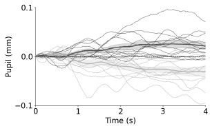
We hope that these additional visualizations make the effects of interest more transparent. Ultimately, however, the LME figure likely provides the information best, albeit at the cost of complexity.
(2) It is not clear why taking the first derivative of pupil size in Figure 5 would isolate the effect of arousal, eliminating those of luminance and contrast changes (in fact, one could argue for the opposite, since arousal effects are generally constant for extended periods of time while contrast effects are typically more local and transient).
First, please note that the results in 2.3.1 cannot be explained by task or context effects such as luminance and contrast: the exact same active color reporting task (same task and context) was presented to synesthetes and non-synesthetes.
Indeed, the reviewer is correct that the first derivative does not eliminate other concurrent pupil-driving effects, that was expressed wrongly in our original text. Indeed, any stimulus-locked effect, such as the luminance and contrast effects, but also the effort effect will reflect similarly in the derivative measure.
We did take the derivative because pupil responses driven by other non-trial related activity, such as increasing tiredness or excitement over the course of trials differ almost by necessity between participants, thus creating variability. However, these effects are most likely happening at a slower timescale and thus show less in the derivative measure. Accordingly in past research, we previously found clearer response-locked effects in the past when using a derivative measure (Douze et al., 2025; Ten Brink et al., 2024). This way, we also hoped to get rid of such variability that happens between participants for this between participant analysis.
Even if we were to use the same baseline corrected analysis, we would arrive at the same conclusion: we here directly compared baseline-corrected pupil sizes by taking individual differences into account (using a LME). In other words, we tested for the same question, but not relying on the derivative. We thus compared baseline-corrected pupil sizes using over-time LMEs. Group (active control vs. synesthete) gained significance between ~1.7s and 3s, aligning with the derivative-based result.
Author response image 2.
t-values of a per-time point LME predicting pupil response from group (synesthete/active control) Group reached significance.
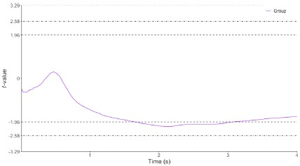
In sum, we deem the derivative more powerful/more appropriate in this context, but the interpretation of findings does not hinge on that analysis choice (as can be seen in the Author response image 2).
We corrected the claims on the derivative as a measure cleaning out other effects that indeed was oversimplified as it stood. We now write:
“Mental effort presents in task-evoked pupil dilations, yet other factors simultaneously affect the pupil, such as luminance and contrast changes at trial onset, as well as slower trends across the session (e.g., fatigue). To reduce the influence of these slower, non-trial-locked fluctuations while retaining the trial-evoked dynamics, we calculated the first derivative of the pupil time course to assess the velocity of pupillary changes (Butterworth filter, 18 Hz, order 3, 2.5 Hz lowpass, following our previous works [60, 61]).”
Douze, B. T., Ten Brink, A. F., Dijkerman, H. C., & Strauch, C. (2025). Pupil responses objectively index pharmacologically altered tactile sensitivity. Cortex, 193, 90-104.
Ten Brink, A. F., Heiner, I., Dijkerman, H. C., & Strauch, C. (2024). Pupil dilation reveals the intensity of touch. Psychophysiology, 61(6), e14538.
(3) It is a pity that responses to physical brightness modulations were only measured in the synesthete group, not in controls, as this would have allowed for ruling out differences in pupil reactivity across the two populations.
The reviewer is correct that this would allow additional comparisons, but argue that light responses in healthy control samples are very well documented and stereotypical. For instance, Bergamin & Kardon (2003) provide very systematic latency estimations, for low-luminance change stimuli in the realm of about 320ms that can accelerate to about 250ms for very strong luminance changes. Our relatively small luminance increments should thus be expected in this range. Indeed, this also well describes the response latencies we observed in synesthetes when exposed to the colored disks. While there is no detailed information about participants in Bergamin & Kardon (2003), data from previous studies shows very similar pupil light response profiles in a healthy student control population that matches our synesthetes well demographically (Strauch, Romein et al., 2022 Figure 2a, exact same lab as for the present study; Koevoet et al., 2025 Figure 3a). See also the further responses, baseline pupil size in millimeters across groups did not differ.
Together, we can safely conclude that pupil light responses in synesthetes are not different from pupil light responses in controls. We agree with the reviewer that this is a sensible point to also make in the manuscript:
“Specifically, pupil size first responded significantly to physical luminance after 330 ms (see Supplementary Figure 7 for per-timepoint LME; in line with response latencies of similar control populations, see Bergamin & Kardon [52], Koevoet et al. [40], and Strauch et al. [53]), but only responded significantly to synesthetic lightness at about 870 ms (see also Figure 3c vs e and Figure 4 for per-timepoint LME)”.
Bergamin, O., & Kardon, R. H. (2003). Latency of the pupil light reflex: sample rate, stimulus intensity, and variation in normal subjects. Investigative Ophthalmology & Visual Science, 44(4), 1546-1554.
Koevoet, D., Naber, M., Strauch, C. & Van der Stigchel, S. Presaccadic Attention Shifts Up-and Downwards: Evidence From the Pupil Light Response. Psychophysiology 62, e70047 (2025).
Strauch, C., Romein, C., Naber, M., Van der Stigchel, S., & Ten Brink, A. F. (2022). The orienting response drives pseudoneglect—Evidence from an objective pupillometric method. Cortex, 151, 259-271.
(4) Another concern is with the visualisation of the pupil traces in Figure 3 (main results); these were heavily pre-processed (per-participant demeaned), losing any feature besides the effect of interest and generating the unrealistic expectation that perception of dark/bright colors generate a net dilation/constriction of the pupil - whereas perception-related modulations of pupil size are always relative and generally small compared to the numerous other effects registered in pupil size. It would be far better to see the actual profiles, preserving the unfolding of dilations and constrictions over time, especially since these are further analysed in Figures 4 and 5.
Indeed, the expectation that any dark synesthetic experience would lead to pupil dilation whereas any bright synesthetic experience would lead to constriction is not warranted – it would only do that relative to the counterfactual of not having that experience.
Many factors affect the pupillary signal at the same time, and often differently across individuals (think of tiredness etc.), making merely baseline corrected traces seemingly noisy. Our visualization highlights that there is a systematic part to that variation that lies in the synesthetic brightness experience.
Visualizing the effects of idiosyncratic experiences, varying within and between participants is challenging. For the theoretical insight brought about through our paper in Figure 4 (synesthesia being sensory in nature), demeaning is favorable in our opinion as it isolates the effect of interest in visualization. However, for methodological reasons and to better show effect sizes etc., there is certainly use in additional transparency. We now thus provide non-demeaned traces in the supplementary material as the reviewer suggested and also refer to these in the main manuscript. Furthermore, all figures are now provided in millimeters, with all pupil related analysis being rerun and updated to this end (without qualitative changes to the results). This should further rectify possibly inflated expectations about the absolute size of effects and allows to put effects into perspective across studies. We now added:
“Pupillary data were transformed from arbitrary eyelink units to millimeters using a conversion factor obtained with an artificial eye (see Hayes & Petrov, 2016).”
Hayes, T. R., & Petrov, A. A. (2016). Mapping and correcting the influence of gaze position on pupil size measurements. Behavior research methods, 48(2), 510-527.
Impact:
Despite these weaknesses, and especially if they are adequately addressed in the review, this work is likely to improve our understanding of synesthesia, providing a new tool to quantify the subjective sensations; an interesting potential extension would be using pupillometry for tracking changes over time of the synesthetic experiences, opening up the possibility to evaluate the importance of learning for this peculiar experience.
We were happy to read our manuscript was evaluated this positively and hope that our replies can address the remaining smaller concerns and make findings more transparent to the readers.
Reviewer #2 (Public review):
Synesthesia is a neurological condition where stimulation of one sensory channel leads to involuntary, automatic, and consistent experience of another, unrelated percept. For example, Sir Francis Galton (1880, Nature) famously described the robust tendency of some individuals (synesthetes) to associate numerals with a distinct color. Ever since, synesthesia has continued to attract a broad interest in the cognitive neurosciences in light of its implications for the study of domains such as perception, consciousness, and brain connectivity, among others.
Strauch, Leenaars, and Rouw measured pupil size in a group of 16 grapheme-color synesthetes and two matched control groups. The participants were presented with gray digits - that is, visual stimuli having identical physical properties in terms of brightness. Each participant subsequently rated the corresponding evoked color and brightness: unlike controls, synesthetes did so in a very consistent and reliable fashion. Accordingly, this was also shown in their pupils: despite the same objective luminance, digits associated with brighter percepts caused their pupils to constrict, and digits associated with darker percepts caused their pupils to dilate more than controls. These results highlight how crossmodal correspondences are deeply rooted in synesthetes, and put forward pupillometry as a particularly appealing biomarker for some phenomenological experience (at least those grounded in "brightness").
Further strengths of the technique are its temporal resolution and its responsiveness to several constructs. Across several tasks, the authors show, for example, that responses to synesthetic light are somewhat slower than responses to real light (i.e., they are likely mediated), but at the same time faster than responses to mental imagery. The role of mental imagery can also be reasonably dismissed when considering the second feature of pupil size: its responsiveness to mental effort and cognitive load. The pupils tend to dilate with demanding, challenging tasks, and this was the case when control participants were asked to report the color of a digit for which they did not consistently experience a synesthetic association. The same task was, instead, seemingly effortless for synesthetes, again speaking in favor of the automaticity of number-color correspondences in their case.
Overall, the findings by Strauch, Leenaars, and Rouw are highly significant for the field and likely to be impactful. The strength of their evidence, when accounting for the relatively small sample size and the inherent variability of both phenomenology (color perception and subjective reporting) and physiology (pupil size), is adequate and sufficiently convincing.
We were glad to read this overall very positive assessment of our work and thank the reviewer for the additional non-public suggestions for improvements.
Reviewer #3 (Public review):
Summary:
In the present study, the authors examined pupillary responses to uncolored stimuli (number graphemes) among number-color synesthetes and non-synesthetes. After seeing a digit, the synesthetes and active control participants were asked to indicate which color they perceived using three dimensions of hue, saturation, and lightness. The lightness values were the primary independent variable for follow-up analyses. To see how the pupil responded to psychologically "bright" and "dark" digits, the authors split the reported lightness values at the median and plotted them. The synesthetes showed a pupillary constriction to digits they perceived as bright and dilation to digits they perceived as dark. Active control participants did not show that effect. In a subsequent block, only the synesthetes were shown the colors they reported perceiving as colored discs. Their pupillary responses were similar. The authors also found that the differences in pupillary responses between light and dark perceptions (with digits) were only slightly delayed in their onset to the perception of a colored disc, and therefore, the color perception accompanying a digit is unlikely to be effortful or a retrieved association, but occurs rather automatically.
Strengths:
The authors employed a well-controlled and designed quasi-experiment comparing colorgrapheme synesthetes to non-synesthetes and showed convincingly that the color perceptions accompanying graphemes alter the physical perception of brightness. They also made a reasoned attempt to rule out the possibility that color associations are occurring effortfully via retrieved associations.
We appreciate the positive assessment and useful suggestions for revision.
Weaknesses:
There are some areas in which the implications of these findings could be elaborated upon. I had the following questions:
(1) Are the pupillary responses among synesthetes, which objectively do not seem to match the degree of physical stimulation entering the retina, in any way maladaptive for eye functioning? I understand the constriction/dilation of the pupil to not only benefit visual acuity but also to protect the retina from damage. Are synesthetes at any risk of retinal damage due to over-dilation of the pupil to brighter stimuli? Or are these effects of a magnitude that is too small to matter? As reported in arbitrary units, it was hard to know how large these effects were in terms of measurable changes in dilation (e.g., millimeters).
This is an interesting point. Some argue that pupil size changes in a mid-range mildly affect optics thus affecting detection performance, contrast perception, and depth of field (Eberhardt et al., 2022, Mathôt & Ivanov 2019, Ruuskanen, Boehler, & Mathôt, 2025), rather than serving a protective role for the retina (Mathôt, 2018). Indeed, any effects reported here were quite small. We agree with the reviewer that this can be made more accessible by reporting effects in millimeters. We thus now adjusted all figures accordingly and write in the methods section:
“Pupillary data were transformed from arbitrary eyelink units to millimeters using a conversion factor obtained with an artificial eye (see Hayes & Petrov, 2016).”
Note that even the largest effects here (those elicited by physical luminance change in block 2 for the synesthetes) only caused differences in pupil size of about 0.3mm. This lies below the maximal pupil dilations observable in response maximal effort (about 0.5mm), for instance, and substantially below the full range of pupil size changes elicited through strong luminance stimulation (several millimeters). We therefore deem the changes in pupil size as obtained in our study too minor to be practically maladaptive for optics/perception.
Eberhardt, L. V., Strauch, C., Hartmann, T. S., & Huckauf, A. (2022). Increasing pupil size is associated with improved detection performance in the periphery. Attention, perception, & psychophysics, 84(1), 138-149.
Hayes, T. R., & Petrov, A. A. (2016). Mapping and correcting the influence of gaze position on pupil size measurements. Behavior research methods, 48(2), 510-527.
Mathôt, S., & Ivanov, Y. (2019). The effect of pupil size and peripheral brightness on detection and discrimination performance. PeerJ, 7, e8220.
Mathôt, S. (2018). Pupillometry: Psychology, physiology, and function. Journal of cognition, 1(1), 16.
Ruuskanen, V., Boehler, C. N., & Mathôt, S. (2025). The Interplay of Spontaneous Pupil-Size Fluctuations and EEG Power in Near-Threshold Detection. Psychophysiology, 62(3), e70035.
(2) Likewise, is the automatic synesthetic merging of two percepts something that could be learned such that natural synesthetes and "artificial" synesthetes would look similar? For example, if a group of non-synesthetic participants were to learn a color-grapheme association to automaticity, would you expect their pupillary responses to the graphemes look similar to the synesthetes'? If so (or if not), what would this tell us anything about the phenomenology of synesthesia?
We find this question most interesting. Likely, different synesthesia researchers wouldn’t even fully agree on the most plausible answers to these questions. Training studies have shown that nonsynesthetes can be trained to associate particular colors to particular graphemes, as revealed in the synesthetic Stroop effect: interference effects of the learned color onto reporting the typeface color of the grapheme. The degree to which non-synesthetes can be trained to become similar to synesthetes is however still topic of debate.
We now discuss as follows:
“Future studies could examine to what degree training a non-synesthete to associate specific colors to particular inducers (e.g., digits), can provide similar patterns of results as genuine synesthesia (Bor et al., 2014, Colizoli et al., 2012, Rothen & Meier, 2014). Could learning produce similar brightness-related pupil effects in non-synesthetes? Similarly, would effort-linked responses diminish with increased training duration? The perhaps most interesting question relates to response latencies: Would a trained participant ever be able to produce brightnessrelated pupil effects as fast as a synesthete?”
Bor, D., Rothen, N., Schwartzman, D. J., Clayton, S., & Seth, A. K. (2014). Adults can be trained to acquire synesthetic experiences. Scientific reports, 4(1), 7089.
Colizoli, O., Murre, J. M., & Rouw, R. (2012). Pseudo-synesthesia through reading books with colored letters. PloS one, 7(6), e39799.
Rothen, N., & Meier, B. (2014). Acquiring synaesthesia: insights from training studies. Frontiers in human neuroscience, 8, 109.
(3) Do the synesthetic perceptions of digit graphemes merge in a sensible way? For example, if a synesthete sees a particular color with the digit 1, and a different color with the digit 9, what do they perceive when they see 19? or 1-9, or 1 9? Is there color blending, or an altogether different color perception?
This is a very interesting question indeed. While each synesthete will have their own specific expression of synesthesia, there are regularities in how a combination of digits evokes synesthetic color. First, if asked about the color of a specific digit, each digit keeps its own color, as the color of a digit is linked to the identity of the digit (Dixon et al., 2006). Context effects are however possible, in particular when context alters the interpretation of the digit (Myles et al., 2003). A particularly common context in a multi-digit number is a dominant first digit, spreading its color to the subsequent digits in the number. However, as the digit color is linked to digit identity, what does ‘not’ happen is a mixing of colors into a qualitatively new color; for example, a yellow "1" and blue "9" do not merge into a green "19".
Dixon, M. J., Smilek, D., Duffy, P. L., Zanna, M. P., & Merikle, P. M. (2006). The role of meaning in grapheme-colour synaesthesia. Cortex, 42(2), 243-252.
Myles, K. M., Dixon, M. J., Smilek, D., & Merikle, P. M. (2003). Seeing double: The role of meaning in alphanumeric-colour synaesthesia. Brain and Cognition, 53(2), 342-345.
Many thanks for the constructive assessment of our work.
Recommendations for the authors:
Reviewer #1 (Recommendations for the authors):
(1) I am not sure I'd use the term 'cross-modal' given that the case considered here (graphemecolor) is purely visual.
The reviewer is absolutely right: the term 'cross-modal' has a historical background rather than reflecting an exact factual accuracy. The term is still commonly used however, as it readily reflects how the induced additional experience is always of a different (sub)type than the inducing experience. There is a cross-over between experiences that might occur within the same sensory modality, or even induce awareness of a particular concept. But key to synesthesia is the crossover experience as the inducer and concurrent are different (sub)types of experiences. For example, seeing a letter can evoke a synesthetic experience of seeing a color, or evoke awareness of a particular gender or personality of that letter, but does not evoke another letter. To remain consistent with literature, we refer to 'cross-modality' when explaining the link to previous literature, but generally switched to using 'cross-over experience':
“Therefore, synesthesia might provide a unique window into how the brain’s constructive processes can generate additional, conscious content, in cross-over experiences, often across modalities, going all the way down to the level of sensory phenomenology.”
We adjusted throughout the manuscript accordingly.
(2) I would not recommend focusing the introduction on the problem of qualia; this is a much more general and complex question than the one addressed in the study; the space of the introduction may be better used to present the actual object of study, giving a better picture of the synesthetic phenomenon and of previous work aimed at characterising it (behavioural, including PA scores and consistency measures, and neuroimaging). It is important to discuss how the pupillometric approach differs from the previously adopted neuroimaging techniques and what it can add to those.
We agree that qualia is a very general and complex question. However, we respectfully disagree that this complex question is not the object of the study. What is remarkable about synesthesia is not the presence of an additional perceptual association per se, but the presence of a specific perceptual experience. As illustration, think of a test where an unconscious color association to the word 'banana' was tested. While a generic 'yellow' could semantically be linked and would likely be obtained in the (e.g. priming) experimental results, a follow-up question of picking on a color wheel the exact shade of yellow to this association, or describing the perceptual sensation of the color, would be non-sensical to the participants.
This sharply contrasts with the current study: synesthetes, but not non-synesthetes, indicate a perceptual sensation of additional colors, and subsequently indeed the sensory properties of this percept (experienced brightness) affects the objective reflection of this sensation (pupil size) in synesthetes but not in non-synesthetes. In our view, the presence of additional qualia is key in understanding what sets synesthetic apart from non-synesthete associations, including so-called cross-modal correspondences (unconscious consistent associations across modalities, common to us all). We even believe that the reported qualia is what makes synesthesia so interesting in the first place. We now more clearly explain this link to qualia better in the introduction.
"The most remarkable aspect of synesthesia is the subjective perceptual phenomenology of the induced colors, setting these sensations apart from color memory, thought, or amodal association. The contrast between synesthetes and non-synesthetes can thus offer an interesting doorway into examining qualia, the subjective perceptual phenomenology or first person (what's-it-like) perspective."
We also improved the explanation of the synesthetic phenomenon, including a more detailed characterisation of behavioural measures (including consistency scores) and added neuroimaging studies. These changes have been incorporated into the text in response to previous comments (point 1- reviewer 1).
Please note that we have chosen not to include more detailed discussion of PA scores. Our results show a trend but do not allow for a conclusive interpretation on PA scores, and we feel that placing greater emphasis on this topic might therefore be confusing or even misleading. Still, it would be a very interesting topic for follow-up research to examine how alterations in characteristics of the synesthetic experience influence pupil responses.
The different synesthesia types all share the defining characteristics of an additional conscious and consistent experience. Synesthetes can verbally report their additional experience, and synesthetic sensations can be measured in behavioral paradigms such as the ’synesthetic Stroop’ effect, or brain activation patterns in sensory cortex [15]. Furthermore, test-retest paradigms show how synesthetic, but not non-synesthetic associations are highly specific and consistent [16-18]. Thus, over the past decades, research has established synesthesia as a ’real’ condition that can reliably be identified using behavior, neurophysiology, and neuroimaging [11, 13, 15–21]. The most remarkable aspect of synesthesia is the subjective perceptual phenomenology of the induced additional sensation, i.e., color in grapheme-color synesthesia. This sets synesthetic sensations apart from (color) memory, thought, or amodal association. Synesthesia can thus offer an interesting doorway into examining qualia, the subjective perceptual phenomenology or first person (what’s-it-like) perspective.
We now discuss the pupillometric approach as it differs from the previously adopted neuroimaging techniques as follows:
“Compared to neuroimaging studies [12,15,51], pupillometry may offer a more direct window into synesthetic phenomenology, as the directionality between pupil light reflex and perceived brightness is straightforward. Finally, improved understanding of the underlying processes can be obtained by contrasting responses to perceived versus actual (physical) brightness, given that the pupil light reflex is a well-characterised reflex arc involving few inferential steps.
This adds to the explanation that was already present on how the current approach differs from previous techniques, and what it can add to those techniques:
"Instead, current paradigms capturing synesthesia employ objective measures, but fail to capture its phenomenology [16, 17, 21, 23]."
(3) There are a few typos and word repetitions.
Many thanks – we identified typos and repetitions after another set of careful reads and hope to have eradicated them completely now.
Reviewer #2 (Recommendations for the authors):
I am overall very supportive of this work, but addressing the following points may enrich it further:
(1) Paragraph 2.2.1. Here, models do not seem to compare synesthetes versus controls but rather assess the effects of interest separately in the two groups. The fact that experimental effects are significant in synesthetes, but not in controls, does not tell us much about differences between groups. Controls (e.g., Figure 3) do show a similar trend, albeit clearly smaller. There is one passage in which this issue appears to be tackled (page 10): "Critically, in an LME ran on synesthetes and controls and using only graphemes and the interaction of group and lightness as predictors, we found lightness to predict pupil size in synesthetes (t = -2.754, p = 0.006), but not controls (t = -1.134, p = 0.257)." But I am not sure that the reported statistics belong to the interaction - they seem to refer to the lightness effect within each group, not the difference.
This is an important point, power for between-group comparisons is inherently limited for n = 16 per group (while still feasible for overall responses, things become trickier when less trials remain). A simple model of pupil ~ grapheme + group * lightness_scaled + (1 | participant) shows no significant interaction (despite one group showing the effect and the other not showing the effect significantly). The additional negative effect for group is in line with the effort-related effect reported later in the manuscript. Where does this leave us? Based on the lightness responses alone, the group difference can be characterized as a quantitative distinction, but the degree in which it is also a qualitative distinction cannot clearly be determined from current data. We revised the manuscript to make sure that such an interaction is not implied/ point to the absence of the significance of that interaction.
The sensory nature of synesthetic color is supported by within-synesthete analyses, where coupling strength parametrically modulates the lightness-pupil relationship in a theoretically predicted manner. Importantly, the effort-related findings provide a complementary and statistically robust group comparison: synesthetes and controls performing the identical colorreporting task showed significantly different pupil dilation rates, directly demonstrating that the two groups differ in how they access color information. Together, these two independent pupillometric signatures, one tracking perceptual quality, one tracking effort, converge on the same conclusion and mutually reinforce the interpretation that synesthetic color constitutes genuine sensory phenomenology.
Author response image 3.
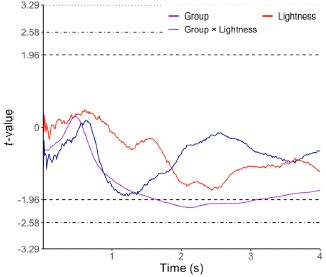
We now make this more explicit in the manuscript as follows:
“We found significant modulations of pupil size by the lightness of the grapheme's synesthetic color - sustained and in the to-be-expected time window. Specifically, the pupil constricted more for brighter reported colors, and dilated more for darker reported colors, as predicted (Average pupil size 800-4000ms, t = -3.601, p < 0.001). In an LME ran for synesthetes and controls and using only graphemes and lightness as predictors, we found lightness to predict pupil size in synesthetes (t = 2.844, p = 0.004), but not controls (t = 0.606, p = 0.544). However, when taking group as interacting factor in a joint LME, there was no interaction of lightness and group (t = -0.949 p = 0.342).”
and
“For controls a separate model was run, now without the PA score as predictor (not assessed for controls). Neither lightness (t = -0.815, p = 0.415), coupling strength (t = 0.438, p = 0.661), nor their interaction gained significance (t = -1.058, p = 0.290; all for average pupil size between 800 ms and 4000 ms). Critically, we also ran a LME with the three-way interaction of coupling strength, group, and lightness (Wilkinson notation: pupil = grapheme + group + lightness * group + coupling strength * lightness * group + (1 | participant)). This analysis revealed a significant three-way interaction between lightness, coupling strength, and group (F = 3.86, p = .021), indicating that the lightness × coupling strength effect on pupil size was not equivalent across groups. Decomposing this interaction by group, the lightness × coupling strength slope was significant in synesthetes (t = 2.59, p = .010) but not in controls (t=-1.01, p=.311), suggesting that reported lightness and its coupling strength were more consistently related to pupil size in synesthetes than in controls. Note however, that this decomposition does not directly test whether the two slopes significantly differ from each other, however. Lastly, pupil size was marginally larger in controls than in synesthetes (t = 1.94, p = .062; see later sections for more in-depth analyses)”
(2) The authors choose to analyze pupil size in arbitrary eye tracker units. This is fine, although I would recommend assessing and reporting whether the average pupil size (e.g., during the baseline) is roughly comparable between groups. The size of the effects may be difficult to compare between groups in the presence of very different baseline pupil size.
Please see Author response image 4 for Baseline pupil sizes per group in millimeters. There were no differences between groups.
Author response image 4.
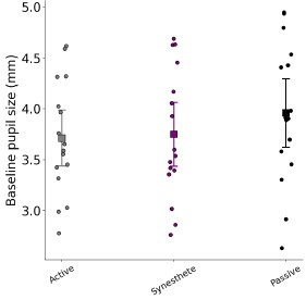
F2, 45) = 0.707, p = 0.499 (One-way Anova).
We now write:
“Baseline pupil sizes did not differ between groups (F(2, 45) = 0.707, p = 0.499).”
We agree with the reviewer that millimeters are a more intuitive measure and updated all figures throughout manuscript and supplementary materials accordingly. We also briefly added to signal processing that this conversion was applied.
“Pupillary data were transformed from arbitrary eyelink units to millimeters using a conversion factor obtained with an artificial eye (see Hayes & Petrov, 2016).”
Hayes, T. R., & Petrov, A. A. (2016). Mapping and correcting the influence of gaze position on pupil size measurements. Behavior research methods, 48(2), 510-527.
(3) If I understand correctly, the main task counted 120 trials overall (12 per digit). It seems, however, that only 3 and 4 participants remained with at least 50 trials (or 25 per median split by lightness) after preprocessing. This appears to be quite a massive data loss: is there a reason behind it? Please also clarify: the overall percentage of discarded trials; whether the median split by lightness was computed on all responses or only on those of the remaining, valid trials.
This is an important point for clarification indeed. The exclusion of participants in Figure 3 applies only to that particular visualization, not to the statistical analyses. The linear mixed effects models (LMEs) used all available valid trials from all participants, with no participant-level exclusions. The figure-specific threshold (≥25 trials per median-split bin) was applied purely for display clarity, as plotting participants with very few trials per bin would produce unreliable/noisy and thus visually misleading traces (as we note in the figure caption and point readers to Supplementary Figure 1, which shows the same visualization without any exclusions).
Since the paradigm required participants to repeat discarded trials until 120 valid trials were collected, all participants thus contributed exactly 120 valid trials to the analyses. There was therefore no data loss at the analysis level for the LME that is central to the claims of the manuscript (albeit more complex to grasp than the t-tests between bins).
Why were there sometimes so little trials per brightness bin?
First, participants differed in how dark or bright (synesthetic or forced-report) colors were overall, meaning that differing proportions thereof would fall above or below the 0.5 cutoff that overall, well represented the sample (but not necessarily every single participant). Note that this median split was not performed per individual but across all color reports to allow an apples-to-apples comparison.
Second, participants often reported colors that differed in Hue and Saturation, but not Lightness. This is in line with synesthetes picking certain colors more often than others, as compared with non-synesthetes (Rouw & Root, 2019; Ward et al., 2025).
We now include a new Supplementary Figure that visualizes responses on the Hue and Saturation dimensions of HSL space for both synesthetes and controls; fully saturated reports appear on the outer edge. We refer to the supplementary figure in the caption of Figure 2 as follows:
"See Supplementary Figure 1 for color reports on the hue and saturation axes.”
Rouw, R., & Root, N. B. (2019). Distinct colours in the ‘synaesthetic colour palette’. Philosophical Transactions of the Royal Society B: Biological Sciences, 374(1787).
Ward, J., Maciel, S., Rouw, R., Simner, J., & Root, N. (2025). Synaesthesia is linked to differences in music preference and musical sophistication and a distinctive pattern of sound-color associations. Psychology of Music, 53(3), 453-473.
Minor points:
(1) "Building on this evidence, we hypothesized that the cross modal color phenomenology in synesthesia can, if truly sensory in nature, could likewise be (...)" -> may need rephrasing (can/could).
Many thanks, fixed.
(2) Caption of Figure 1: "Block 2 (synesthetes only): a colored disk and gray central patch, matching the average indicated color per digit, and the number and luminance of pixels of said digit were presented to assess externally triggered light responses." -> I find this sentence a bit hard to follow; perhaps consider rephrasing it.
Agreed, we rephrased to:
Block 2 (synesthetes only): a colored disk was presented, colored according to the synesthete's average indicated color for that digit. At its center sat a gray patch matching the luminance and pixel area of the original digit from Block 1, together allowing assessment of externally triggered light responses.
(3) Figure 2 b: Consider truncating the y-axis to 1 if that improves the visualization.
We adjusted the axis accordingly and added a bit more detail in the caption for the interpretation of the measure.
(4) Caption of Figure 3 points to "see Supplementary Figure 1", but it should probably be SF2.
Many thanks for spotting, all references to supplementary figures have been checked and are corrected now.
Elvio Blini
Reviewer #3 (Recommendations for the authors):
(1) As a minor comment, there are some terms that felt overused in the manuscript. For example, the words "extraordinary" and "exceptional" were used multiple times throughout. I believe I understand the authors to mean them in their descriptive sense (i.e., outside the realm of typical experience), but in context, those words make it seem like they are touting their own experiment as "exceptional" or "extraordinary," which I don't believe was their intention.
We agree. We removed words such as exceptional and extraordinary when they do not directly refer to the sensation throughout the manuscript (which is indeed how we intended to use it). We hope that this removes unnecessary and convoluting hyperbole.
(2) It seemed counterintuitive to me that the color consistency score would be reverse-coded. In this case, the scores actually seem to indicate inconsistency, rather than consistency. Perhaps the raw scores can be inverted for a more intuitive interpretation that aligns with the terminology. I understand that they were following a previous publication in their method (Rothen et al., 2013).
This manner of coding is counter-intuitive indeed. However, there are both logical and practical reasons to this approach. Importantly, this is indeed the standard way of reporting color consistency in synesthesia research (Carmichael et al., 2015; Eagleman et al., 2007; Root et al., 2025; Rothen et al., 2013). The calculation is based on a simple logic; a higher number reflects a larger distance in color space. An additional advantage is the clear and intuitive zero- reference: a score of zero implies choosing the exact same color. Finally, it intuitively reflects the distinction between synesthetes and non-synesthetes; there is by definition little variation across synesthetes (visualized at the bottom of the graph), then a 'cut-off line' (if consistency is used as diagnostic tool), and then the height of the range shows how large the range in consistency is, in that particular sample of non-synesthetes. In a way we therefore inherit a confusing definition/standard, but changing it would lead to new confusion instead. We now specifically clarify this in the caption as follows:
“Note that higher consistency is reflected in lower color distance, hence lower values [17].”
Carmichael, D.A., Down, M.P., Shillcock, R.C., Eagleman, D.M., Simner, J., 2015. Validating a standardised test battery for synesthesia: does the synesthesia battery reliably detect synesthesia? Conscious. Cogn. 33, 375–385
Eagleman, D.M., Kagan, A.D., Nelson, S.S., Sagaram, D., Sarma, A.K., 2007. A standardized test battery for the study of synesthesia. J. Neurosci. Methods 159 (1), 139–145.
Root, N., Chkhaidze, A., Melero, H., Sidoro -Dorso, A., Volberg, G., Zhang, Y., & Rouw, R. (2025). How “diagnostic” criteria interact to shape synesthetic behavior: The role of self-report and test–retest consistency in synesthesia research. Consciousness and Cognition, 129, 103819.
Rothen, N., Seth, A.K., Witzel, C., Ward, J., 2013. Diagnosing synaesthesia with online colour pickers: maximising sensitivity and specificity. J. Neurosci. Methods 215 (1), 156–160.
-
eLife Assessment
This study used pupillometry to provide an objective assessment of a form of synesthesia in which people see additional color when reading numbers. It provides convincing evidence that subjective color ratings are matched by changes in pupil size that recapitulate brightness-mediated changes when exposed to the real color. The work provides a valuable contribution to the literature on both synesthetic perception and the use of pupillometry to probe perception and related psychological processes.
-
Reviewer #1 (Public review):
Summary:
Knowing that small pupil-size variations accompany brightness variations (even when these are illusory), the authors asked whether pupil constrictions would accompany the synesthetic perception of a brighter color (compared with a darker one), induced by the presentation of a black-white character. This grapheme-colour synesthesia is only experienced by a few participants, sixteen of whom were enrolled in this study. The results reliably showed that a relative pupil constriction would "betray" the perception of a brighter color in these participants, while no such effect would be observed in control participants who were asked to report a color in association with each grapheme, even though they did not perceive any.
Strengths:
The main strength of the study lies in its combination of psychophysics …
Reviewer #1 (Public review):
Summary:
Knowing that small pupil-size variations accompany brightness variations (even when these are illusory), the authors asked whether pupil constrictions would accompany the synesthetic perception of a brighter color (compared with a darker one), induced by the presentation of a black-white character. This grapheme-colour synesthesia is only experienced by a few participants, sixteen of whom were enrolled in this study. The results reliably showed that a relative pupil constriction would "betray" the perception of a brighter color in these participants, while no such effect would be observed in control participants who were asked to report a color in association with each grapheme, even though they did not perceive any.
Strengths:
The main strength of the study lies in its combination of psychophysics (brightness ratings) and pupillometry, which allowed for showing clear-cut results.
Weaknesses:
Some relatively minor weaknesses concern the ancillary analyses, which tackle secondary questions and are not entirely convincing.
(1) The linear mixed model approach is a powerful way to identify important variables, but it does not clarify whether the key factors are between-subject or between-trial variations. Some variables are inherently defined at a subject level (e.g., PA scores), others are not. I would strongly recommend an alternative visualisation of the results to examine inter-individual variability.
(2) It is not clear why taking the first derivative of pupil size in Figure 5 would isolate the effect of arousal, eliminating those of luminance and contrast changes (in fact, one could argue for the opposite, since arousal effects are generally constant for extended periods of time while contrast effects are typically more local and transient).
(3) It is a pity that responses to physical brightness modulations were only measured in the synesthete group, not in controls, as this would have allowed for ruling out differences in pupil reactivity across the two populations.
(4) Another concern is with the visualisation of the pupil traces in Figure 3 (main results); these were heavily pre-processed (per-participant demeaned), losing any feature besides the effect of interest and generating the unrealistic expectation that perception of dark/bright colors generate a net dilation/constriction of the pupil - whereas perception-related modulations of pupil size are always relative and generally small compared to the numerous other effects registered in pupil size. It would be far better to see the actual profiles, preserving the unfolding of dilations and constrictions over time, especially since these are further analysed in Figures 4 and 5.
Impact:
Despite these weaknesses, and especially if they are adequately addressed in the review, this work is likely to improve our understanding of synesthesia, providing a new tool to quantify the subjective sensations; an interesting potential extension would be using pupillometry for tracking changes over time of the synesthetic experiences, opening up the possibility to evaluate the importance of learning for this peculiar experience.
-
Reviewer #2 (Public review):
Synesthesia is a neurological condition where stimulation of one sensory channel leads to involuntary, automatic, and consistent experience of another, unrelated percept. For example, Sir Francis Galton (1880, Nature) famously described the robust tendency of some individuals (synesthetes) to associate numerals with a distinct color. Ever since, synesthesia has continued to attract a broad interest in the cognitive neurosciences in light of its implications for the study of domains such as perception, consciousness, and brain connectivity, among others.
Strauch, Leenaars, and Rouw measured pupil size in a group of 16 grapheme-color synesthetes and two matched control groups. The participants were presented with gray digits - that is, visual stimuli having identical physical properties in terms of brightness. …
Reviewer #2 (Public review):
Synesthesia is a neurological condition where stimulation of one sensory channel leads to involuntary, automatic, and consistent experience of another, unrelated percept. For example, Sir Francis Galton (1880, Nature) famously described the robust tendency of some individuals (synesthetes) to associate numerals with a distinct color. Ever since, synesthesia has continued to attract a broad interest in the cognitive neurosciences in light of its implications for the study of domains such as perception, consciousness, and brain connectivity, among others.
Strauch, Leenaars, and Rouw measured pupil size in a group of 16 grapheme-color synesthetes and two matched control groups. The participants were presented with gray digits - that is, visual stimuli having identical physical properties in terms of brightness. Each participant subsequently rated the corresponding evoked color and brightness: unlike controls, synesthetes did so in a very consistent and reliable fashion. Accordingly, this was also shown in their pupils: despite the same objective luminance, digits associated with brighter percepts caused their pupils to constrict, and digits associated with darker percepts caused their pupils to dilate more than controls. These results highlight how crossmodal correspondences are deeply rooted in synesthetes, and put forward pupillometry as a particularly appealing biomarker for some phenomenological experience (at least those grounded in "brightness").
Further strengths of the technique are its temporal resolution and its responsiveness to several constructs. Across several tasks, the authors show, for example, that responses to synesthetic light are somewhat slower than responses to real light (i.e., they are likely mediated), but at the same time faster than responses to mental imagery. The role of mental imagery can also be reasonably dismissed when considering the second feature of pupil size: its responsiveness to mental effort and cognitive load. The pupils tend to dilate with demanding, challenging tasks, and this was the case when control participants were asked to report the color of a digit for which they did not consistently experience a synesthetic association. The same task was, instead, seemingly effortless for synesthetes, again speaking in favor of the automaticity of number-color correspondences in their case.
Overall, the findings by Strauch, Leenaars, and Rouw are highly significant for the field and likely to be impactful. The strength of their evidence, when accounting for the relatively small sample size and the inherent variability of both phenomenology (color perception and subjective reporting) and physiology (pupil size), is adequate and sufficiently convincing.
-
Reviewer #3 (Public review):
Summary:
In the present study, the authors examined pupillary responses to uncolored stimuli (number graphemes) among number-color synesthetes and non-synesthetes. After seeing a digit, the synesthetes and active control participants were asked to indicate which color they perceived using three dimensions of hue, saturation, and lightness. The lightness values were the primary independent variable for follow-up analyses. To see how the pupil responded to psychologically "bright" and "dark" digits, the authors split the reported lightness values at the median and plotted them. The synesthetes showed a pupillary constriction to digits they perceived as bright and dilation to digits they perceived as dark. Active control participants did not show that effect. In a subsequent block, only the synesthetes were …
Reviewer #3 (Public review):
Summary:
In the present study, the authors examined pupillary responses to uncolored stimuli (number graphemes) among number-color synesthetes and non-synesthetes. After seeing a digit, the synesthetes and active control participants were asked to indicate which color they perceived using three dimensions of hue, saturation, and lightness. The lightness values were the primary independent variable for follow-up analyses. To see how the pupil responded to psychologically "bright" and "dark" digits, the authors split the reported lightness values at the median and plotted them. The synesthetes showed a pupillary constriction to digits they perceived as bright and dilation to digits they perceived as dark. Active control participants did not show that effect. In a subsequent block, only the synesthetes were shown the colors they reported perceiving as colored discs. Their pupillary responses were similar. The authors also found that the differences in pupillary responses between light and dark perceptions (with digits) were only slightly delayed in their onset to the perception of a colored disc, and therefore, the color perception accompanying a digit is unlikely to be effortful or a retrieved association, but occurs rather automatically.
Strengths:
The authors employed a well-controlled and designed quasi-experiment comparing color-grapheme synesthetes to non-synesthetes and showed convincingly that the color perceptions accompanying graphemes alter the physical perception of brightness. They also made a reasoned attempt to rule out the possibility that color associations are occurring effortfully via retrieved associations.
Weaknesses:
There are some areas in which the implications of these findings could be elaborated upon. I had the following questions:
(1) Are the pupillary responses among synesthetes, which objectively do not seem to match the degree of physical stimulation entering the retina, in any way maladaptive for eye functioning? I understand the constriction/dilation of the pupil to not only benefit visual acuity but also to protect the retina from damage. Are synesthetes at any risk of retinal damage due to over-dilation of the pupil to brighter stimuli? Or are these effects of a magnitude that is too small to matter? As reported in arbitrary units, it was hard to know how large these effects were in terms of measurable changes in dilation (e.g., millimeters).
(2) Likewise, is the automatic synesthetic merging of two percepts something that could be learned such that natural synesthetes and "artificial" synesthetes would look similar? For example, if a group of non-synesthetic participants were to learn a color-grapheme association to automaticity, would you expect their pupillary responses to the graphemes look similar to the synesthetes'? If so (or if not), what would this tell us anything about the phenomenology of synesthesia?
(3) Do the synesthetic perceptions of digit graphemes merge in a sensible way? For example, if a synesthete sees a particular color with the digit 1, and a different color with the digit 9, what do they perceive when they see 19? or 1-9, or 1 9? Is there color blending, or an altogether different color perception?
-
-
