Symmetric brain-liver circuits mediate lateralized regulation of hepatic glucose output in mice
Curation statements for this article:-
Curated by eLife

eLife Assessment
This important study combines anatomical tracing, tissue clearing, and functional manipulations to demonstrate lateralized brainstem control of hepatic glucose metabolism and identify a site of sympathetic nerve crossover supplying the liver. The evidence supporting the anatomical organization of hepatic sympathetic innervation is compelling, and the functional studies provide solid support for a role of asymmetric sympathetic outflow in regulating glucose homeostasis. While some uncertainty remains regarding the contribution of sensory innervation and the extent to which these findings generalize beyond mice, the work provides an invaluable advance in understanding neural regulation of liver metabolism.
This article has been Reviewed by the following groups
Listed in
- Evaluated articles (eLife)
Abstract
Neural lateralization is well recognized in the control of contralateral somatic movement, yet its relevance to visceral organ regulation remains poorly understood. This study aimed to determine whether the central nervous system exerts lateralized control over hepatic glucose metabolism and to localize the site of peripheral sympathetic crossover to the liver. Pseudorabies virus (PRV) tracing revealed bilateral projections from the lateral paragigantocellular nucleus (LPGi) with preferential innervation of the contralateral hepatic lobes. Unilateral LPGi activation elevated systemic glucose by enhancing glycogenolysis and gluconeogenesis specifically in contralateral lobes, whereas bilateral activation produced additive effects. Following unilateral hepatic denervation, contralateral LPGi activation induced metabolic compensation in the remaining innervated lobes, characterized by increased norepinephrine release, glucose production, and glycogen depletion. Whole-mount tissue clearing and dual viral tracing localized the sympathetic crossover to the porta hepatis. Developmental analysis showed that lobar-specific innervation along the vasculature emerges by postnatal week 2. These findings demonstrate that the brainstem can exert lobe-specific, lateralized control of hepatic glucose metabolism via bilaterally projecting brain – liver sympathetic pathways. This contralateral regulation arises from a peripheral decussation at the porta hepatis, and the compensatory activation observed after denervation reveals an intrinsic neuroadaptive mechanism that helps safeguard systemic glucose homeostasis.
Article activity feed
-

-
-

eLife Assessment
This important study combines anatomical tracing, tissue clearing, and functional manipulations to demonstrate lateralized brainstem control of hepatic glucose metabolism and identify a site of sympathetic nerve crossover supplying the liver. The evidence supporting the anatomical organization of hepatic sympathetic innervation is compelling, and the functional studies provide solid support for a role of asymmetric sympathetic outflow in regulating glucose homeostasis. While some uncertainty remains regarding the contribution of sensory innervation and the extent to which these findings generalize beyond mice, the work provides an invaluable advance in understanding neural regulation of liver metabolism.
-
Reviewer #2 (Public review):
[Editors' note: this version has been assessed by the Reviewing Editor without further input from the original reviewers. The authors have addressed the comments raised in the previous round of review.]
Summary:
The manuscript by Wang and colleagues aims to determine whether hepatic glucose metabolism is differentially regulated by the left and right sides of the LPGi and to reveal decussation of hepatic sympathetic nerves.
The authors used tissue clearing to identify sympathetic fibers in the liver lobes, then injected PRV into the hepatic lobes. Five days post-injection, PRV-labeled neurons in the LPGi were identified. The results indicated contralateral dominance of premotor neurons and partial innervation of more than one lobe. The authors then activated each side of the LPGi, resulting in a greater …
Reviewer #2 (Public review):
[Editors' note: this version has been assessed by the Reviewing Editor without further input from the original reviewers. The authors have addressed the comments raised in the previous round of review.]
Summary:
The manuscript by Wang and colleagues aims to determine whether hepatic glucose metabolism is differentially regulated by the left and right sides of the LPGi and to reveal decussation of hepatic sympathetic nerves.
The authors used tissue clearing to identify sympathetic fibers in the liver lobes, then injected PRV into the hepatic lobes. Five days post-injection, PRV-labeled neurons in the LPGi were identified. The results indicated contralateral dominance of premotor neurons and partial innervation of more than one lobe. The authors then activated each side of the LPGi, resulting in a greater increase in blood glucose levels after right-sided activation than after left-sided activation, and in changes in protein expression in the liver lobes. These data suggested lobe-specific modulation of HGP. Chemical denervation of a particular lobe did not affect glucose levels due to compensation by the other lobes. In addition, nerve bundles decussate in the hepatic portal region.
Strengths:
The manuscript is timely and relevant. It is important to understand the sympathetic regulation of the liver and the contribution of each lobe to hepatic glucose production. The authors use state-of-the-art methodology.
Weaknesses:
(1) Image clarity was improved in some cases, but not in others. For example, Figure 3I, showing c-Fos expression, is not convincing due to the image quality and lack of orientation.
(2) The methods section states that 8-week-old male mice were used in the experiments without specifying the experiments (e.g., brain injection with AAVs or PRV organ inoculation). The authors should include these details.
(3) The authors should use the exact location of pre- and postganglionic neurons, as they often refer to neurons in the sympathetic chain. Their findings should be compared with the existing literature on the location of preganglionic cells.
(4) Figure legends should be revised and matched with the text.
-
Reviewer #4 (Public review):
Summary of General Strengths & Weaknesses:
The studies here are highly informative for anatomical tracing and sympathetic nerve function in the liver in relation to glucose levels, but because they are conducted in a single species, it is challenging to translate them to humans or determine whether these neural circuits are evolutionarily conserved. Dual-labeling anatomical studies are elegant, and the addition of chemogenetic and optogenetic studies provides mechanistically informative. Denervation studies lack proper controls, and sensory innervation in the liver is overlooked.
Specific Weaknesses - Major:
(1) The species name should be included in the title.
(2) Tyrosine hydroxylase was used to mark sympathetic fibers in the liver, but this marker also labels a portion of sensory fibers that need to be …
Reviewer #4 (Public review):
Summary of General Strengths & Weaknesses:
The studies here are highly informative for anatomical tracing and sympathetic nerve function in the liver in relation to glucose levels, but because they are conducted in a single species, it is challenging to translate them to humans or determine whether these neural circuits are evolutionarily conserved. Dual-labeling anatomical studies are elegant, and the addition of chemogenetic and optogenetic studies provides mechanistically informative. Denervation studies lack proper controls, and sensory innervation in the liver is overlooked.
Specific Weaknesses - Major:
(1) The species name should be included in the title.
(2) Tyrosine hydroxylase was used to mark sympathetic fibers in the liver, but this marker also labels a portion of sensory fibers that need to be ruled out in whole-mount imaging data.
(3) Chemogenetic and optogenetic data demonstrating hyperglycemia should be described in the context of prior work demonstrating liver nerve involvement in these processes. The Discussion currently mentions this only briefly, but comparing methods and observations would be helpful.
(4) Sympathetic denervation with 6-OHDA can drive compensatory increases in tissue sensory innervation, and this should be measured in the liver denervation studies to implicate potential crosstalk, especially given the increase in LPGi cFOS that may be due to afferent nerve activity. Compensatory sympathetic drive may not be the only culprit, though that is clearly assumed. The sensory or parasympathetic/vagal innervation of the liver is altogether ignored in this paper and could be better described in general.
Comments on the revised version.
Across all reviewer comments, the revised resubmission has adequately addressed all concerns.
-
Author response:
The following is the authors’ response to the previous reviews
Public Reviews:
Reviewer #2 (Public review):
Summary:
The manuscript by Wang and colleagues aims to determine whether hepatic glucose metabolism is differentially regulated by the left and right sides of the LPGi and to reveal decussation of hepatic sympathetic nerves.
The authors used tissue clearing to identify sympathetic fibers in the liver lobes, then injected PRV into the hepatic lobes. Five days post-injection, PRV-labeled neurons in the LPGi, which were identified. The results indicated contralateral dominance of premotor neurons and partial innervation of more than one lobe. Then the authors activated each side of the LPGi, resulting in a greater increase in blood glucose levels after right-sided activation than after left-sided activation, …
Author response:
The following is the authors’ response to the previous reviews
Public Reviews:
Reviewer #2 (Public review):
Summary:
The manuscript by Wang and colleagues aims to determine whether hepatic glucose metabolism is differentially regulated by the left and right sides of the LPGi and to reveal decussation of hepatic sympathetic nerves.
The authors used tissue clearing to identify sympathetic fibers in the liver lobes, then injected PRV into the hepatic lobes. Five days post-injection, PRV-labeled neurons in the LPGi, which were identified. The results indicated contralateral dominance of premotor neurons and partial innervation of more than one lobe. Then the authors activated each side of the LPGi, resulting in a greater increase in blood glucose levels after right-sided activation than after left-sided activation, and in changes in protein expression in the liver lobes. These data suggested lobe-specific modulation of HGP. Chemical denervation of a particular lobe did not affect glucose levels due to compensation by the other lobes. In addition, nerve bundles decussate in the hepatic portal region.
Strengths:
The manuscript is timely and relevant. It is important to understand the sympathetic regulation of the liver and the contribution of each lobe to hepatic glucose production. The authors use state-of-the-art methodology.
Weaknesses:
(1) Image clarity was improved in some cases, but not in others. For example, Figure 3I, showing c-Fos expression, is not convincing due to the image quality and lack of orientation.
We sincerely apologize for the insufficient image clarity and anatomical orientation in the original Figure 3I. To resolve this issue, we have performed the following revisions in the revised Figure 3I:
(1) Replaced the original panels with the high-resolution confocal images showing clear c-FOS immunofluorescence in the LPGi.
(2) Included explicit anatomical orientation indicators (Bregma −6.75 mm) to clearly demarcate the boundaries of the LPGi.
(3) Added ROI outlines surrounding the LPGi region.
(2) The methods section states that 8-weeks-old male mice were used in the experiments without specifying the experiments (e.g., brain injection with AAVs or PRV organ inoculation). The authors should include these details.
We thank the reviewer pointing out this oversight. We have updated the Methods section under "Animals" and specific procedure subsections to clearly state the exact age of animals.
(1) For retrograde trans-synaptic PRV tracing, 8-week-old mice received intrahepatic viral injections and were sacrificed 5 days post-injection.
(2) For chemogenetic and optogenetic manipulations, stereotaxic AAV injections were performed at 8 weeks of age. Mice were allowed 4 weeks for viral expression and recovery before undergoing metabolic tests or light stimulation at 12 weeks of age.
(3) For chemical denervation (6-OHDA), 8-week-old mice were injected into targeted lobes and examined 7 days post-denervation.
(4) For postnatal innervation mapping, neonatal mice at postnatal week 0 (P0), week 1 (P7), and week 2 (P14) were harvested for tissue clearing.
(3) The authors should use the exact location of pre- and postganglionic neurons as they often refer to neurons in the sympathetic chain. Their findings should be compared with the existing literature on the location of preganglionic cells.
We appreciate the reviewer for this feedback. We agree that our original description lacked precise anatomical localization regarding the pre- and postganglionic neurons, and it was inaccurate to state that descending fibers pass through the sympathetic chain (SyC).
Based on our whole-mount tissue clearing data, we observed that the preganglionic neurons of the brain-liver sympathetic circuit are primarily located in the T6–T12 segments of the thoracic spinal cord. Accordingly, we have revised the text in Results 4 to specify these exact locations.
Manuscript Revision (Results 4):
"Using whole-mount clearing, we visualized the brain–liver sympathetic circuit and found that preganglionic neurons in the thoracic spinal cord (T6–T12) send descending fibers via the splanchnic nerves to innervate postganglionic neurons in the CG-SMG (Figure 4A)."
Furthermore, following your valuable suggestion to compare our findings with existing literature, we reviewed a recent study published in Nature Communications (Harima, Yukiko et al. Parallel labeled-line organization of sympathetic outflow for selective organ regulation in mice. Nat Commun. 2024;15(1):10478). In that study, researchers injected retrogradely transducible AAVs directly into the CG-SMG and traced the preganglionic neurons predominantly to the T8–T13 segments. Their results are largely consistent with our findings. Interestingly, the broader anatomical range observed in our trans-synaptic liver-to-brain mapping (T6–T12) compared to their CG-SMG-specific tracing (T8–T13) reveals a slight discrepancy. This observation suggests an intriguing anatomical hypothesis: a subset of sympathetic preganglionic nerves may bypass the CG-SMG relay entirely and project directly to the liver.
(4) Figure legends should be revised and matched with the text.
We apologize for the oversight. We have conducted a comprehensive audit of all figure and legends to ensure precise matching between the main text and the figures.
Specifically, we have corrected a typographical error in the Figure 1 Legend where panel (C) was mistakenly labeled as a second panel (B), and we fixed a spelling error ("LPG" corrected to "LPGi"). Additionally, we corrected a miscitation in Results (Section 3) regarding Figure 3. In the original text, Figure 3C was incorrectly grouped with blood glucose data, whereas it actually displays the Western blot validation of sympathetic denervation.
We have revised the corresponding sections in the manuscript as follows:
Manuscript Revision (Figure 1 Legend):
“(C) Quantification of PRV-labeled neurons in left and right LPGi across different hepatic lobes: left lateral, median, right posterior, right anterior, caudate, and porta hepatis (n = 3).
(D) Sankey diagram showing projection patterns from left and right LPGi to individual hepatic lobes. (E and F) Representative slices of EGFP+ and mRFP+ neurons in left (top) and right (bottom) LPGi following PRV-EGFP (right anterior lobe) and PRV-mRFP (median lobe) injections. Proportions of EGFP+, mRFP+, and co-labeled neurons in left and right LPGi (F, n = 3). Scale bars, 100 μm.”
Manuscript Revision (Results 3):
“Despite the absence of directly sympathetic input to denervated lobes, systemic blood glucose levels were unchanged compared with controls (Figures 3A-3B, Figure S5A), indicating functional compensation through the remaining intact liver.”
Reviewer #4 (Public review):
Summary of General Strengths & Weaknesses:
The studies here are highly informative for anatomical tracing and sympathetic nerve function in the liver in relation to glucose levels, but because they are conducted in a single species, it is challenging to translate them to humans or determine whether these neural circuits are evolutionarily conserved. Dual-labeling anatomical studies are elegant, and the addition of chemogenetic and optogenetic studies provides mechanistically informative. Denervation studies lack proper controls, and sensory innervation in the liver is overlooked.
We sincerely thank the reviewer for their time and evaluation. We respectfully note that these comments mirror those raised during the previous round of review. We would like to kindly direct the reviewer to the extensive revisions we implemented in our previous resubmission, which directly and comprehensively addressed these exact concerns. These revisions remain intact in the current version of the manuscript. Below, we briefly summarize how each point was previously addressed for your convenience.
Specific Weaknesses - Major:
(1) The species name should be included in the title.
As addressed in our previous revision, we fully agree with this suggestion. We updated the title of the manuscript to explicitly include the species: "Symmetric brain-liver circuits mediate lateralized regulation of hepatic glucose output in mice." We also clarified the species used throughout the main text to ensure accuracy.
(2) Tyrosine hydroxylase was used to mark sympathetic fibers in the liver, but this marker also labels a portion of sensory fibers that need to be ruled out in whole-mount imaging data.
As detailed in our previous response, we acknowledge this important limitation. In our prior revision, we addressed this concern through both additional data analysis and text revisions:
(1) We provided SyGlass 3D reconstruction data demonstrating that the TH-positive nerve fibers originate from the celiac-superior mesenteric ganglia (CG-SMG), a well-established sympathetic ganglion (Figure S5F).
(2) In parallel, we collected dorsal root ganglia (DRG) from spinal segments T1-6 and T7-12 five days after intrahepatic PRV injection. While the T7-12 DRG segments are historically known to contain the sensory neurons that innervate the liver (Anat Rec A Discov Mol Cell Evol Biol. 2004; Auton Neurosci. 2024), we detected only a remarkably sparse number of PRV-positive neurons in these segments. This effectively functionally distinguishes this efferent pathway from primary sensory afferents (Supplementary figure B).
(3) We explicitly added this methodological limitation to the Discussion section (paragraph 6) of the current manuscript, noting that more selective approaches, such as genetic targeting of sympathetic lineages, will be important for future validation."
(3) Chemogenetic and optogenetic data demonstrating hyperglycemia should be described in the context of prior work demonstrating liver nerve involvement in these processes. There is only a brief mention in the Discussion currently, but comparing methods and observations would be helpful.
As outlined in our previous response, we incorporated this crucial context into our revised manuscript. Specifically, we expanded the Discussion section (paragraph 3) to contrast our precise cell-type-specific chemogenetic and optogenetic approaches with historical studies that relied on coarse electrical stimulation. This addition highlights how our current methodology reveals the contralateral and lobe-specific architecture of brain-liver sympathetic control that was previously obscured.
(4) Sympathetic denervation with 6-OHDA can drive compensatory increases in tissue sensory innervation, and this should be measured in the liver denervation studies to implicate potential crosstalk, especially given the increase in LPGi cFOS that may be due to afferent nerve activity. Compensatory sympathetic drive may not be the only culprit, though that is clearly assumed. The sensory or parasympathetic/vagal innervation of the liver is altogether ignored in this paper and could be better described in general.
We appreciate this insightful physiological perspective, which we addressed comprehensively in our previous revision. As we previously agreed, the central nervous system integrates a broad range of afferent signals, and compensatory sensory or parasympathetic mechanisms likely contribute to the observed LPGi activation following hepatic sympathetic denervation.
To address this, we significantly expanded our Discussion section (paragraph 4) in the prior revision. We explicitly proposed a model wherein hepatic glucose production is regulated by an integrated afferent-central-efferent loop, acknowledging that our current study primarily resolves the efferent component. We clearly noted the lack of direct assessment of sensory or parasympathetic innervation as a limitation and highlighted this dynamic crosstalk as a critical avenue for future investigation.
Comments on the revised version.
Across all reviewer comments, the revised resubmission has adequately addressed all concerns.
Recommendations for the authors:
Reviewer #4 (Recommendations for the authors):
No further recommendations aside from tempering the CGRP language, as marking all sensory fibers.
We appreciate the reviewer for pointing out this important anatomical distinction. We entirely agree that CGRP specifically labels peptidergic sensory afferents and does not represent the entirety of the sensory nervous system.
We have carefully reviewed the entire manuscript and tempered our language accordingly. Wherever CGRP is mentioned, we have clarified that it serves as a marker for peptidergic sensory fibers, rather than functioning as a pan-sensory marker.
Manuscript Revision (Results 1):
"Unlike the NTS, a well-established hepatic sensory center served here as a positive control, the LPGi contained few CGRP-positive cell bodies (Figure S1G), indicating a lack of peptidergic sensory projections."
-
eLife Assessment
This fundamental study, supported by convincing evidence, identifies the specific neuronal components that enable HIF-1 signaling in specific serotonergic neurons to regulate effectors in peripheral tissues. Specifically, stabilizing HIF-1 in ADF and NSM serotonergic neurons is sufficient to extend lifespan and healthspan in C. elegans, and these physiological benefits require several oxygen-sensing neurons, as well as neurotransmitters (GABA and tyramine), and a neuropeptide (NLP-17). The strengths of the study include rigorous experimental design, high quality data, the use of multiple genetic tools, and functional dissection using the well-validated C. elegans model.
-
Reviewer #1 (Public review):
[Editors' note: this version has been assessed by the Reviewing Editor without further input from the original reviewers. The authors have addressed the comments raised in the previous round of review.]
Summary:
In this study by Kitto et al., the authors set out to identify specific signaling components regulating the hypoxic response from the neurons to the periphery and which components are required for lifespan extension. Their previous work had shown that expression of a stabilized HIF-1 mutant in the nervous system extends lifespan through the serotonin receptor SER-7 and leads to the induction of fmo-2 in the intestine. In the current study, they mapped the precise neural circuits required for this response, as well as the signaling mediators. Their work reveals that neurotransmitters GABA and …
Reviewer #1 (Public review):
[Editors' note: this version has been assessed by the Reviewing Editor without further input from the original reviewers. The authors have addressed the comments raised in the previous round of review.]
Summary:
In this study by Kitto et al., the authors set out to identify specific signaling components regulating the hypoxic response from the neurons to the periphery and which components are required for lifespan extension. Their previous work had shown that expression of a stabilized HIF-1 mutant in the nervous system extends lifespan through the serotonin receptor SER-7 and leads to the induction of fmo-2 in the intestine. In the current study, they mapped the precise neural circuits required for this response, as well as the signaling mediators. Their work reveals that neurotransmitters GABA and tyramine, and the neuropeptide NLP-17, act downstream of neuronal HIF-1 to convey a "hypoxic signal" to peripheral tissues. Through cell-type-specific expression studies, targeted knockouts, and comprehensive lifespan analysis, the authors provide robust evidence to support their conclusions. The insights gained from the study are both moving the field forward as they advance our understanding of neuro-peripheral hypoxic signaling, but they also lay the groundwork for potential therapeutic strategies aimed at the modulation of such signaling pathways.
Strengths:
(1) This study provides new evidence further delineating signaling components required for hypoxic signaling-mediated longevity, from the nervous system to the periphery. Using a rigorous approach where they express stabilized HIF-1 mutant selectively in ADF, NSM, and HSN serotonergic neurons, followed by cell-type-specific tph-1 knockouts to pinpoint ADF-dependent serotonin signaling as essential for both lifespan extension and intestinal fmo-2 induction.
This was followed by generating 11 transgenic lines that drive SER-7 expression under distinct neuron-specific promoters, to systematically tease out in which of 27 candidate neurons SER-7 functions to mediate hypoxia-induced longevity. This ultimately highlighted the RIS interneuron as the required signaling hub.
(2) As the intestine lacks direct neuronal innervation, the authors employ neuron-specific RNAi (TU3311 strain) and dense core vesicle analyses to identify that the neuropeptide NLP-17 is required to transmit the hypoxic signal from RIS to induce fmo-2 in the intestine.
(3) Overall, the paper is very well written. The experiments were carried out carefully and thoroughly, and the conclusions drawn are also well supported by the results they are showing.
-
Reviewer #2 (Public review):
Summary:
The authors aimed to identify the specific neurons, neurotransmitters, and neuropeptides that mediate the longevity effects of the hypoxic response in C. elegans. By genetically dissecting the pathway downstream of HIF-1, they define a neural circuit involving ADF serotonergic neurons, the SER-7 receptor in the RIS interneuron, tyraminergic signaling from RIM, and neuropeptide NLP-17, ultimately linking neuronal hypoxic sensing to pro-longevity signaling in the intestine.
Strengths:
The study employs a diverse genetic toolkit, including neuron-specific transgenes, tissue-specific knockouts and rescues, RNAi knockdowns, allowing the authors to pinpoint causality, sufficiency, and necessity with high resolution. The comprehensive mapping of cell-nonautonomous signaling adds depth to our understanding …
Reviewer #2 (Public review):
Summary:
The authors aimed to identify the specific neurons, neurotransmitters, and neuropeptides that mediate the longevity effects of the hypoxic response in C. elegans. By genetically dissecting the pathway downstream of HIF-1, they define a neural circuit involving ADF serotonergic neurons, the SER-7 receptor in the RIS interneuron, tyraminergic signaling from RIM, and neuropeptide NLP-17, ultimately linking neuronal hypoxic sensing to pro-longevity signaling in the intestine.
Strengths:
The study employs a diverse genetic toolkit, including neuron-specific transgenes, tissue-specific knockouts and rescues, RNAi knockdowns, allowing the authors to pinpoint causality, sufficiency, and necessity with high resolution. The comprehensive mapping of cell-nonautonomous signaling adds depth to our understanding of how HIF and serotonin signaling interface with aging pathways. The conclusions are supported by consistent survival assays and fmo-2 gene expression analyses.
-
Reviewer #3 (Public review):
Summary:
This study found that ADF serotonergic neurons have a significant role in extending lifespan mediated by HIF-1, as well as serotonin receptor SER-7 in the GABAergic RIS interneurons. The author focuses on the sufficiency and necessity of components from the central nervous system and how they contribute to aging upon hypoxia.
Previous work from the lab has identified that the stabilization of HIF-1 in neurons is sufficient to extend lifespan through the serotonin receptor, SER-7, which subsequently activates fmo-2 in the intestine and leads to lifespan extension. Building on this, the author sought to determine which serotonergic neurons are involved and found that serotonin signaling in ADF neurons is required for lifespan extension mediated by HIF-1.
The author next tested which subset of neurons …
Reviewer #3 (Public review):
Summary:
This study found that ADF serotonergic neurons have a significant role in extending lifespan mediated by HIF-1, as well as serotonin receptor SER-7 in the GABAergic RIS interneurons. The author focuses on the sufficiency and necessity of components from the central nervous system and how they contribute to aging upon hypoxia.
Previous work from the lab has identified that the stabilization of HIF-1 in neurons is sufficient to extend lifespan through the serotonin receptor, SER-7, which subsequently activates fmo-2 in the intestine and leads to lifespan extension. Building on this, the author sought to determine which serotonergic neurons are involved and found that serotonin signaling in ADF neurons is required for lifespan extension mediated by HIF-1.
The author next tested which subset of neurons requires Ser-7 expression to rescue hypoxic response. They found that ser-7 expression in multiple neurons is sufficient to induce fmo-2, with the top candidate being the RIS neuron. Ablation of the RIS neuron did not extend lifespan, suggesting that ser-7 expression in the RIS neuron is required for lifespan extension, positioning it as a key component in the longevity signaling pathway.
The author also investigated neurotransmitters and found that GABA and tyramine are important components in this circuit. They showed that the tyramine receptor called tyra-3 is required for vhl-1-mediated longevity. Given that tyra-3 is expressed in oxygen- and carbon dioxide-sensing neurons, the author demonstrated that these sensing neurons work downstream of serotonin signaling. Lastly, the author screened neuropeptide/receptor binding pairs and identified NLP-17 as playing a role in hypoxia-mediated longevity.
Originality and Significance:
This research is significant in that it uncovers components that are sufficient and necessary for lifespan extension via the hypoxic response. It provides comprehensive data supporting longevity induced by HIF-1-mediated hypoxic response, in conjunction with fmo-2, a longevity gene, as demonstrated in previous work from the lab. Moreover, it provides a number of new transgenic worm tools for C. elegans and aging communities.
Conclusions:
This study provides insights into how hypoxic response regulates aging in a cell non-autonomous manner, outlining a potential circuit involving neurons, neurotransmitters, and neuropeptides.
-
Author response:
The following is the authors’ response to the original reviews.
We thank the reviewers and editors for their time and valuable input into improving our manuscript. The reviewers recognized the value of this work while identifying places where further explanation and/or additional experiments could strengthen the manuscript. We greatly appreciate this feedback and have addressed reviewer comments through additional experiments and necessary textual edits.
In response to the reviewer comments, our principal data-driven changes to revise the manuscript include: 1) testing how stabilizing HIF-1 in the ADF and NSM neurons affects healthspan; 2) measuring for interactions between HIF-1 stabilization in serotonergic neurons and the mt-UPR; and 3) measuring nlp-17 expression downstream of HIF-1 stabilization in serotonergic …
Author response:
The following is the authors’ response to the original reviews.
We thank the reviewers and editors for their time and valuable input into improving our manuscript. The reviewers recognized the value of this work while identifying places where further explanation and/or additional experiments could strengthen the manuscript. We greatly appreciate this feedback and have addressed reviewer comments through additional experiments and necessary textual edits.
In response to the reviewer comments, our principal data-driven changes to revise the manuscript include: 1) testing how stabilizing HIF-1 in the ADF and NSM neurons affects healthspan; 2) measuring for interactions between HIF-1 stabilization in serotonergic neurons and the mt-UPR; and 3) measuring nlp-17 expression downstream of HIF-1 stabilization in serotonergic neurons. We also made textual changes including: 1) clarifying that our study focuses specifically on the vhl-1-mediated genetic hypoxic response; 2) summarizing which parts of the working model are experimentally validated vs. speculative; and 3) expanding our discussion of future directions needed to understand the epistasis of the many signals acting in this circuit. Our responses to each reviewer comment are below.
Public Reviews:
Reviewer #1 (Public review):
Summary:
In this study by Kitto et al., the authors set out to identify specific signaling components regulating the hypoxic response from the neurons to the periphery and which components are required for lifespan extension. Their previous work had shown that expression of a stabilized HIF-1 mutant in the nervous system extends lifespan through the serotonin receptor SER-7 and leads to the induction of fmo-2 in the intestine. In the current study, they mapped the precise neural circuits required for this response, as well as the signaling mediators. Their work reveals that neurotransmitters GABA and tyramine, and the neuropeptide NLP-17, act downstream of neuronal HIF-1 to convey a "hypoxic signal" to peripheral tissues. Through cell-type-specific expression studies, targeted knockouts, and comprehensive lifespan analysis, the authors provide robust evidence to support their conclusions. The insights gained from the study are both moving the field forward as they advance our understanding of neuro-peripheral hypoxic signaling, but they also lay the groundwork for potential therapeutic strategies aimed at the modulation of such signaling pathways.
We appreciate the reviewer’s positive assessment of the topic and general interest in this work.
Strengths:
(1) This study provides new evidence further delineating signaling components required for hypoxic signaling-mediated longevity, from the nervous system to the periphery. Using a rigorous approach where they express stabilized HIF-1 mutant selectively in ADF, NSM, and HSN serotonergic neurons, followed by cell-type-specific tph-1 knockouts to pinpoint ADF-dependent serotonin signaling as essential for both lifespan extension and intestinal fmo-2 induction.
This was followed by generating 11 transgenic lines that drive SER-7 expression under distinct neuron-specific promoters, to systematically tease out in which of 27 candidate neurons SER-7 functions to mediate hypoxia-induced longevity. This ultimately highlighted the RIS interneuron as the required signaling hub.
(2) As the intestine lacks direct neuronal innervation, the authors employ neuron-specific RNAi (TU3311 strain) and dense core vesicle analyses to identify that the neuropeptide NLP-17 is required to transmit the hypoxic signal from RIS to induce fmo-2 in the intestine.
(3) Overall, the paper is very well written. The experiments were carried out carefully and thoroughly, and the conclusions drawn are also well supported by the results they are showing.
Weaknesses:
Overall, I don't see many weaknesses. One point relates to their read-outs, which rely heavily on lifespan measurements and fmo-2 induction without evaluating other physiological processes that serotonin or NLP-17 might affect. For translational relevance, it would be valuable to assess or mention potential adverse effects, such as changes in reproduction, pharyngeal pumping, or proteostasis capacity (proteostasis capacity specifically in the tissue showing fmo-2 upregulation).
We thank the reviewer for the positive review and fully agree and acknowledge that the primary readouts used in this work, fmo-2 induction and lifespan, may not reflect other important elements of health and physiology. To address this weakness, we have performed three measurements of healthspan (pumping, thrashing, and maximum velocity), in the ADF and NSM HIF-1 stabilized strains at young adulthood and at middle age. We focused on examining these HIF-1 stabilized strains rather than our nlp-17 knockout animals because stabilizing HIF-1 in the NSM or ADF neurons is sufficient to extend lifespan. nlp-17 is necessary, but it remains unclear whether nlp-17 signaling is sufficient to extend lifespan. Our new data show that both ADF- and NSM-specific HIF-1 stabilization had no effect on pumping rate in young (day 1 of adulthood) worms. In aged animals (day 12 of adulthood), however, NSM-, but not ADF-, specific HIF-1 stabilization rescued the pumping rate decline in hif-1 knockout compared to WT worms (new Fig. S1C). Similarly in the new thrashing data, NSM-, but not ADF-, specific HIF-1 stabilization rescued the thrashing rate decline in the hif-1 knockout young and aged worms (new Fig. S1D). Lastly, our new data show that ADF and NSM HIF-1 stabilization had no effect on average or maximum movement speed at days 1 and 5 of adulthood (new Fig. S1E-F). Together, these results indicate that genetic activation of the hypoxic response in NSM neurons but not in the ADF neurons could improve healthspan.
While we did not examine reproductive capacity in these strains, we have expanded our discussion section to mention the importance of fully characterizing other elements of physiology, health, and behavior in future work.
“Another limitation of this work is that it uses lifespan as the main readout for organismal health. While genetic manipulations that extend lifespan often improve stress resistance and healthspan [8,80], longevity manipulations can also have adverse effects on reproduction [81,82] and behavior [83,84]. In this study, we find that HIF-1 stabilization in the ADF neurons does not prevent the deleterious effects of hif-1 knockout on mobility, but that HIF-1 stabilization in the NSM neurons may attenuate age-related decline in pumping and thrashing (Fig. S1). However, future work should examine whether other modifications to this pathway, such as manipulations to RIM, RIS, or NLP-17 signaling, influence healthspan in addition to lifespan. It will be important for future studies to determine whether various components of this pathway affect both longevity and the response to different types of stressors like oxidative stress, proteotoxic stress, and infection, as HIF-1 activity also interacts with multiple stress responses [41,42,39,40,43].”
While lifespan assays and fmo-2 expression do provide strong evidence, incorporating additional markers of stress resistance could strengthen the link between hypoxic signaling and organismal health as well.
We also measured hsp-6 expression via qPCR in the serotonergic neuron-specific HIF-1 stabilized strains to determine whether these conditions that lead to upregulated fmo-2 may also affect the mt-UPR. Interestingly, we find that stabilizing HIF-1 in either the ADF or NSM serotonergic neurons decreases hsp-6 expression relative to WT worms (new Fig. S1G). This could suggest either that the mt-UPR response is impaired in these worms, or that HIF-1 stabilization decreases proteotoxic stress leading to a lower basal level of hsp-6. Although this method of measurement did not allow us to interrogate whether these changes occur in the specific tissues where fmo-2 is upregulated, we have expanded our discussion to emphasize that further investigation of cell- and tissue-specificity within the hypoxic response should be a focus of future work.
Finally, we agree that it is important to examine whether activating the hypoxic response promotes stress resistance in addition to longevity. We did not focus on this element of the hypoxic response in this work because HIF activity is known to promote adaptive stress-responses to some stressors like infection [7,8] and oxidative stress [9,10], while simultaneously impairing the proteosasis stress response [11]. We have added this important information to our introduction section, and have expanded our discussion section to emphasize that a key future direction will be to test which specific components of this pathway also facilitate stress resistance.
Introduction Section Modification:
“However, the physiological changes induced by the hypoxic response are broad and involve adaptations such as increased vascularization, metabolic rewiring, and changes in cell survival pathways. In mammals, some of these same adaptations can be detrimental, as mutations in components of the hypoxic response have been linked to conditions like cancer and cardiovascular disease [12-15]. Additionally, HIF activity is essential to promote some forms of stress resistance to infection [9,10] and oxidative stressors [7,8] but can have detrimental effects on proteostasis [11].”
Discussion Section Modification:
“Another limitation of this work is that it uses lifespan as the main readout for organismal health. While genetic manipulations that extend lifespan often improve stress resistance and healthspan [1,2], longevity manipulations can also have adverse effects on reproduction [3,4] and behavior [5,6]. In this study, we find that HIF-1 stabilization in the ADF or NSM neurons has little effect on mobility in young and aged animals. However, future work should examine whether other modifications to this pathway, such as manipulations to RIM, RIS, or NLP-17 signaling, influence healthspan in addition to lifespan. It will be important for future studies to determine whether various components of this pathway affect both longevity and the response to different types of stressors like oxidative stress, proteotoxic stress, and infection, as HIF-1 activity also interacts with multiple stress responses [7,8,9-11].”
Reviewer #2 (Public review):
Summary:
The authors aimed to identify the specific neurons, neurotransmitters, and neuropeptides that mediate the longevity effects of the hypoxic response in C. elegans. By genetically dissecting the pathway downstream of HIF-1, they define a neural circuit involving ADF serotonergic neurons, the SER-7 receptor in the RIS interneuron, tyraminergic signaling from RIM, and neuropeptide NLP-17, ultimately linking neuronal hypoxic sensing to pro-longevity signaling in the intestine.
Strengths:
The study employs a diverse genetic toolkit, including neuron-specific transgenes, tissue-specific knockouts and rescues, RNAi knockdowns, allowing the authors to pinpoint causality, sufficiency, and necessity with high resolution. The comprehensive mapping of cell-nonautonomous signaling adds depth to our understanding of how HIF and serotonin signaling interface with aging pathways. The conclusions are supported by consistent survival assays and fmo-2 gene expression analyses.
Weaknesses:
A key limitation is the lack of clear evidence showing epistasis of so many identified molecular/neuronal components downstream of HIF-1 and serotonin. Thus, the mechanisms of how a diverse set of molecules/neurons coordinate and mediate neuronal HIF-1 effects on intestinal fmo-2 and longevity remain murky.
We thank the reviewer for these important points. We agree that the epistatic relationships between ADF serotonin, RIM tyramine, RIS GABA, and neuropeptide NLP-17 signaling remain unclear within this pathway. Determining the epistatic relationships of each signal within this complex pathway will require: 1) generating genetic manipulations to each identified signaling component that may mimic vhl-1 knockout to promote longevity; 2) crossing these new strains into multiple genetic knockouts we identified as required for vhl-1 mediated longevity; and 3) measuring the lifespans of each double and triple mutant. We are very interested in testing the epistatic relationships between all of these molecules and neurons, and believe this extensive follow-up exploration will generate significant future results.
To better address these limitations of the current work, we have added a summary table to Fig. 7 as well as a paragraph to our discussion section. This table and paragraph better explain which components of our working model have been tested for necessity, sufficiency, and epistasis, and which components of this model remain unclear (Fig. 7). See updated Fig. 7 with added table.
Updated discussion section detailing this limitation:
“While many individual neurosignaling components are essential for genetic activation of the hypoxic response to extend lifespan, their epistasis is unclear (Fig. 7B). Most components of the pathway identified in this work act downstream of vhl-1 depletion, and upstream of fmo-2 induction (summarized in Fig. 7A-B). However, the order of each signal between these two endpoints is only predicted based on C. elegans neural wiring and the overlap between various identified signals and cells. For example, we hypothesize in our working model that GABA may be produced by the RIS neuron in this circuit because RIS is the primary GABAergic neuron required for vhl-1-mediated longevity. Alternatively, it is possible that GABA is produced by a different cell that either acts in series or in parallel with RIS signaling. In order to determine the order of each signaling component, future studies should generate genetic manipulations to each signaling component that may mimic vhl-1 knockout to promote longevity, cross these new strains into knockouts of other signals required for vhl-1 mediated longevity; and measure the lifespans of each double and triple mutant. This approach would also narrow down which signals are downstream of the genetic activation of the hypoxic response, and which are sufficient to extend lifespan upstream of the hypoxic response in a normoxic environment. One notable target for further exploration is the SER-7 expressing RIS neuron, which plays a role in sleep [16] and stress resistance [17], and can extend lifespan when optogenetically activated under normoxic conditions [18].”
Some rescue strategies may inadvertently cause non-physiological expression.
This is a great point. We bring attention to this limitation in the discussion section. Our cell-specific knockouts (ADF tph-1 KO, Fig. 1E) or ablations (RIS ablation, Fig. 2C) data showed that the neurons identified using rescue strains (i.e., tph-1 in the ADF and ser-7 in RIS) are likely not false positives. However, we did not generate a RIM-specific knockout or ablation strain to confirm our tdc-1 results and have added this limitation to the discussion of these results.
Discussion of rescue strategy limitations and the need for a RIM-specific knockout:
“The circuit-mapping approaches employed in this work are also impacted by limitations in cell-specific genetic modifications and in the use of RNAi knockdown. For example, cell-specific rescue constructs can sometimes lead to unintended rescues in other cell types due to cell-nonautonomous signaling. Because all serotonin-producing neurons also express the serotonin reuptake transporter mod-5, serotonin produced by one cell in our rescue strains could be taken up by other serotonin-producing neurons, leading to unintended signaling effects. This may also be true of the uv1 and RIM tyraminergic rescue strains, although little is known about tyramine reuptake in C. elegans. Two cells identified via tissue-specific rescue experiments (ADF and RIS, Fig. 1G and Fig. 2D) were also found to be necessary via cell-specific knockout (ADF tph-1 KO, Fig. 1E; RIS ablation, Fig. 2C), decreasing the likelihood of a false positive from the rescue strain technique. However, the role of the RIM neuron was identified via a tdc-1 rescue strain and was not validated using a RIM-specific knockout (Fig. 7B). Therefore, the contribution of RIM signaling to this circuit is less well-validated, and a RIM-specific tdc-1 knockout strain should be examined in future work.”
Additionally, environmental hypoxia was not tested in parallel, so the claim on "hypoxia response" throughout the manuscript is not justified by genetic manipulation alone, and the translational relevance of the genetic manipulations remains somewhat uncertain.
We thank the reviewer for identifying the need for additional specificity in our language. We agree that it is critical to make it clear that this paper only examines genetic mimics of hypoxia, rather than environmental hypoxia, and have replaced every occurrence of “the hypoxic response” with either 1) “genetic activation of the hypoxic response”, or 2) describing the specific manipulation used in that experiment (e.g., “vhl-1 mediated longevity”). We also agree that examining the similarities and differences between genetic and environmental activation is a key next step towards evaluating the translational potential of this pathway. We discuss this in Discussion and are excited to interrogate these differences in future work.
“While this study identifies many neural signals required for vhl-1 knockdown or knockout to extend lifespan, one key limitation of this work is the potential differences between genetic and environmental methods of inducing the hypoxic response. While vhl-1 knockdown or knockout leads to HIF-1 stabilization by blocking its proteasomal degradation, it also results in hydroxylated HIF-1. This contrasts with environmental hypoxia, in which HIF-1 remains stable because it cannot be hydroxylated. While HIF-1 is stabilized and localized to the nucleus in both cases, there are differences in transcriptional outcomes between stable hydroxylated and unhydroxylated states [19,20]. Additionally, we did not explore the effects of alternative genetic activators of the hypoxic response such as HIF-1 hydroxylase PHD/EGL mutants, which may provide further insight into how different manipulations of the hypoxic response impact longevity. Future work should interrogate the similarities and differences between the circuits driving longevity in response to environmental hypoxia, vhl-1 knockdown, HIF-1 stabilization, and PHD/EGL knockdown.”
Reviewer #3 (Public review):
Summary:
This study found that ADF serotonergic neurons have a significant role in extending lifespan mediated by HIF-1, as well as serotonin receptor SER-7 in the GABAergic RIS interneurons. The author focuses on the sufficiency and necessity of components from the central nervous system and how they contribute to aging upon hypoxia.
Previous work from the lab has identified that the stabilization of HIF-1 in neurons is sufficient to extend lifespan through the serotonin receptor, SER-7, which subsequently activates fmo-2 in the intestine and leads to lifespan extension. Building on this, the author sought to determine which serotonergic neurons are involved and found that serotonin signaling in ADF neurons is required for lifespan extension mediated by HIF-1.
The author next tested which subset of neurons requires Ser-7 expression to rescue hypoxic response. They found that ser-7 expression in multiple neurons is sufficient to induce fmo-2, with the top candidate being the RIS neuron. Ablation of the RIS neuron did not extend lifespan, suggesting that ser-7 expression in the RIS neuron is required for lifespan extension, positioning it as a key component in the longevity signaling pathway.
The author also investigated neurotransmitters and found that GABA and tyramine are important components in this circuit. They showed that the tyramine receptor called tyra-3 is required for vhl-1-mediated longevity. Given that tyra-3 is expressed in oxygen- and carbon dioxide-sensing neurons, the author demonstrated that these sensing neurons work downstream of serotonin signaling. Lastly, the author screened neuropeptide/receptor binding pairs and identified NLP-17 as playing a role in hypoxia-mediated longevity.
Originality and Significance:
This research is significant in that it uncovers components that are sufficient and necessary for lifespan extension via the hypoxic response. It provides comprehensive data supporting longevity induced by HIF-1-mediated hypoxic response, in conjunction with fmo-2, a longevity gene, as demonstrated in previous work from the lab. Moreover, it provides a number of new transgenic worm tools for C. elegans and aging communities.
We thank the reviewer for the positive assessment of the manuscript. We appreciate all suggestions for further improving the work and have made changes based on these suggestions (see details below).
Data and Methodology:
(1) The experiments were thoroughly conducted, especially the generations of strains using different neuron-type promoters and crossing into mutant strains to demonstrate sufficiency and necessity.
(2) Some figure legends from the text do not match what the data show. (Figure 6E, F, G).
We have made changes to the legends accordingly to make sure figures and legends are consistent.
(3) The lifespan graph legends are confusing and could use some revamping for better clarification.
We have updated the lifespan graph legends to provide the statistics in the same section as the labels, indicating which line is which condition (see updated Figure 1E). We hope this change better clarifies the lifespan graph legends.
Conclusions:
This study provides insights into how hypoxic response regulates aging in a cell non-autonomous manner, outlining a potential circuit involving neurons, neurotransmitters, and neuropeptides.
Recommendations for the authors:
Reviewing Editor Comments:
As suggested by the Reviewers 2 & 3, including environmental hypoxia will broaden the impact of the study. If not, the authors should consider clarifying their response as "genetic activation of the hypoxic response" (see Reviewer 2 below).
We thank the editor and reviewers 2 and 3 for this clarification. We agree that it is critical to make it clear that this paper only examines genetic mimics of hypoxia, rather than environmental hypoxia, and have replaced every occurrence of “the hypoxic response” with either 1) “genetic activation of the hypoxic response”, or 2) describing the specific manipulation used in that experiment (e.g., “vhl-1 mediated longevity”). We also agree that examining the similarities and differences between genetic and environmental activation is a key next step towards evaluating the translational potential of this pathway. We have also expanded our discussion of this important future direction.
“While this study identifies many neural signals required for vhl-1 knockdown or knockout to extend lifespan, one key limitation of this work is the potential differences between genetic and environmental methods of inducing the hypoxic response. While vhl-1 knockdown or knockout leads to HIF-1 stabilization by blocking its proteasomal degradation, it also results in hydroxylated HIF-1. This contrasts with environmental hypoxia, in which HIF-1 remains stable because it cannot be hydroxylated. While HIF-1 is stabilized and localized to the nucleus in both cases, there are differences in transcriptional outcomes between stable hydroxylated and unhydroxylated states [19,20]. Additionally, we did not explore the effects of alternative genetic activators of the hypoxic response such as HIF-1 hydroxylase PHD/EGL mutants, which may provide further insight into how different manipulations of the hypoxic response impact longevity. Future work should interrogate the similarities and differences between the circuits driving longevity in response to environmental hypoxia, vhl-1 knockdown, HIF-1 stabilization, and PHD/EGL knockdown.”
Suggested minor changes by Reviewer 3 should also be made to improve the readability of the paper.
We have made changes suggested by Reviewer 3 to improve the readability of the paper.
Reviewer #2 (Recommendations for the authors):
(1) Suggestions for additional experiments or analyses:
(a) To clarify the hierarchical relationships among the components of the identified circuit, epistasis experiments between serotonin, tyramine, NLP-17, and oxygen-sensing neurons (e.g., double mutants or sequential rescues) would strengthen the proposed model and help determine whether these signals act in parallel or downstream of each other.
We thank the reviewer for identifying this important caveat. As described in the public review response, we completely agree that understanding the epistasis of serotonin, tyramine, NLP-17, and oxygen-sensing neuron signaling within this pathway is important to fully test our working model. We hope to address these questions about epistasis and interactions between different signals in upcoming projects.
To clarify that the order of many of these signals remains speculative in our working model, we have added a table to Fig. 7, that summarizes what is known and unknown about the epistasis of these pathway components. We have also expanded our discussion of this limitation. See updated Fig. 7 with added table.
Updated discussion section detailing this limitation:
“While many individual neurosignaling components are essential for genetic activation of the hypoxic response to extend lifespan, their epistasis is unclear (Fig. 7B). Most components of the pathway identified in this work act downstream of vhl-1 depletion, and upstream of fmo-2 induction (summarized in Fig. 7A-B). However, the order of each signal between these two endpoints is only predicted based on C. elegans neural wiring and the overlap between various identified signals and cells. For example, we hypothesize in our working model that GABA may be produced by the RIS neuron in this circuit because RIS is the primary GABAergic neuron we found to be required for vhl-1-mediated longevity. Alternatively, it is possible that GABA is produced by a different cell that either acts in series or in parallel with RIS signaling. In order to determine the order of each signaling component, future studies should generate genetic manipulations to each signaling component that may mimic vhl-1 knockout to promote longevity, cross these new strains into knockouts of other signals required for vhl-1 mediated longevity; and measure the lifespans of each double and triple mutant.”
(b) Testing whether environmental hypoxia (e.g., 0.5-1% O₂ exposure) elicits similar neuronal requirements and fmo-2 induction as the genetic HIF-1 stabilization would validate that the described pathway is relevant to the actual hypoxic response and improve translational relevance.
We thank the reviewer for identifying the need for clarification. As also discussed in the public review section, we agree that it is important to emphasize that this paper exclusively examines genetic mimetics of hypoxia, rather than actual exposure to a hypoxic environment. To clarify this point, we have replaced every occurrence of “the hypoxic response” with either 1) “genetic activation of the hypoxic response”, or 2) describing the specific manipulation used in that experiment (ie “vhl-1 mediated longevity”). We also agree that examining the similarities and differences between genetic and environmental activation is a key next step towards evaluating the translational potential of this pathway. We discuss this in discussion and are excited to interrogate these differences in future work.
“While this study identifies many neural signals required for vhl-1 knockdown or knockout to extend lifespan, one key limitation of this work is the potential differences between genetic and environmental methods of inducing the hypoxic response. While vhl-1 knockdown or knockout leads to HIF-1 stabilization by blocking its proteasomal degradation, it also results in hydroxylated HIF-1. This contrasts with environmental hypoxia, in which HIF-1 remains stable because it cannot be hydroxylated. While HIF-1 is stabilized and localized to the nucleus in both cases, there are differences in transcriptional outcomes between stable hydroxylated and unhydroxylated states [19,20]. Additionally, we did not explore the effects of alternative genetic activators of the hypoxic response such as HIF-1 hydroxylase PHD/EGL mutants, which may provide further insight into how different manipulations of the hypoxic response impact longevity. Future work should interrogate the similarities and differences between the circuits driving longevity in response to environmental hypoxia, vhl-1 knockdown, HIF-1 stabilization, and PHD/EGL knockdown.”
(c) Functional readouts beyond lifespan and fmo-2 induction (e.g., neuronal activity monitoring or optogenetic modulation of key neurons) could help clarify how information flows through the circuit.
We thank the reviewer for this valuable suggestion. We are hoping to be able to implement these experimental techniques in future work. We agree that in combination with genetic epistasis analyses, direct measurements of neuronal signaling will greatly improve our understanding of the directionality and interactions between signals in this circuit. Our discussion section recommends employing these techniques in future studies.
“Finally, while the use of RNAi knockdown and genetic knockouts establishes the necessity of many signals within the vhl-1-mediated longevity circuit, the exact directionality of these signals remains unclear. It is possible that increased, decreased, or pulsatile changes in signaling through these bioamines and neuropeptides are required for genetic activation of the hypoxic response to extend lifespan. Work on C. elegans reversal behavior has also revealed an antagonistic relationship between RIM and RIS activity facilitated by both chemical (neuropeptide and tyramine) and electrical (gap junction) signaling [16,21]. This known interaction should also be interrogated in the context of how these cells may communicate following genetic induction of the hypoxic response. Future work in this area could use tools to measure or modify neuronal activity, such as calcium imaging or optogenetics, to begin answering these questions.”
(2) Recommendations for improving the writing and presentation:
(a) The manuscript is well written overall, but clarity would be improved by explicitly stating in the abstract and introduction that the study is based on genetic activation of the hypoxic response, rather than environmental hypoxia.
We appreciate this valuable suggestion and have updated the abstract and introduction to clarify that this work examines genetic activation of the hypoxic response.
Updated sentences from the abstract:
“Here, we interrogate the cell-nonautonomous signaling pathway downstream of genetic activation of the hypoxic response.”
“Together, these insights develop a circuit for how genetic induction of the hypoxic response cell-nonautonomously modulates ageing and suggests valuable targets for modulating ageing in mammals.”
Updated sentences from the introduction:
“In this study, we uncover key neural components of the longevity circuit initiated by genetic induction of the hypoxic response. Within this circuit, we identify individual cells, signals, and receptors necessary and/or sufficient to extend lifespan downstream of genetic activation of the hypoxic response. More specifically, we find serotonin signaling in the ADF serotonergic neurons is both necessary and sufficient to extend lifespan through genetic activation of the hypoxic response. This pathway signals through the serotonin receptor SER-7 in the RIS interneuron. We further demonstrate additional neurotransmitters (GABA and tyramine), and a neuropeptide (NLP-17) are critical for mediating these longevity effects. Finally, we identify that oxygen sensing neurons (URX, AQR, PQR and BAG) act downstream of neuronal HIF-1 in this circuit. Our insights into this longevity pathway provide a mechanistic understanding of how genetic activation of the hypoxic response delays aging and improves health.”
(b) In the discussion, clearly delineating which parts of the proposed pathway are firmly established versus inferred would aid interpretation.
We thank the reviewer for this idea, and have added the following text to the discussion:
“Evidence for the necessity, sufficiency, and epistatic relationships between each signal are summarized in new Fig. 7B. In brief, all signaling molecules presented in this work are necessary for vhl-1 to extend lifespan. Rescuing ADF serotonin production, RIS ser-7 expression, and RIM tyramine production is sufficient for vhl-1 to extend lifespan. The sufficiency of oxygen sensing neurons BAG and UPA/PQR/AQR, and the neuronal signals of GABA, NLP-17, and TYRA-3 to restore vhl-1 mediated longevity remains unclear. All signals act downstream of vhl-1. ADF HIF-1 stabilization and SER-7 signaling act upstream of fmo-2 induction, and the oxygen sensing neurons (BAG, UPA/PQR/AQR) act upstream of or in parallel to ADF HIF-1 stabilization.”
(c) Adding a summary table or schematic that visually distinguishes necessity vs. sufficiency for each component (e.g., ADF, RIS, RIM, NLP-17) would make the overall model more accessible.
We thank the reviewer for this great suggestion and have added a table to new Fig. 7B that summarizes what is known about necessity for vhl-1, sufficiency for vhl-1, and sufficiency to extend lifespan independently of vhl-1 for each signal (table included in response to Reviewer 2, comment 1a).
(3) Minor corrections and clarifications:
(a) Define or replace "hypoxic response" with "genetically induced hypoxic response" where appropriate to avoid conflating genetic manipulations with actual environmental hypoxia.
We appreciate this valuable suggestion and have replaced “the hypoxic response” and with “genetic activation of the hypoxic response” or “genetically induced hypoxic response” throughout the manuscript to clarify this point.
(b) All genes and alleles should be italicized per worm nomenclature.
We thank the reviewer for this comment and have reviewed the manuscript to italicize all gene names and alleles. In some locations, the protein is referred to instead of the gene using the conventional uppercase non-italicized format.
Reviewer #3 (Recommendations for the authors):
(1) Using vhl-1 RNAi as the sole approach to demonstrate hypoxic response appears somewhat limited, as vhl-1 is also involved in HIF-1 independent processes that can influence lifespan in C. elegans. Including additional downstream effectors of HIF-1, such as egl-9, or having HIF-1 nondegradable strain as validation could strengthen the findings.
We thank the reviewer for this valuable comment and agree that an important next step is to test whether these signals are also required for other genetic (HIF-1 stabilized, egl-9) and environmental activators of the hypoxic response to extend lifespan. We have worked to clarify that this paper focuses primarily on vhl-1 mediated longevity throughout the text and have also included this limitation in our discussion section (for details, please see response to Reviewer 2 public review).
(2) Investigating how the healthspan is affected by serotonergic neuron-specific hypoxic responses would be interesting and could enhance understanding of the physiological mechanisms underlying lifespan extension.
We appreciate this suggestion, and have performed three measurements of healthspan (pumping, thrashing, and maximum velocity), in the ADF and NSM HIF-1 stabilized strains at young adulthood and at middle age. We found that both ADF- and NSM-specific HIF-1 stabilization had no effect on pumping rate in young (day 1 of adulthood) worms. In aged animals (day 12 of adulthood), however, NSM-, but not ADF-, specific HIF-1 stabilization rescued the pumping rate decline in hif-1 knockout compared to WT worms (new Fig. S1C). Similarly, NSM-, but not ADF-, specific HIF-1 stabilization rescued the thrashing rate decline in the hif-1 knockout young and aged worms (new Fig. S1D). ADF and NSM HIF-1 stabilization also had no effect on average or maximum movement speed at days 1 and 5 of adulthood (new Fig. S1E-F). Together, these results indicate that genetic activation of the hypoxic response in NSM neurons but not in the ADF neurons could improve healthspan.
(3) While the experiments were thoroughly performed, the connections between components such as NLP-17, GABA, and tyramine in regulating aging appear critical for establishing a "cell non-autonomous circuit." Additionally, how the potentially antagonistic roles of RIM and RIS neurons influence this axis could be interesting to further explore.
We thank the reviewer for identifying this important caveat. As described in the public review response to Reviewer 2, we completely agree that understanding the epistasis of serotonin, tyramine, NLP-17, and oxygen-sensing neuron signaling within this pathway is important to fully test our working model. Our current data showed that intestinal fmo-2 is required for neuronal HIF-1 stabilization to extend lifespan, indicating information must be communicated between the nervous system and the intestine through serotonin, tyramine, NLP-17 and responsible neurons using a “cell non-autonomous circuit” [22]. However, we will continue to address questions about epistasis and interactions between different signals in upcoming projects to fully establish the circuit.
With respect to RIM and RIS, we value this suggestion and agree that there could be interesting signaling occurring between RIM and RIS in this circuit, as is observed in initiation of reversal behaviors [16,21]. We have updated the discussion section to mention this interesting antagonistic relationship between RIM and RIS signaling in the context of reversal behaviors:
“Finally, while the use of RNAi knockdown and genetic knockouts establishes the necessity of many signals within the vhl-1-mediated longevity circuit, the exact directionality of these signals remains unclear. It is possible that increased, decreased, or pulsatile changes in signaling through these bioamines and neuropeptides are required for genetic activation of the hypoxic response to extend lifespan. Work on C. elegans reversal behavior has also revealed an antagonistic relationship between RIM and RIS activity facilitated by both chemical (neuropeptide and tyramine) and electrical (gap junction) signaling [16,21]. This known interaction should also be interrogated in the context of how these cells may communicate following genetic induction of the hypoxic response. Future work in this area could use tools to measure or modify neuronal activity, such as calcium imaging or optogenetics, to begin answering these questions.”
(4) Does serotonergic neuron-specific rescue impact the mitochondrial unfolded protein response (mtUPR), given that serotonin signaling has been shown to modulate mtUPR?
We appreciate this question and suggestion. To determine whether activating the hypoxic response in serotonergic neurons modifies the mt-UPR, we measured hsp-6 expression via qPCR in the ADF and NSM-specific HIF-1 stabilized strains. Interestingly, we find that stabilizing HIF-1 in either the ADF or NSM serotonergic neurons decreases hsp-6 expression relative to WT worms (new Fig. S1G. This could suggest either that the mt-UPR response is impaired in these worms, or that HIF-1 stabilization decreases proteotoxic stress leading to a lower basal level of hsp-6. Although this method of measurement did not allow us to interrogate whether these changes occur in the specific tissues where fmo-2 is upregulated, we have expanded our discussion of these results to emphasize that further investigation of this response should be a focus of future work.
(5) To remain consistent with the flow of Figure 1, the authors should include fmo-2 expression in NSM:HIF-1S in addition to the ADF:HIF-1S (Figure 1I).
We appreciate this suggestion and have added NSM::HIF-1S data to Fig. 1I. We found that stabilizing HIF-1 in either the ADF or the NSM has a similar effect on fmo-2 induction.
(6) It would greatly strengthen the RIS observation if the authors demonstrated that ablation of another neural subtype from their screen does not abolish lifespan extension by vhl-1 RNAi. This would be a good supplemental figure, but it is not necessary for the overall story.
We thank the reviewer for this suggestion and agree that ablating a ser-7 expressing neuron that was not a hit from our screen would be an excellent additional control. We did find that ablating a non-ser-7-expressing neuron (the RIC, Fig. 3E) did not affect vhl-1 mediated longevity, suggesting that impairing the signaling of any interneuron is not sufficient to disrupt the phenotype. In addition, ablating a ser-7 expressing neuron other than RIS would provide much stronger support for this finding. We have suggested this approach to further validate this working model in the future directions section of our discussion.
“Finally, additional genetic controls could better support the role of RIS-specific ser-7 expression in genetic activation of the hypoxic response. For example, a ser-7 expressing neuron that was not a hit in our screen could also be ablated and tested for necessity in vhl-1 mediated longevity. This experiment would test whether the ability of RIS ablation to attenuate vhl-1-mediated longevity is not a false positive driven by any disruption to ser-7 expression.”
(7) For consistency with the rest of the manuscript, it would strengthen the hypothesis if modulating the expression of nlp-17 or its receptors impacted the intestinal activation of fmo-2 transcription.
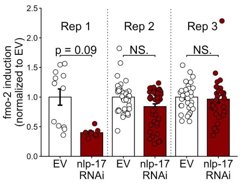
This is a great point. We attempted this experiment, but were unable to achieve consistent results (see Author response image 1). This result could be due to indirect effects of the overexpression of nlp-17 signaling modulating fmo-2 induction in a complicated circuit, variability in expression of its receptor, or other complexities within the circuit.
Author response image 1.
(8) It would strengthen the manuscript to determine whether serotonergic, GABA, and/or tyramine signaling activate the expression or secretion of this nlp-17 neuropeptide.
We thank the reviewer for this great idea of experiment. To address this suggestion, we performed qPCR to measure nlp-17 mRNA in WT, hif-1 KO, ADF HIF-1 stabilized, and NSM HIF-1 stabilized strains. Compared to WT and hif-1 KO controls, we observed no change in nlp-17 expression when HIF-1 was stabilized in the ADF or NSM serotonergic neurons (new Fig. S5F). This could suggest either that nlp-17 signaling acts in parallel to serotonergic signaling following genetic activation of the hypoxic response. Alternatively, neuronal HIF-1 stabilization may modify nlp-17 splicing or translation without resulting in detectable differences in mRNA levels. Together, these data indicate NLP-17 signaling is required for longevity following genetic activation of the hypoxic response, although whether this peptide is synthesized or released in response to hypoxic response remains unclear.
We agree that it is also important to connect nlp-17 expression and/or secretion to other components of this pathway. However, we believe the most effective experiment to confirm a connection between GABA and tyramine signaling and nlp-17 in the context of hypoxia would be to measure nlp-17 expression in strains that manipulate GABA and/or tyramine signaling in a manner that mimics vhl-1 knockdown and extends lifespan. Because we have not yet validated hypoxic-response mimetics for these specific signals, we hope to first generate these strains and then measure their effect on nlp-17 expression in future work. The importance of identifying manipulations to GABA and tyramine signaling that promote longevity has been added to our discussion section.
“While many individual neurosignaling components are essential for genetic activation of the hypoxic response to extend lifespan, their epistasis is unclear (Fig. 7B). Most components of the pathway identified in this work act downstream of vhl-1, and upstream of fmo-2 induction (summarized in Fig. 7A-B). However, the order of each signal between these two endpoints is only predicted based on C. elegans neural wiring and the overlap between various identified signals and cells. For example, we hypothesize in our working model that GABA may be produced by the RIS neuron in this circuit because RIS is the primary GABAergic neuron required for vhl-1-mediated longevity. Alternatively, it is possible that GABA is produced by a different cell that either acts in series or in parallel with RIS signaling. In order to determine the order of each signaling component, future studies should generate genetic manipulations to each signaling component that may mimic vhl-1 knockout to promote longevity, cross these new strains into knockouts of other signals required for vhl-1 mediated longevity; and measure the lifespans of each double and triple mutant. This approach would also narrow down which signals are downstream of the genetic activation of the hypoxic response, and which are sufficient to extend lifespan upstream of the hypoxic response in a normoxic environment. One notable target for further exploration is the SER-7 expressing RIS neuron, which plays a role in sleep [16] and stress resistance [17], and can extend lifespan when optogenetically activated under normoxic conditions [18].”
References:
(1) Calabrese, E. J., Dhawan, G., Kapoor, R., Iavicoli, I. & Calabrese, V. What is hormesis and its relevance to healthy aging and longevity? Biogerontology 16, 693-707 (2015). https://doi.org/10.1007/s10522-015-9601-0
(2) Zhou, I. K., Pincus, Z. & Slack, J. F. Longevity and stress in Caenorhabditis elegans. Aging 3, 733-753 (2011). https://doi.org/10.18632/aging.100367
(3) Yuan, R., Hascup, E., Hascup, K. & Bartke, A. Relationships among Development, Growth, Body Size, Reproduction, Aging, and Longevity - Trade-Offs and Pace-Of-Life. Biochemistry (Mosc) 88, 1692-1703 (2023). https://doi.org/10.1134/S0006297923110020
(4) Mautz, B. S., Lind, M. I. & Maklakov, A. A. Dietary Restriction Improves Fitness of Aging Parents But Reduces Fitness of Their Offspring in Nematodes. J Gerontol A Biol Sci Med Sci 75, 843-848 (2020). https://doi.org/10.1093/gerona/glz276
(5) Duric, V., Clayton, S., Leong, L. M. & Yuan, L.-L. Comorbidity Factors and Brain Mechanisms Linking Chronic Stress and Systemic Illness. Neural Plasticity 2016, 1-16 (2016). https://doi.org/https://doi.org/10.1155/2016/5460732
(6) Mariotti, A. The Effects of Chronic Stress On Health: New Insights Into the Molecular Mechanisms of Brain–Body Communication. Future Science OA 1 (2015). https://doi.org/https://doi.org/10.4155/fso.15.21
(7) Bellier, A., Chen, C.-S., Kao, C.-Y., Cinar, H. N. & Aroian, R. V. Hypoxia and the Hypoxic Response Pathway Protect against Pore-Forming Toxins in C. elegans. PLoS Pathog 5, e1000689 (2009).
(8) Palazon, A., Goldrath, W. A., Nizet, V. & Johnson, S. R. HIF Transcription Factors, Inflammation, and Immunity. Immunity 41, 518-528 (2014). https://doi.org/https://doi.org/10.1016/j.immuni.2014.09.008
(9) Vora, M. et al. The hypoxia response pathway promotes PEP carboxykinase and gluconeogenesis in C. elegans. Nature Communications 13 (2022). https://doi.org/https://doi.org/10.1038/s41467-022-33849-x
(10) Nakazawa, S. M., Keith, B. & Simon, C. M. Oxygen availability and metabolic adaptations. Nature Reviews Cancer 16, 663-673 (2016). https://doi.org/https://doi.org/10.1038/nrc.2016.84
(11) Fawcett, M. E., Hoyt, M. J., Johnson, K. J. & Miller, L. D. Hypoxia disrupts proteostasis in Caenorhabditis elegans. Aging Cell 14, 92-101 (2015). https://doi.org/https://doi.org/10.1111/acel.12301
(12) Ohh, M., Taber, C. C., Ferens, F. G. & Tarade, D. Hypoxia-inducible factor underlies von Hippel-Lindau disease stigmata. Elife 11 (2022). https://doi.org/10.7554/eLife.80774
(13) Wind, J. J. & Lonser, R. R. Management of von Hippel-Lindau disease-associated CNS lesions. Expert Rev Neurother 11, 1433-1441 (2011). https://doi.org/10.1586/ern.11.124
(14) Lonser, R. R. et al. von Hippel-Lindau disease. Lancet 361, 2059-2067 (2003). https://doi.org/10.1016/S0140-6736(03)13643-4
(15) Kaelin, W. G. Molecular basis of the VHL hereditary cancer syndrome. Nat Rev Cancer 2, 673-682 (2002).
(16) Costa, S. W. et al. A GABAergic and peptidergic sleep neuron as a locomotion stop neuron with compartmentalized Ca2+ dynamics. Nature Communications 10 (2019). https://doi.org/10.1038/s41467-019-12098-5
(17) Wu, Y., Masurat, F., Preis, J. & Bringmann, H. Sleep Counteracts Aging Phenotypes to Survive Starvation-Induced Developmental Arrest in C. elegans. Curr Biol 28, 3610-3624.e3618 (2018). https://doi.org/10.1016/j.cub.2018.10.009
(18) Busack, I. & Bringmann, H. A sleep-active neuron can promote survival while sleep behavior is disturbed. PLOS Genetics 19, e1010665 (2023). https://doi.org/10.1371/journal.pgen.1010665
(19) Powell-Coffman, J. A. & Coffman, C. R. Apoptosis: Lack of oxygen aids cell survival. Nature 465, 554-555 (2010). https://doi.org/10.1038/465554a
(20) Kruempel, J. C. P. et al. Hypoxic response regulators RHY-1 and EGL-9/PHD promote longevity through a VHL-1-independent transcriptional response. Geroscience 42, 1621-1633 (2020). https://doi.org/10.1007/s11357-020-00194-0
(21) Bach, M., Bergs, A., Mulcahy, B., Zhen, M. & Gottschalk, A. (2023).
(22) Leiser, S. F. et al. Cell nonautonomous activation of flavin-containing monooxygenase promotes longevity and health span. Science (2015). https://doi.org/10.1126/science.aac9257
-

-
eLife Assessment
This important study combines anatomical tracing, tissue clearing, and functional manipulations to demonstrate lateralized brainstem control of hepatic glucose metabolism and identify a site of sympathetic nerve crossover supplying the liver. The evidence supporting the anatomical organization of hepatic sympathetic innervation is compelling, and the functional studies provide solid support for a role of asymmetric sympathetic outflow in regulating glucose homeostasis. While some uncertainty remains regarding the contribution of sensory innervation and the extent to which these findings generalize beyond mice, the work provides an invaluable advance in understanding neural regulation of liver metabolism.
-
Reviewer #2 (Public review):
Summary:
The manuscript by Wang and colleagues aims to determine whether hepatic glucose metabolism is differentially regulated by the left and right sides of the LPGi and to reveal decussation of hepatic sympathetic nerves.
The authors used tissue clearing to identify sympathetic fibers in the liver lobes, then injected PRV into the hepatic lobes. Five days post-injection, PRV-labeled neurons in the LPGi, which were identified. The results indicated contralateral dominance of premotor neurons and partial innervation of more than one lobe. Then the authors activated each side of the LPGi, resulting in a greater increase in blood glucose levels after right-sided activation than after left-sided activation, and in changes in protein expression in the liver lobes. These data suggested lobe-specific modulation …
Reviewer #2 (Public review):
Summary:
The manuscript by Wang and colleagues aims to determine whether hepatic glucose metabolism is differentially regulated by the left and right sides of the LPGi and to reveal decussation of hepatic sympathetic nerves.
The authors used tissue clearing to identify sympathetic fibers in the liver lobes, then injected PRV into the hepatic lobes. Five days post-injection, PRV-labeled neurons in the LPGi, which were identified. The results indicated contralateral dominance of premotor neurons and partial innervation of more than one lobe. Then the authors activated each side of the LPGi, resulting in a greater increase in blood glucose levels after right-sided activation than after left-sided activation, and in changes in protein expression in the liver lobes. These data suggested lobe-specific modulation of HGP. Chemical denervation of a particular lobe did not affect glucose levels due to compensation by the other lobes. In addition, nerve bundles decussate in the hepatic portal region.
Strengths:
The manuscript is timely and relevant. It is important to understand the sympathetic regulation of the liver and the contribution of each lobe to hepatic glucose production. The authors use state-of-the-art methodology.
Weaknesses:
(1) Image clarity was improved in some cases, but not in others. For example, Figure 3I, showing c-Fos expression, is not convincing due to the image quality and lack of orientation.
(2) The methods section states that 8-weeks-old male mice were used in the experiments without specifying the experiments (e.g., brain injection with AAVs or PRV organ inoculation). The authors should include these details.
(3) The authors should use the exact location of pre- and postganglionic neurons as they often refer to neurons in the sympathetic chain. Their findings should be compared with the existing literature on the location of preganglionic cells.
(4) Figure legends should be revised and matched with the text.
-
Reviewer #4 (Public review):
Summary of General Strengths & Weaknesses:
The studies here are highly informative for anatomical tracing and sympathetic nerve function in the liver in relation to glucose levels, but because they are conducted in a single species, it is challenging to translate them to humans or determine whether these neural circuits are evolutionarily conserved. Dual-labeling anatomical studies are elegant, and the addition of chemogenetic and optogenetic studies provides mechanistically informative. Denervation studies lack proper controls, and sensory innervation in the liver is overlooked.
Specific Weaknesses - Major:
(1) The species name should be included in the title.
(2) Tyrosine hydroxylase was used to mark sympathetic fibers in the liver, but this marker also labels a portion of sensory fibers that need to be …
Reviewer #4 (Public review):
Summary of General Strengths & Weaknesses:
The studies here are highly informative for anatomical tracing and sympathetic nerve function in the liver in relation to glucose levels, but because they are conducted in a single species, it is challenging to translate them to humans or determine whether these neural circuits are evolutionarily conserved. Dual-labeling anatomical studies are elegant, and the addition of chemogenetic and optogenetic studies provides mechanistically informative. Denervation studies lack proper controls, and sensory innervation in the liver is overlooked.
Specific Weaknesses - Major:
(1) The species name should be included in the title.
(2) Tyrosine hydroxylase was used to mark sympathetic fibers in the liver, but this marker also labels a portion of sensory fibers that need to be ruled out in whole-mount imaging data.
(3) Chemogenetic and optogenetic data demonstrating hyperglycemia should be described in the context of prior work demonstrating liver nerve involvement in these processes. There is only a brief mention in the Discussion currently, but comparing methods and observations would be helpful.
(4) Sympathetic denervation with 6-OHDA can drive compensatory increases in tissue sensory innervation, and this should be measured in the liver denervation studies to implicate potential crosstalk, especially given the increase in LPGi cFOS that may be due to afferent nerve activity. Compensatory sympathetic drive may not be the only culprit, though that is clearly assumed. The sensory or parasympathetic/vagal innervation of the liver is altogether ignored in this paper and could be better described in general.
Comments on the revised version.
Across all reviewer comments, the revised resubmission has adequately addressed all concerns.
-
Author response:
The following is the authors’ response to the original reviews.
Public Reviews:
Reviewer #1 (Public review):
Summary:
This manuscript by Wang et al. reports the potential involvement of an asymmetric neurocircuit in the sympathetic control of liver glucose metabolism.
Strengths:
The concept that the contralateral brain-liver neurocircuit preferentially regulates each liver lobe may be interesting.
Weaknesses:
However, the experimental evidence presented did not support the study's central conclusion.
We thank the reviewer for recognizing the conceptual novelty of our work and for constructive comments aimed at enhancing its rigor and clarity. In response, we carried out targeted experiments to address the points raised, including: (i) further characterization of LPGi projections to vagal and sympathetic circuits; (ii) …
Author response:
The following is the authors’ response to the original reviews.
Public Reviews:
Reviewer #1 (Public review):
Summary:
This manuscript by Wang et al. reports the potential involvement of an asymmetric neurocircuit in the sympathetic control of liver glucose metabolism.
Strengths:
The concept that the contralateral brain-liver neurocircuit preferentially regulates each liver lobe may be interesting.
Weaknesses:
However, the experimental evidence presented did not support the study's central conclusion.
We thank the reviewer for recognizing the conceptual novelty of our work and for constructive comments aimed at enhancing its rigor and clarity. In response, we carried out targeted experiments to address the points raised, including: (i) further characterization of LPGi projections to vagal and sympathetic circuits; (ii) evaluation of potential pancreatic involvement; and (iii) validation of the specificity of chemogenetic activation within the proposed circuit. All new experiments, figures, and text have been incorporated, and corresponding revisions are highlighted for ease of review.
(1) Pseudorabies virus (PRV) tracing experiment:
The liver not only possesses sympathetic innervations but also vagal sensory innervations. The experimental setup failed to distinguish whether the PRV-labeling of LPGi (Lateral Paragigantocellular Nucleus) is derived from sympathetic or vagal sensory inputs to the liver.
Thank you for raising this important point. We fully agree that the liver receives both sympathetic and vagal sensory innervation, and we acknowledge that PRV-based tracing alone does not definitively distinguish between these two pathways. This represented a limitation of the original experimental design.
Based on established anatomical literature as well as our experimental observations, vagal sensory neuron cell bodies reside in the nodose ganglion (NG), and their central projections terminate predominantly in the nucleus of the solitary tract (NTS) (Nature. 2023;623(7986):387-396; Curr Biol. 2020;30(20):3986-3998.e5.), which is located in the dorsomedial medulla. In contrast, the LPGi, together with other sympathetic-related nuclei, is predominantly distributed in the ventral medulla (Cell Metab. 2025;37(11):2264-2279.e10; Nat Commun. 2022;13(1):5079).
To determine whether the LPGi contains neurons that modulate the liver via vagal sensory pathways, we performed two complementary experiments.
First, we conducted CGRP immunohistochemistry on brainstem sections, using the NTS, a well-established visceral sensory centre, as a positive control. While abundant CGRP-positive cell bodies were detected in the NTS as expected, few to no CGRP-positive cell bodies were observed in the LPGi (Figure S1G). These results strongly support that the LPGi neurons labeled in our PRV tracing predominantly belong to sympathetic efferent circuits rather than vagal sensory pathways.
Second, to examine whether LPGi neurons send axonal projections to sensory ganglia, we injected hSyn-Cre combined with DIO-Axon-EGFP into the LPGi and examined both the dorsal root ganglia (DRG) and nodose ganglia (NG). No Axon-EGFP-positive signals were detected in either ganglion (Figures S1H-S1J), indicating that LPGi neurons do not directly innervate sensory ganglia. In other words, PRV cannot retrogradely trace to the LPGi via the NG or DRG.
These additions have been incorporated into the revised manuscript, with the Result 1 clearly documenting that these findings confirm that the LPGi specifically regulates sympathetic, rather than vagal sensory, inputs to the liver.
(2) Impact on pancreas:
The celiac ganglia not only provide sympathetic innervations to the liver but also to the pancreas, the central endocrine organ for glucose metabolism. The chemogenetic manipulation of LPGi failed to consider a direct impact on the secretion of insulin and glucagon from the pancreas.
Thank you for this important comment. We agree that the celiac ganglia (CG) provide sympathetic innervation not only to the liver but also to the pancreas, which plays a central role in glucose homeostasis through the secretion of both insulin and glucagon. Therefore, the potential pancreatic implications associated with LPGi chemogenetic manipulation are worth careful consideration.
To address this concern, we measured circulating glucagon and insulin levels following chemogenetic manipulation of the LPGiGAD1 neurons. We found that neither glucagon nor insulin levels changed significantly under our experimental conditions, which indicated that the hyperglycemic effect induced by LPGi activation is unlikely to be mediated by changes in pancreatic hormone secretion (Figure S2G).
These additions have been incorporated into the revised manuscript, with the Result 2 clearly documenting that these findings confirm that the hyperglycemic effect induced by LPGi activation is unlikely to be mediated indirectly via altered pancreatic endocrine output.
(3) Neuroanatomy of the brain-liver neurocircuit:
The current study and its conclusion are based on a speculative brain-liver sympathetic circuit without the necessary anatomical information downstream of LPGi.
Thank you for raising this important point. A clear anatomical definition of the downstream pathways linking the brain to the liver was essential for interpreting the proposed brain-liver sympathetic circuit.
The present study (Figure 4A) provides direct anatomical evidence supporting the organization of the brain–liver sympathetic neurocircuit. These observations are consistent with our recent detailed characterization of the brain-liver sympathetic circuit published in Cell Metabolism (Cell Metab. 2025;37(11):2264–2279). In that study, we showed that LPGi GABAergic neurons inhibit GABAergic neurons in the caudal ventrolateral medulla (CVLM). Disinhibition of CVLM reduced GABAergic suppression of rostral ventrolateral medulla (RVLM) neurons, which are key excitatory drivers of sympathetic tone. RVLM neurons project to sympathetic preganglionic neurons in the sympathetic chain (Syc). These neurons synapse with postganglionic sympathetic neurons in ganglia such as the celiac-superior mesenteric ganglion (CG-SMG). Postganglionic sympathetic fibers then innervate the liver, releasing norepinephrine (NE) to activate hepatic β2-adrenergic receptors and stimulate hepatic glucose production (HGP).
Together, these data establish a coherent anatomical basis for the proposed brain-liver sympathetic pathway and clarify the downstream organization relevant to the functional experiments presented in figure 4A and Author response image 1..
Author response image 1.
Tracing scheme (Left) and whole-mount imaging (Right) of PRV-labeled brain-liver neurocircuit. Scale bars, 3,000 (whole mount) or 1,000 (optical sections) μm.
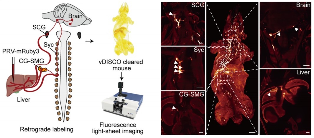
(4) Local manipulation of the celiac ganglia:
The left and right ganglia of mice are not separate from each other but rather anatomically connected. The claim that the local injection of AAV in the left or right ganglion without affecting the other side is against this basic anatomical feature.
Thank you for raising this important anatomical point. We fully acknowledge that the left and right CG in mice are interconnected, and that unilateral viral injection could theoretically affect the contralateral side. The CG-SMG complex serves as a major sympathetic hub that regulates visceral organ functions. Recent transcriptomic, anatomical, and functional studies have revealed that the CG-SMG is not a homogeneous structure but is composed of molecularly and functionally distinct neuronal populations. These populations exhibit specialized projection patterns and regulate different aspects of gastrointestinal physiology, supporting a model of modular sympathetic control. (Nature. 2025 Jan;637(8047):895-902). Therefore, we were aware of this phenomenon during the initial stages of these experiments.
To minimize unintended spread to the contralateral CG, we took two complementary approaches.
First, we optimized the injection strategy by using an extremely small injection volume (100 nL per site), with a very slow infusion rate (50 nL/min), and fine glass micropipettes. With these refinements, contralateral viral spread was rarely observed.
Second, and importantly, all animals included in the final analyses were subjected to post hoc anatomical verification. After completion of the experiments, CGs were collected, sectioned, and examined for viral expression. As shown in Supplementary Figure 5F, only mice in which viral expression was strictly confined to the targeted CG, with no detectable infection in the contralateral ganglion, were included in the presented data.
Together, these measures ensure that our local manipulation of the intended CG produced the reported effects. We have revised the Methods section to more explicitly detail these technical precautions, and the legend for Figure S5F clearly states its role in validating injection specificity.
Reviewer #2 (Public review):
Summary:
The manuscript by Wang and colleagues aims to determine whether the left and right LPGi differentially regulate hepatic glucose metabolism and to reveal decussation of hepatic sympathetic nerves.
The authors used tissue clearing to identify sympathetic fibers in the liver lobes, then injected PRV into the hepatic lobes. Five days post-injection, PRV-labeled neurons in the LPGi were identified. The results indicated contralateral dominance of premotor neurons and partial innervation of more than one lobe. Then the authors activated each side of the LPGi, resulting in a greater increase in blood glucose levels after right-sided activation than after left-sided activation, as well as changes in protein expression in the liver lobes. These data suggested modulation of HGP (hepatic glucose production) in a lobe-specific manner. Chemical denervation of a particular lobe did not affect glucose levels due to compensation by the other lobes. In addition, nerve bundles decussate in the hepatic portal region.
We thank the reviewer for the thorough and constructive evaluation of our manuscript. In direct response, we undertook comprehensive revisions to enhance the rigor and clarity of the study, including: (i) correcting ambiguous or misleading terminology about anatomical resolution and sympathetic circuit organization; (ii) expanding the Methods section with complete experimental details, improved image presentation, and explicit justification of our viral and genetic approaches; and (iii) strengthening data interpretation by addressing issues related to sparse PRV labeling, projection heterogeneity, and the functional implications of double-labeled neurons.
Strengths:
The manuscript is timely and relevant. It is important to understand the sympathetic regulation of the liver and the contribution of each lobe to hepatic glucose production. The authors use state-of-the-art methodology.
Weaknesses:
(1) The wording/terminology used in the manuscript is misleading, and it is not used in the proper context. For instance, the goal of the study is "to investigate whether cerebral hemispheres differentially regulate hepatic glucose metabolism..." (see abstract); however, the authors focus on the brainstem (a single structure without hemispheres). Similarly, symmetric is not the best word for the projections.
We thank the reviewer for raising these critical points regarding terminology and conceptual framing. We acknowledge that certain phrases in our original manuscript may have been overly broad or ambiguous, particularly in describing the scope of sympathetic heterogeneity and the specificity of neural projections. Due to practical constraints and the scope of our study, our investigation focused on the brainstem, which represents the final common pathway for these lateralized commands. We acknowledge that terms referring to the cerebral hemispheres do not accurately describe our study. We have revised the manuscript to ensure accurate and consistent terminology.
Below are specific examples:
Original 1: This study aims to investigate whether cerebral hemispheres differentially regulate hepatic glucose metabolism and localize the site of sympathetic crossover to the liver.
Revised 1: “This study aimed to determine whether the central nervous system exerts lateralized control over hepatic glucose metabolism and to localize the site of peripheral sympathetic crossover to the liver.”
Original 2: These findings demonstrate that the brain exerts lobe-specific, lateralized control of hepatic glucose metabolism via symmetric brain-liver sympathetic pathways.
Revised 2: “These findings demonstrate that the brainstem can exert lobe-specific, lateralized control of hepatic glucose metabolism via bilaterally projecting brain–liver sympathetic pathways.”
Original 3: The cerebral hemispheres exhibit pronounced functional asymmetry, [1,2] a phenomenon traditionally associated with cognitive and motor processes such as language, voluntary movement, and spatial navigation.[3,4]
Revised 3: “Pronounced functional lateralization within the central nervous system (CNS) is a well-documented phenomenon, [1,2] traditionally associated with cognitive and motor processes such as language, voluntary movement, and spatial navigation [3,4].
(2) Sparse labeling of liver-related neurons was shown in the LPGi (Figure 1). It would be ideal to have lower magnification images to show the area. Higher quality images would be necessary, as it is difficult to identify brainstem areas. The low number of labeled neurons in the LPGi after five days of inoculation is surprising. Previous findings showed extensive labeling in the ventral brainstem at four days post-inoculation (Desmoulins et al., 2025). Unfortunately, it is not possible to compare the injection paradigm/methods because the PRV inoculation is missing from the methods section. If the PRV is different from the previously published viral tracers, time-dependent studies to determine the order of neurons and the time course of infection would be necessary.
We sincerely thank the reviewer for these detailed and constructive comments regarding the PRV tracing experiments. We fully agree that careful presentation and interpretation of the anatomical data are essential for ensuring rigor and transparency. We address each point in detail below.
(1) Image magnification and anatomical context of LPGi labeling
We agree that the original images did not sufficiently convey the broader anatomical context of the LPGi. Due to fluorescence quenching in previous sections, we repeated the PRV retrograde tracing experiment and performed statistical analysis. In the revised manuscript, we replaced the original panels in Figure 1 and Figure S1 with new images that include lower-magnification overviews of the brainstem, alongside higher-magnification views of the LPGi (Figure 1). These images clearly delineate the LPGi with respect to established anatomical landmarks and atlas boundaries. Image contrast and resolution were optimized to allow unambiguous identification of PRV-labeled neurons and surrounding structures.
(2) Sparse LPGi labeling at 5 days post-injection and methodological details
We apologize for the omission of the detailed PRV injection protocol in the original Methods section. We deliberately used small-volume, local injections (1 µL per liver lobe) to minimize viral spread and to restrict labeling to circuits specifically connected to the targeted hepatic region. This sparse labeling was consistent with the use of small, spatially restricted injections designed to minimize off-target spread and preferentially label higher-order upstream neurons. This information has now been added, including the PRV strain, viral titer, injection volume, precise injection coordinates, and surgical procedures. All new figures, legends, and Method details have been incorporated, with changes clearly highlighted for ease of review.
These additions have been incorporated into the revised manuscript, in Figure 1B-D, Figure S1C-F. Furthermore, we also added details of the Methods.
(3) Not all LPGi cells are liver-related. Was the entire LPGi population stimulated, or was it done in a cell-type-specific manner? What was the strain, sex, and age of the mice? What was the rationale for using the particular viral constructs?
We thank the reviewer for this insightful and important question. We agree that not all neurons within the LPGi are liver-related, and we apologize that our rationale was not clearly articulated in the original manuscript.
(1) Our decision to target GABAergic neurons in the LPGi using GAD1-Cre mice was based on prior experimental evidence rather than an assumption about the entire LPGi population. In our previous study (Cell Metab. 2025;37(11):2264-2279.e10), we performed single-cell RNA sequencing on retrogradely labeled LPGi neurons following liver tracing. These analyses revealed that the majority of liver-projecting LPGi neurons are GABAergic in nature. Based on these findings, we chose to selectively manipulate GABAergic neurons in the LPGi rather than the entire LPGi neuronal population, to achieve greater cellular specificity and to minimize potential confounding effects arising from heterogeneous neuron types within this region. We regret that this rationale was not clearly described in the original submission and have now revised the manuscript to explicitly state this reasoning (Results section 2, paragraph 2: “Prior single-nucleus RNA sequencing and immunofluorescence analyses demonstrated that liver-projecting LPGi neurons are predominantly GABAergic.”).
(2) In addition, we apologize for the omission of mouse strain, sex, and age information in the Methods section. These details have been fully added.
(3) We selected AAV-based viral vectors, specifically the AAV9 serotype, due to their well-established efficiency in transducing neurons in the brainstem, relatively low toxicity, and widespread use in circuit-level chemogenetic and optogenetic studies. When combined with Cre-dependent viral constructs in GAD1-Cre mice, this approach enabled selective and reliable manipulation of LPGi GABAergic neurons.
(4) The authors should consider the effect of stimulation of double-labeled neurons (innervating more than one lobe) and potential confounding effects regarding other physiological functions.
We thank the reviewer for raising this important point. We agree that neurons innervating more than one liver lobe could, in principle, introduce potential confounding effects and may reflect higher-order integrative autonomic neurons.
This consideration is consistent with a key finding of the cited study: the CG-SMG contains molecularly distinct sympathetic neuron populations (e.g., RXFP1+ vs. SHOX2+) that exhibit complementary organ projections and separate, non‑overlapping functions. Specifically, RXFP1+ neurons innervate secretory organs (pancreas, bile duct) to regulate secretion, while SHOX2+ neurons innervate the gastrointestinal tract to control motility. This functional segregation supports the concept of specialized autonomic modules rather than a uniform, “fight-or-flight” response, reinforcing the need for careful interpretation of circuit-specific manipulations. (Nature. 2025;637(8047):895-902; Neuron. 2026;114(3):463-478.e7).
In our PRV tracing experiments, the proportion of double-labeled neurons was relatively small, suggesting that the majority of labeled LPGi neurons preferentially associate with individual hepatic lobes. Nevertheless, we recognize that activation of this minority population could contribute to broader physiological effects beyond strictly lobe-specific regulation. We have therefore added a paragraph in the second paragraph of the Discussion (Paragraph 2: “A small subset of LPGi neurons was double-labeled after bilateral PRV injections, suggesting a fraction of these neurons projects bilaterally to both sides of the liver. Such neurons may support interlobar coordination.”).
(5) The authors state that "central projections directly descend along the sympathetic chain to the celiac-superior mesenteric ganglia". What they mean is unclear. Do the authors refer to pre-ganglionic neurons or premotor neurons? How does it fit with the previous literature?
We thank the reviewer for pointing out this imprecise wording. We agree that the original phrasing was anatomically inaccurate and potentially confusing. The pathways we intended to describe involve brainstem premotor neurons that project to sympathetic preganglionic neurons in the spinal cord. These preganglionic neurons then innervate neurons in the CG-SMG, which in turn provide postganglionic input to the liver.
We have revised the manuscript to clearly distinguish premotor from preganglionic neurons (Results section 4, paragraph 1: “Using whole-mount clearing, we visualized the brain–liver sympathetic circuit and found that preganglionic neurons in the spinal cord send descending fibers through the sympathetic chain (SyC) to innervate postganglionic neurons in the CG-SMG (Figure 4A). Further whole-mount TH immunostaining showed that TH-positive sympathetic cell bodies within the CG-SMG project to the liver along the hepatic vasculature (Figure 4B and Figure S5F). These observations suggest that the nerve bundles likely decussate at the porta hepatis before entering the individual hepatic lobes.”).
(6) How was the chemical denervation completed for the individual lobes?
We thank the reviewer for raising this important methodological concern. We agree that potential diffusion of 6-OHDA is a critical issue when performing lobe-specific chemical denervation, and we apologize that our original description did not sufficiently clarify how this was controlled.
In the revised Methods section, we provided a detailed description of the denervation procedure, including the injection volume and concentration of 6-OHDA, as well as the physical separation and isolation of individual hepatic lobes during application to minimize diffusion to adjacent tissue.
To directly assess the specificity of the chemical denervation, we included immunofluorescence and Western blot analyses demonstrating a selective reduction of sympathetic markers in the targeted lobe (Figure 3C), with minimal effects on non-targeted lobes. These results support the effectiveness and relative spatial confinement of the 6-OHDA treatment under our experimental conditions.
We thank the reviewer for highlighting this point, which has helped us improve both the clarity and rigor of the manuscript.
(7) The Western Blot images look like they are from different blots, but there are no details provided regarding protein amount (loading) or housekeeping. What was the reason to switch beta-actin and alpha-tubulin? In Figures 3F -G, the GS expression is not a good representative image. Were chemiluminescence or fluorescence antibodies used? Were the membranes reused?
We thank the reviewer for this careful and detailed evaluation of the Western blot data. We apologize that insufficient methodological detail was provided in the original submission.
(1) We would like to clarify that the protein bands shown within each panel were derived from the same membrane. To improve transparency, we provided full, uncropped images of the corresponding membranes in the supplementary materials. In addition, detailed information regarding protein loading amounts, gel conditions, and housekeeping controls has also been added to the Methods section.
(2) The use of different loading controls (β-actin or α-tubulin) reflects a technical consideration rather than an experimental inconsistency. In our experiments, the molecular weight of the TH (62kDa) was too close to that of α-tubulin (55kDa), and β-actin (42kDa) was therefore used to avoid band overlap and to ensure accurate quantification.
(3) Regarding the GS signal shown in Figures 3F–G, we agree that the original representative image was suboptimal. This appears to be related to antibody performance rather than sample quality. To address this, we repeated the Western blot from Figures 3F–G using a newly validated antibody. The original tissue samples had been aliquoted and stored at −80 °C, allowing reliable re-analysis.
(4) All Western blot experiments were detected using chemiluminescence, and membrane stripping and reprobing procedures are now explicitly described in the Methods section.
We thank the reviewer for highlighting these issues, which significantly improve the rigor and clarity of our data presentation. All new figures and legends have been incorporated, with changes clearly highlighted for ease of review.
(8) Key references using PRV for liver innervation studies are missing (Stanley et al, 2010 [PMID: 20351287]; Torres et al., 2021 [PMID: 34231420]; Desmoulins et al., 2025 [PMID: 39647176]).
We thank the reviewer for pointing out these important and highly relevant references that were inadvertently omitted in our initial submission. The studies by Stanley et al. (Proc Natl Acad Sci U S A, 2010), Torres et al. (Am J Physiol Regul Integr Comp Physiol, 2021), and Desmoulins et al. (Auton Neurosci, 2025) represent key PRV-based retrograde tracing work that has mapped central neural circuits innervating the liver and thus provide essential context for our anatomical analyses.
We agree that the inclusion of these studies is necessary to properly situate our findings within the existing literature. Accordingly, we incorporated citations to these references in the revised manuscript and discussed their relationship to our results.
Reviewer #3 (Public review):
Summary:
This study found a lobe-specific, lateralized control of hepatic glucose metabolism by the brain and provides anatomical evidence for sympathetic crossover at the porta hepatis. The findings are particularly insightful to the researchers in the field of liver metabolism, regeneration, and tumors.
Strengths:
Increasing evidence suggests spatial heterogeneity of the liver across many aspects of metabolism and regenerative capacity. The current study has provided interesting findings: neuronal innervation of the liver also shows anatomical differences across lobes. The findings could be particularly useful for understanding liver pathophysiology and treatment, such as metabolic interventions or transplantation.
Weaknesses:
Inclusion of detailed method and Discussion:
We sincerely thank the reviewer for the positive and constructive feedback, which significantly enhances both the methodological rigor and the broader biological interpretation of our study. In direct response, we revised the Discussion to elaborate on the potential physiological advantages of a lateralized and lobe-specific pattern of liver innervation. Furthermore, we expanded the Methods section to include a comprehensive description of the quantitative analysis applied to PRV-labeled neurons. Together, these revisions strengthened the manuscript’s clarity, depth, and relevance to researchers in hepatic metabolism, regeneration, and disease.
(1) The quantitative results of PRV-labeled neurons are presented, and please include the specific quantitative methods.
We thank the reviewer for this helpful suggestion. We have added a detailed description of the quantitative methods used to analyze PRV-labeled neurons in the revised Methods section. We have now provided detailed information in the Methods section, including the criteria used for cell counting, the anatomical boundaries of the brain regions analyzed, the delineation of regions of interest, and the normalization procedures applied to derive the reported neuron counts. These additions have been incorporated into the revised Methods, with all changes clearly indicated for ease of review.
(2) The Discussion can be expanded to include potential biological advantages of this complex lateralized innervation pattern.
We appreciated the reviewer’s suggestion. We have expanded the Discussion to include a paragraph addressing the potential biological significance of lateralized liver innervation. We highlight that this asymmetric organization could allow for more precise, lobe-specific regulation of hepatic metabolism, enable integration of distinct physiological signals, and potentially provide robustness against perturbations. The additional discussion content has been highlighted in the revised version as indicated (Discussion section, paragraph 3: “Bilateral LPGi activation produced additive effects, indicating that both sides of the brainstem can cooperatively regulate hepatic metabolism in a spatially segregated manner. This pattern suggests that hepatic glucose output can be modulated in a lobe-specific, rather than uniform whole-organ, manner.”).
Reviewer #4 (Public review):
Summary:
The studies here are highly informative in terms of anatomical tracing and sympathetic nerve function in the liver related to glucose levels, but given that they are performed in a single species, it is challenging to translated them to humans, or to determine whether these neural circuits are evolutionarily conserved. Dual-labeling anatomical studies are elegant, and the addition of chemogenetic and optogenetic studies is mechanistically informative. Denervation studies lack appropriate controls, and the role of sensory innervation in the liver is overlooked.
We sincerely appreciate the reviewer's thoughtful evaluation and fully agree that findings derived from a single-species model must be interpreted with caution in relation to human physiology. In direct response, we revised the manuscript to explicitly clarify that all experimental data were obtained in mice and to provide a discussion of the limitations regarding direct extrapolation to humans. Concurrently, we expanded the Discussion section by integrating our findings with recent human and translational studies, including a multicenter clinical trial demonstrating that catheter-based endovascular denervation of the celiac and hepatic arteries significantly improved glycemic control in patients with poorly controlled type 2 diabetes, without major adverse events (Signal Transduct Target Ther. 2025;10(1):371). While our current work focuses on defining the anatomical organization and functional asymmetry of this circuit in mice, the clinical findings suggest that the core principles, sympathetic control of hepatic glucose metabolism via CG-liver pathways, may be conserved and of translational relevance. Additionally, we clarified the interpretation of TH labeling and expanded the discussion of hepatic sensory and parasympathetic innervation, acknowledging their important roles in liver-brain communication and identifying them as key directions for future research. Collectively, these revisions provide a more balanced, clinically informed, and rigorous framework for interpreting our findings.
Specific Weaknesses - Major:
(1) The species name should be included in the title.
We thank the reviewer for this suggestion. We agree that the species should be clearly indicated. The findings presented in this study were obtained in mice using tissue clearing and whole-organ imaging approaches. Due to technical limitations, these observations are currently restricted to the mouse strain. We have updated the title (Symmetric brain-liver circuits mediate lateralized regulation of hepatic glucose output in mice) and clarified the species used throughout the manuscript.
(2) Tyrosine hydroxylase was used to mark sympathetic fibers in the liver, but this marker also hits a portion of sensory fibers that need to be ruled out in whole-mount imaging data
We thank the reviewer for pointing this out. We acknowledge that TH labels not only sympathetic fibers but also a subset of sensory fibers. We have added a limitation of this point in the revised manuscript. In addition, using SyGlass (2.4.0) three-dimensional reconstruction, we observed TH-positive nerve fibers originating from the CG-SMG extending along the porta hepatis and penetrating into the liver parenchyma. Given that the CG-SMG is a well-established sympathetic ganglion innervating visceral organs (Nature. 2025 Jan;637(8047):895-902.), these nerve fibers can be definitively identified as sympathetic. In parallel, we collected DRG from spinal segments T1-6 and T7-12 five days after intrahepatic PRV injection. While T7-12 DRG are known to contain sensory neurons innervating the liver, only a sparse number of PRV-positive neurons were detected in these segments (Anat Rec A Discov Mol Cell Evol Biol. 2004 Sep;280(1):827-35. Auton Neurosci. 2024 Jun;253:103174). The additional figure and discussion content have been highlighted in the revised version as indicated (Discussion section, paragraph 6: “Third, although whole-mount TH immunostaining with three-dimensional reconstruction revealed sympathetic nerve bundles projecting from the CG to the liver, TH is not entirely specific and can also label a subset of sensory neurons. More selective approaches, such as genetic targeting of sympathetic lineages, will be important for further validation.”).
Author response image 2.
Representative immunofluorescence images of PRV-labeled neurons (EGFP) in DRG from the spinal segments T1-6 (bottom) and T7-12 (top) following PRV injections into the liver lobes. Scale bars, 200μm
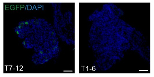
(3) Chemogenetic and optogenetic data demonstrating hyperglycemia should be described in the context of prior work demonstrating liver nerve involvement in these processes. There is only a brief mention in the Discussion currently, but comparing methods and observations would be helpful.
We thank the reviewer for this suggestion. Previous studies largely relied on electrical stimulation to modulate liver innervation, which provides relatively coarse control of neural activity (Eur J Biochem. 1992;207(2):399-411). By contrast, our use of chemogenetic and optogenetic approaches allows selective, cell-type-specific manipulation of LPGi neurons. We revised the Discussion to place our functional data in the context of prior work, highlighting how these more precise approaches improve understanding of the contribution of liver-innervating neurons to hyperglycemia. The newly added discussion has been clearly labeled in the response to facilitate your review (Discussion section, paragraph 3: “This spatial organization is likely obscured by conventional electrical stimulation, which indiscriminately activates heterogeneous sympathetic fibers. By contrast, chemogenetic and optogenetic approaches permit selective, cell type-specific manipulation of LPGi neurons, thereby revealing the contralateral and lobe-specific architecture of brain-liver sympathetic control”).
(4) Sympathetic denervation with 6-OHDA can drive compensatory increases to tissue sensory innervation, and this should be measured in the liver denervation studies to implicate potential crosstalk, especially given the increase in LPGi cFOS that may be due to afferent nerve activity. Compensatory sympathetic drive may not be the only culprit, though it is clearly assumed to be. The sensory or parasympathetic/vagal innervation of the liver is altogether ignored in this paper and could be better described in general.
We thank the reviewer for this insightful and important comment, which highlights a potential alternative interpretation of our findings. We agree that chemical sympathetic denervation with 6-OHDA may induce compensatory changes in non-sympathetic inputs, including sensory and parasympathetic (vagal) innervation of the liver.
Conceptually, we agree with the reviewer’s perspective that the central nervous system operates as a highly integrated homeostatic regulatory system, continuously receiving and integrating a broad range of afferent signals. These inputs include, as noted by the reviewer, hepatic sensory and vagal afferents (Science. 2024;386(6722):673-677), as well as centrally derived interoceptive signals such as brain glucose, temperature sensing, even the pulsation of cerebral vascular system (Cell Metab. 2025;37(11):2264-2279.e10.; Cell Metab. 2022;34(6):888-901.e5; Science. 2024;383(6682):eadk8511). The CNS integrates these diverse signals and generates coordinated efferent outputs to maintain systemic homeostasis.
From this viewpoint, the changes in c-FOS activity that we observe in the LPGi likely represent only a limited snapshot of this broader integrative process, rather than evidence of a single dominant pathway. We acknowledge that compensatory sensory or parasympathetic mechanisms, in addition to altered sympathetic drive, contributed to the observed LPGi activation following hepatic sympathetic denervation.
We further acknowledge that, due to limitations in scope and experimental focus, we did not directly assess sensory or parasympathetic innervation of the liver in the present study. As appropriately pointed out by the reviewer, a more comprehensive characterization of hepatic neural inputs would provide a more complete picture of the underlying neurocircuitry. To address this, we expanded the Discussion and explicitly noted this limitation, including a more balanced discussion of potential crosstalk among sympathetic, sensory, and parasympathetic pathways and how these may collectively influence LPGi activity. For your convenience, the newly added discussion text has been distinctly marked in the manuscript (Discussion section, paragraph 4: “Although enhanced sympathetic output appears to mediate much of this compensation, our findings suggest that the underlying regulation extends beyond a purely descending pathway. In particular, c-FOS activation in the contralateral LPGi after unilateral 6-OHDA-mediated denervation suggests that the loss of peripheral input may be sensed through an ascending neural pathway, centrally integrated, and translated into compensatory sympathetic output to the intact hepatic lobes. These results therefore support a model in which hepatic glucose production is regulated by an integrated afferent-central-efferent loop, with our current analyses primarily resolving its efferent component.”).
Recommendations for the authors:
Reviewer #2 (Recommendations for the authors):
Although the findings are interesting, this reviewer has major concerns about the experimental design, methodology, results, and interpretation of the data. Experimental details are lacking, including basic information (age, sex, strain of mice, procedures, magnification, etc.).
We thank the reviewer for this important recommendation. We agree that comprehensive reporting of experimental details is essential for rigor and reproducibility.
In the revised manuscript, we added complete information regarding mouse strain, sex, age, and sample size for each experiment. In addition, detailed descriptions of surgical procedures, viral constructs, injection parameters, imaging magnification, and analysis methods have been incorporated into the Methods section.
These revisions ensured that all experiments are described with sufficient technical detail and clarity to allow accurate interpretation and replication of our findings. Experimental details have been incorporated, and corresponding revisions are highlighted for ease of review.
Reviewer #3 (Recommendations for the authors):
Addressing a few questions might help:
(1) The study found that liver-associated LPGi neurons are predominantly GABAergic. It would be informative to molecularly characterize the PRV-traced, liver-projecting LPGi neurons to determine their neurochemical phenotypes.
We thank the reviewer for this insightful suggestion. We agree that molecular characterization of liver-projecting LPGi neurons is important for understanding their functional identity.
This issue has been addressed in detail in our recent study (Cell Metab. 2025;37(11):2264-2279.e10), in which we performed single-cell RNA sequencing on retrogradely traced LPGi neurons connected to the liver. These analyses demonstrated that the majority of liver-projecting LPGi neurons are GABAergic, with a defined transcriptional profile distinct from neighboring non–liver-related populations.
Based on these findings, the current study selectively targeted GABAergic LPGi neurons using GAD1-Cre mice. We have explicitly cited these molecular results in the revised manuscript to clarify the neurochemical identity of the PRV-traced LPGi neurons. New text has been incorporated, and corresponding revisions are highlighted for ease of review.
(2) Is it possible to do a local microinjection of a sodium channel blocker (e.g., lidocaine) or an adrenergic receptor antagonist into the porta hepatis? That would potentially provide additional evidence for the porta hepatis as the functional crossover point.
We appreciated the reviewer’s thoughtful suggestion. Although pharmacological blockade at the porta hepatis can modulate local neural activity, this approach is inherently limited in its ability to distinguish between ipsilateral and contralateral inputs. Consequently, it may not provide definitive evidence for neural crossover at this specific site.
In our view, the anatomical evidence provided by whole-mount tissue clearing, dual-labeled tracing, and direct visualization of decussating nerve bundles at the porta hepatis offers a more definitive demonstration of sympathetic crossover. Pharmacological blockade would affect both crossed and uncrossed fibers simultaneously and therefore would not specifically resolve the anatomical organization of this decussation.
Nevertheless, we agree that functional interrogation of the porta hepatis represents an interesting direction for future work, and we acknowledge this possibility in the Discussion (Paragraph 6: “Fourth, although our data support a peripheral decussation at the porta hepatis, direct validation of this crossover site was not feasible with local pharmacological blockade, as currently available approaches lack sufficient spatial specificity and would likely perturb multiple neural components. Future studies employing more selective inhibitory strategies will be required to directly test this possibility.”).
(3) It is possible to investigate the effects of unilateral LPGi manipulation or ablation of one side of CG/SMG on liver metabolism, such as hyperglycemia?
We thank the reviewer for this important suggestion. Because unilateral LPGi manipulation was already examined in our study (Figure 2D), we focused here on unilateral ablation of the CG to further assess lateralized sympathetic control of hepatic metabolism. We successfully performed unilateral CG ablation without LPGi manipulation, but observed no significant change in blood glucose compared with the sham group (Author response image 3A and 3B). To determine whether glucose homeostasis was nonetheless affected, we further performed glucose tolerance tests (GTT) and insulin tolerance tests (ITT) (Author response image 3C and 3D). Neither test showed significant impairment after unilateral ablation, suggesting that compensatory neural mechanisms and/or hormonal homeostatic regulation may be recruited to preserve systemic glucose homeostasis.
Author response image 3.
(A) Blood glucose levels in mice subjected to left- or right-sided CG ablation via 6-OHDA treatment (n = 6). (B) Representative images of ablation of CG. Scale bars, 100 μm. (C and D) Blood glucose levels during GTT (C, n = 6) and ITT (D, n = 6) in mice with left- or right-sided CG ablation.
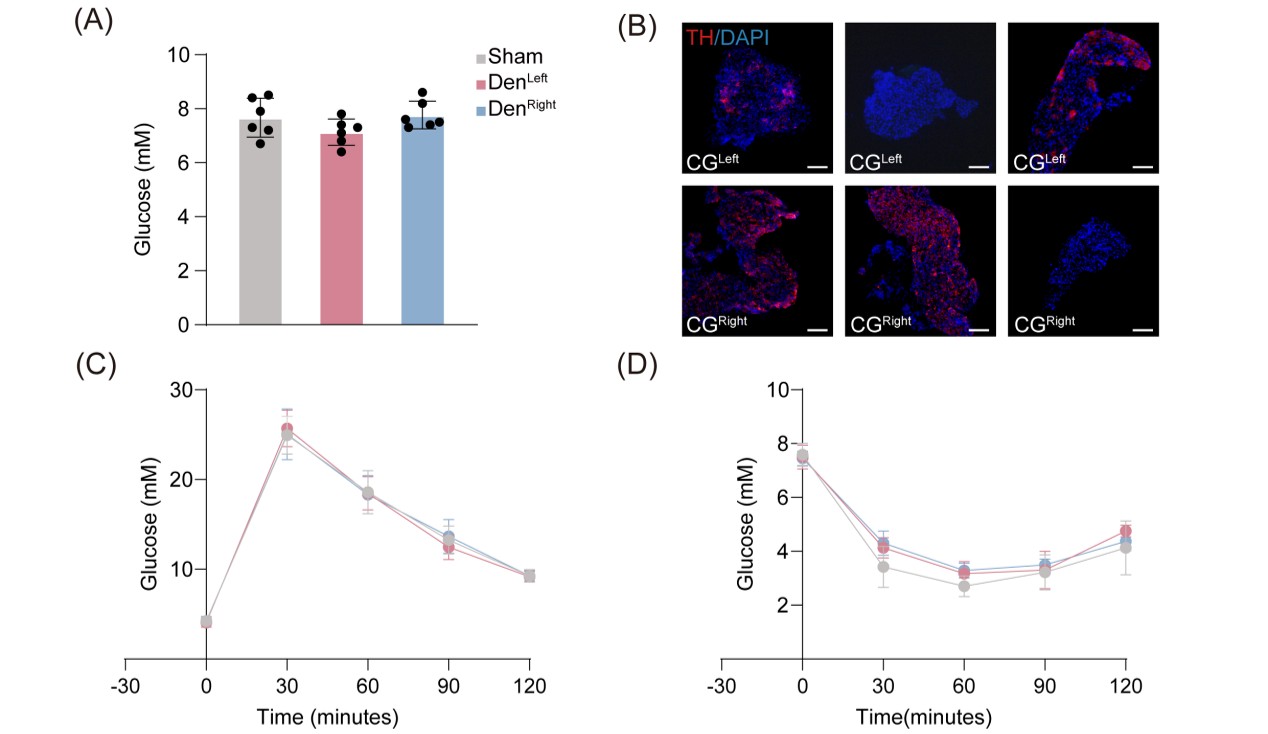
Reviewer #4 (Recommendations for the authors):
In the abstract and elsewhere, the use of the term 'sympathetic release' is unclear - do you mean release of nerve products, such as the neurotransmitter norepinephrine? This should be more clearly defined.
We thank the reviewer for pointing out this ambiguity. We agree that the term “sympathetic release” was imprecise. In the revised manuscript, we explicitly referred to the release of sympathetic neurotransmitters, primarily norepinephrine, from postganglionic sympathetic fibers.
We revised the wording throughout the manuscript to ensure accurate and consistent terminology and to avoid potential confusion regarding the underlying neurobiological mechanisms.
Original: “Following unilateral hepatic denervation, contralateral LPGi activation induced metabolic compensation in the remaining innervated lobes, characterized by increased sympathetic release, glucose production, and glycogen depletion.”
Revised: “Following unilateral hepatic denervation, contralateral LPGi activation induced metabolic compensation in the remaining innervated lobes, characterized by increased norepinephrine release, glucose production, and glycogen depletion.”
-
-
Authors used C57BL/6J from #InternationalMouseSt in their study. Including #RRIDs will make this less ambiguous. SciScore made a table with this resource, see “Automated Services” module (download as csv, xml or #jats) #STMpublishing #RRID
-
-
eLife Assessment
This manuscript proposes a lateralized, lobe-specific brain-liver sympathetic neurocircuit regulating hepatic glucose metabolism and presents anatomical evidence for sympathetic crossover at the porta hepatis using viral tracing and neuromodulation approaches. While the topic is of important significance and the methodologies are, in principle, state-of-the-art, significant concerns regarding experimental design, incomplete methodological reporting, sparse and ambiguous labeling, and overi-nterpretation of the data substantially weaken support for the study's central conclusions, thereby limiting the study's completeness. The work will be of interest to biologists, clinicians, and physiologists.
-
Reviewer #1 (Public review):
Summary:
This manuscript by Wang et al. reports the potential involvement of an asymmetric neurocircuit in the sympathetic control of liver glucose metabolism.
Strengths:
The concept that the contralateral brain-liver neurocircuit preferentially regulates each liver lobe may be interesting.
Weaknesses:
However, the experimental evidence presented did not support the study's central conclusion.
(1) Pseudorabies virus (PRV) tracing experiment:
The liver not only possesses sympathetic innervations but also vagal sensory innervations. The experimental setup failed to distinguish whether the PRV-labeling of LPGi (Lateral Paragigantocellular Nucleus) is derived from sympathetic or vagal sensory inputs to the liver.(2) Impact on pancreas:
The celiac ganglia not only provide sympathetic innervations to the liver …Reviewer #1 (Public review):
Summary:
This manuscript by Wang et al. reports the potential involvement of an asymmetric neurocircuit in the sympathetic control of liver glucose metabolism.
Strengths:
The concept that the contralateral brain-liver neurocircuit preferentially regulates each liver lobe may be interesting.
Weaknesses:
However, the experimental evidence presented did not support the study's central conclusion.
(1) Pseudorabies virus (PRV) tracing experiment:
The liver not only possesses sympathetic innervations but also vagal sensory innervations. The experimental setup failed to distinguish whether the PRV-labeling of LPGi (Lateral Paragigantocellular Nucleus) is derived from sympathetic or vagal sensory inputs to the liver.(2) Impact on pancreas:
The celiac ganglia not only provide sympathetic innervations to the liver but also to the pancreas, the central endocrine organ for glucose metabolism. The chemogenetic manipulation of LPGi failed to consider a direct impact on the secretion of insulin and glucagon from the pancreas.(3) Neuroanatomy of the brain-liver neurocircuit:
The current study and its conclusion are based on a speculative brain-liver sympathetic circuit without the necessary anatomical information downstream of LPGi.(4) Local manipulation of the celiac ganglia:
The left and right ganglia of mice are not separate from each other but rather anatomically connected. The claim that the local injection of AAV in the left or right ganglion without affecting the other side is against this basic anatomical feature. -
Reviewer #2 (Public review):
Summary:
The manuscript by Wang and colleagues aims to determine whether the left and right LPGi differentially regulate hepatic glucose metabolism and to reveal decussation of hepatic sympathetic nerves.
The authors used tissue clearing to identify sympathetic fibers in the liver lobes, then injected PRV into the hepatic lobes. Five days post-injection, PRV-labeled neurons in the LPGi were identified. The results indicated contralateral dominance of premotor neurons and partial innervation of more than one lobe. Then the authors activated each side of the LPGi, resulting in a greater increase in blood glucose levels after right-sided activation than after left-sided activation, as well as changes in protein expression in the liver lobes. These data suggested modulation of HGP (hepatic glucose production) in …
Reviewer #2 (Public review):
Summary:
The manuscript by Wang and colleagues aims to determine whether the left and right LPGi differentially regulate hepatic glucose metabolism and to reveal decussation of hepatic sympathetic nerves.
The authors used tissue clearing to identify sympathetic fibers in the liver lobes, then injected PRV into the hepatic lobes. Five days post-injection, PRV-labeled neurons in the LPGi were identified. The results indicated contralateral dominance of premotor neurons and partial innervation of more than one lobe. Then the authors activated each side of the LPGi, resulting in a greater increase in blood glucose levels after right-sided activation than after left-sided activation, as well as changes in protein expression in the liver lobes. These data suggested modulation of HGP (hepatic glucose production) in a lobe-specific manner. Chemical denervation of a particular lobe did not affect glucose levels due to compensation by the other lobes. In addition, nerve bundles decussate in the hepatic portal region.
Strengths:
The manuscript is timely and relevant. It is important to understand the sympathetic regulation of the liver and the contribution of each lobe to hepatic glucose production. The authors use state-of-the-art methodology.
Weaknesses:
(1) The wording/terminology used in the manuscript is misleading, and it is not used in the proper context. For instance, the goal of the study is "to investigate whether cerebral hemispheres differentially regulate hepatic glucose metabolism..." (see abstract); however, the authors focus on the brainstem (a single structure without hemispheres). Similarly, symmetric is not the best word for the projections.
(2) Sparse labeling of liver-related neurons was shown in the LPGi (Figure 1). It would be ideal to have lower magnification images to show the area. Higher quality images would be necessary, as it is difficult to identify brainstem areas. The low number of labeled neurons in the LPGi after five days of inoculation is surprising. Previous findings showed extensive labeling in the ventral brainstem at four days post-inoculation (Desmoulins et al., 2025). Unfortunately, it is not possible to compare the injection paradigm/methods because the PRV inoculation is missing from the methods section. If the PRV is different from the previously published viral tracers, time-dependent studies to determine the order of neurons and the time course of infection would be necessary.
(3) Not all LPGi cells are liver-related. Was the entire LPGi population stimulated, or was it done in a cell-type-specific manner? What was the strain, sex, and age of the mice? What was the rationale for using the particular viral constructs?
(4) The authors should consider the effect of stimulation of double-labeled neurons (innervating more than one lobe) and potential confounding effects regarding other physiological functions.
(5) The authors state that "central projections directly descend along the sympathetic chain to the celiac-superior mesenteric ganglia". What they mean is unclear. Do the authors refer to pre-ganglionic neurons or premotor neurons? How does it fit with the previous literature?
(6) How was the chemical denervation completed for the individual lobes?
(7) The Western Blot images look like they are from different blots, but there are no details provided regarding protein amount (loading) or housekeeping. What was the reason to switch beta-actin and alpha-tubulin? In Figures 3F -G, the GS expression is not a good representative image. Were chemiluminescence or fluorescence antibodies used? Were the membranes reused?
(8) Key references using PRV for liver innervation studies are missing (Stanley et al, 2010 [PMID: 20351287]; Torres et al., 2021 [PMID: 34231420]; Desmoulins et al., 2025 [PMID: 39647176]).
-
Reviewer #3 (Public review):
Summary:
This study found a lobe-specific, lateralized control of hepatic glucose metabolism by the brain and provides anatomical evidence for sympathetic crossover at the porta hepatis. The findings are particularly insightful to the researchers in the field of liver metabolism, regeneration, and tumors.
Strengths:
Increasing evidence suggests spatial heterogeneity of the liver across many aspects of metabolism and regenerative capacity. The current study has provided interesting findings: neuronal innervation of the liver also shows anatomical differences across lobes. The findings could be particularly useful for understanding liver pathophysiology and treatment, such as metabolic interventions or transplantation.
Weaknesses:
Inclusion of detailed method and Discussion:
(1) The quantitative results of …
Reviewer #3 (Public review):
Summary:
This study found a lobe-specific, lateralized control of hepatic glucose metabolism by the brain and provides anatomical evidence for sympathetic crossover at the porta hepatis. The findings are particularly insightful to the researchers in the field of liver metabolism, regeneration, and tumors.
Strengths:
Increasing evidence suggests spatial heterogeneity of the liver across many aspects of metabolism and regenerative capacity. The current study has provided interesting findings: neuronal innervation of the liver also shows anatomical differences across lobes. The findings could be particularly useful for understanding liver pathophysiology and treatment, such as metabolic interventions or transplantation.
Weaknesses:
Inclusion of detailed method and Discussion:
(1) The quantitative results of PRV-labeled neurons are presented, and please include the specific quantitative methods.
(2) The Discussion can be expanded to include potential biological advantages of this complex lateralized innervation pattern.
-
Reviewer #4 (Public review):
Summary:
The studies here are highly informative in terms of anatomical tracing and sympathetic nerve function in the liver related to glucose levels, but given that they are performed in a single species, it is challenging to translated them to humans, or to determine whether these neural circuits are evolutionarily conserved. Dual-labeling anatomical studies are elegant, and the addition of chemogenetic and optogenetic studies is mechanistically informative. Denervation studies lack appropriate controls, and the role of sensory innervation in the liver is overlooked.
Specific Weaknesses - Major:
(1) The species name should be included in the title.
(2) Tyrosine hydroxylase was used to mark sympathetic fibers in the liver, but this marker also hits a portion of sensory fibers that need to be ruled out in …
Reviewer #4 (Public review):
Summary:
The studies here are highly informative in terms of anatomical tracing and sympathetic nerve function in the liver related to glucose levels, but given that they are performed in a single species, it is challenging to translated them to humans, or to determine whether these neural circuits are evolutionarily conserved. Dual-labeling anatomical studies are elegant, and the addition of chemogenetic and optogenetic studies is mechanistically informative. Denervation studies lack appropriate controls, and the role of sensory innervation in the liver is overlooked.
Specific Weaknesses - Major:
(1) The species name should be included in the title.
(2) Tyrosine hydroxylase was used to mark sympathetic fibers in the liver, but this marker also hits a portion of sensory fibers that need to be ruled out in whole-mount imaging data
(3) Chemogenetic and optogenetic data demonstrating hyperglycemia should be described in the context of prior work demonstrating liver nerve involvement in these processes. There is only a brief mention in the Discussion currently, but comparing methods and observations would be helpful.
(4) Sympathetic denervation with 6-OHDA can drive compensatory increases to tissue sensory innervation, and this should be measured in the liver denervation studies to implicate potential crosstalk, especially given the increase in LPGi cFOS that may be due to afferent nerve activity. Compensatory sympathetic drive may not be the only culprit, though it is clearly assumed to be. The sensory or parasympathetic/vagal innervation of the liver is altogether ignored in this paper and could be better described in general.
-
Author response:
Public Reviews:
Reviewer #1 (Public review):
Summary:
This manuscript by Wang et al. reports the potential involvement of an asymmetric neurocircuit in the sympathetic control of liver glucose metabolism.
Strengths:
The concept that the contralateral brain-liver neurocircuit preferentially regulates each liver lobe may be interesting.
Weaknesses:
However, the experimental evidence presented did not support the study's central conclusion.
We sincerely thank the reviewer for recognizing the conceptual novelty of our work and for constructive comments aimed at enhancing its rigor and clarity. In response, we will carry out targeted experiments to address the points raised, including: (i) further characterization of LPGi projections to vagal and sympathetic circuits; (ii) evaluation of potential pancreatic …
Author response:
Public Reviews:
Reviewer #1 (Public review):
Summary:
This manuscript by Wang et al. reports the potential involvement of an asymmetric neurocircuit in the sympathetic control of liver glucose metabolism.
Strengths:
The concept that the contralateral brain-liver neurocircuit preferentially regulates each liver lobe may be interesting.
Weaknesses:
However, the experimental evidence presented did not support the study's central conclusion.
We sincerely thank the reviewer for recognizing the conceptual novelty of our work and for constructive comments aimed at enhancing its rigor and clarity. In response, we will carry out targeted experiments to address the points raised, including: (i) further characterization of LPGi projections to vagal and sympathetic circuits; (ii) evaluation of potential pancreatic involvement; and (ii) validation of the specificity of chemogenetic activation within the proposed circuit. We anticipate completing the revised version within 8 weeks.
(1) Pseudorabies virus (PRV) tracing experiment:
The liver not only possesses sympathetic innervations but also vagal sensory innervations. The experimental setup failed to distinguish whether the PRV-labeling of LPGi (Lateral Paragigantocellular Nucleus) is derived from sympathetic or vagal sensory inputs to the liver.
Thank you for raising this important point. We fully agree that the liver receives both sympathetic and vagal sensory innervation, and we acknowledge that PRV-based tracing alone does not definitively distinguish between these two pathways. This represents a limitation of the original experimental design.
Based on established anatomical literature as well as our experimental observations, vagal sensory neuron cell bodies reside in the nodose ganglion (NG), and their central projections terminate predominantly in the nucleus of the solitary tract (NTS) (Nature. 2023;623(7986):387-396; Curr Biol. 2020;30(20):3986-3998.e5.), which is located in the dorsomedial medulla. In contrast, the LPGi, together with other sympathetic-related nuclei, is predominantly distributed in the ventral medulla (Cell Metab. 2025;37(11):2264-2279.e10; Nat Commun. 2022;13(1):5079.).
To directly assess the contribution of vagal sensory pathways, we will perform an additional PRV tracing experiment using two groups of mice: one with bilateral nodose ganglion (NG) removal and a sham-operated control group. Identical PRV injections will be delivered to the liver in both groups, and PRV labeling in the LPGi will be quantitatively compared. Preservation of LPGi labeling following NG ablation would indicate that PRV transmission occurs primarily via sympathetic, rather than vagal sensory, pathways. These data will be incorporated into the revised manuscript and are expected to be completed within 3 weeks.
(2) Impact on pancreas:
The celiac ganglia not only provide sympathetic innervations to the liver but also to the pancreas, the central endocrine organ for glucose metabolism. The chemogenetic manipulation of LPGi failed to consider a direct impact on the secretion of insulin and glucagon from the pancreas.
Thank you for this important comment. We agree that the celiac ganglia (CG) provide sympathetic innervation not only to the liver but also to the pancreas, which plays a central role in glucose homeostasis through the secretion of both insulin and glucagon. Therefore, the potential pancreatic implications associated with LPGi chemogenetic manipulation worth careful consideration.
To address this concern, we examined circulating glucagon levels following chemogenetic manipulation of the LPGi. As shown in the Supplementary Figure below, plasma glucagon (GCG) concentrations were not significantly altered at 30, 60, 90, or 120 minutes compared with control mice (n = 6), indicating that LPGi manipulation does not measurably affect glucagon secretion under our experimental conditions.
We acknowledge that insulin secretion was not assessed in the study, which represents an important limitation given the pancreatic innervation of the CG. To further strengthen our interpretation, we are performing additional experiments in newly prepared mice to measure circulating insulin levels following LPGi manipulation. These data together with Author response image 1 below will be included in the revised manuscript upon completion.
Author response image 1.
Plasma concentrations of GCG in mice following LPGi GABAergic neurons activation.
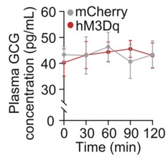
(3) Neuroanatomy of the brain-liver neurocircuit:
The current study and its conclusion are based on a speculative brain-liver sympathetic circuit without the necessary anatomical information downstream of LPGi.Thank you for raising this important point. A clear anatomical definition of the downstream pathways linking the brain to the liver is essential for interpreting the proposed brain-liver sympathetic circuit.
However, the present study (Figure 4A) provides direct anatomical evidence supporting the organization of the brain–liver sympathetic neurocircuit. These observations are consistent with our recent detailed characterization of the brain-liver sympathetic circuit published in Cell Metabolism (Cell Metab. 2025;37(11):2264–2279), LPGi GABAergic neurons inhibit GABAergic neurons in the caudal ventrolateral medulla (CVLM). Disinhibition of CVLM reduces GABAergic suppression of rostral ventrolateral medulla (RVLM) neurons, which are key excitatory drivers of sympathetic tone. RVLM neurons project to sympathetic preganglionic neurons in the sympathetic chain (Syc). These neurons synapse with postganglionic sympathetic neurons in ganglia such as the celiac-superior mesenteric ganglion (CG-SMG). Postganglionic sympathetic fibers then innervate the liver, releasing NE to activate hepatic β2-adrenergic receptors and stimulate HGP.
Together, these data establish a coherent anatomical basis for the proposed brain-liver sympathetic pathway and clarify the downstream organization relevant to the functional experiments presented here.
Author response image 2.
Tracing scheme (Left) and whole-mount imaging (Right) of PRV-labeled brain-liver neurocircuit. Scale bars, 3,000 (whole mount) or 1,000 (optical sections) μm.
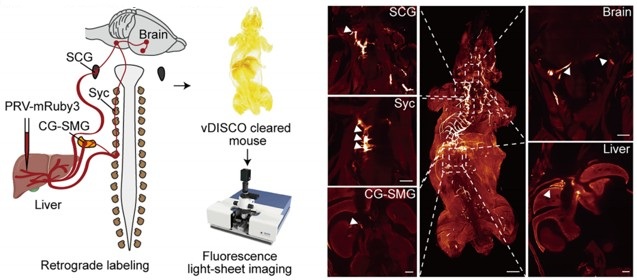
(4) Local manipulation of the celiac ganglia:
The left and right ganglia of mice are not separate from each other but rather anatomically connected. The claim that the local injection of AAV in the left or right ganglion without affecting the other side is against this basic anatomical feature.Thank you for raising this important anatomical point. We fully acknowledge that the left and right celiac ganglia (CG) in mice are interconnected, and that unilateral viral injection could theoretically affect the contralateral side. The celiac–superior mesenteric ganglion (CG-SMG) complex serves as a major sympathetic hub that regulates visceral organ functions. Recent transcriptomic, anatomical, and functional studies have revealed that the CG-SMG is not a homogeneous structure but is composed of molecularly and functionally distinct neuronal populations. These populations exhibit specialized projection patterns and regulate different aspects of gastrointestinal physiology, supporting a model of modular sympathetic control. (Nature. 2025 Jan;637(8047):895-902). Therefore, we were aware of this phenomenon during the initial stages of these experiments.
To minimize unintended spread to the contralateral CG, we took two complementary approaches.
First, we optimized the injection strategy by using an extremely small injection volume (100 nL per site), with a very slow infusion rate (50 nL/min), and fine glass micropipettes. With these refinements, contralateral viral spread was rarely observed.
Second, and importantly, all animals included in the final analyses were subjected to post hoc anatomical verification. After completion of the experiments, CG were collected, sectioned, and examined for viral expression. As shown in Supplementary Figure 5F, only mice in which viral expression was strictly confined to the targeted CG, with no detectable infection in the contralateral ganglion, were included in the presented data.
Together, these measures ensure that the reported effects are attributable to local manipulation of the intended CG. We will ensure that the Methods section more explicitly details these technical precautions and that the legend for Figure S5F clearly states its role in validating injection specificity.
Reviewer #2 (Public review):
Summary:
The manuscript by Wang and colleagues aims to determine whether the left and right LPGi differentially regulate hepatic glucose metabolism and to reveal decussation of hepatic sympathetic nerves.
The authors used tissue clearing to identify sympathetic fibers in the liver lobes, then injected PRV into the hepatic lobes. Five days post-injection, PRV-labeled neurons in the LPGi were identified. The results indicated contralateral dominance of premotor neurons and partial innervation of more than one lobe. Then the authors activated each side of the LPGi, resulting in a greater increase in blood glucose levels after right-sided activation than after left-sided activation, as well as changes in protein expression in the liver lobes. These data suggested modulation of HGP (hepatic glucose production) in a lobe-specific manner. Chemical denervation of a particular lobe did not affect glucose levels due to compensation by the other lobes. In addition, nerve bundles decussate in the hepatic portal region.
We thank the reviewer for the thorough and constructive evaluation of our manuscript. In direct response, we will undertake comprehensive revisions to enhance the rigor and clarity of the study, including: (i) correcting ambiguous or misleading terminology pertaining to anatomical resolution and sympathetic circuit organization; (ii) expanding the Methods section with complete experimental details, improved image presentation, and explicit justification of our viral and genetic approaches; and (iii) strengthening data interpretation by addressing issues related to sparse PRV labeling, projection heterogeneity, and the functional implications of double-labeled neurons. All revisions are expected to be completed within 8 weeks.
Strengths:
The manuscript is timely and relevant. It is important to understand the sympathetic regulation of the liver and the contribution of each lobe to hepatic glucose production. The authors use state-of-the-art methodology.
Weaknesses:
(1) The wording/terminology used in the manuscript is misleading, and it is not used in the proper context. For instance, the goal of the study is "to investigate whether cerebral hemispheres differentially regulate hepatic glucose metabolism..." (see abstract); however, the authors focus on the brainstem (a single structure without hemispheres). Similarly, symmetric is not the best word for the projections.
We thank the reviewer for raising these critical points regarding terminology and conceptual framing. We acknowledge that certain phrases in our original manuscript may have been overly broad or ambiguous, particularly in describing the scope of sympathetic heterogeneity and the specificity of neural projections. Due to practical constraints and the scope of our study, our investigation is focused on the brainstem, which represents the final common pathway for these lateralized commands. We acknowledge that terms referring to the cerebral hemispheres do not accurately describe our study.
We are revising the manuscript to ensure accurate and consistent terminology and will submit the revised version with these corrections.
(2) Sparse labeling of liver-related neurons was shown in the LPGi (Figure 1). It would be ideal to have lower magnification images to show the area. Higher quality images would be necessary, as it is difficult to identify brainstem areas. The low number of labeled neurons in the LPGi after five days of inoculation is surprising. Previous findings showed extensive labeling in the ventral brainstem at four days post-inoculation (Desmoulins et al., 2025). Unfortunately, it is not possible to compare the injection paradigm/methods because the PRV inoculation is missing from the methods section. If the PRV is different from the previously published viral tracers, time-dependent studies to determine the order of neurons and the time course of infection would be necessary.
We sincerely thank the reviewer for these detailed and constructive comments regarding the PRV tracing experiments. We fully agree that careful presentation and interpretation of the anatomical data are essential for ensuring rigor and transparency. We address each point in detail below.
(1) Image magnification and anatomical context of LPGi labeling
We agree that the original images did not sufficiently convey the broader anatomical context of the LPGi. In the revised manuscript, we will replace the original panels in Figure 1 with new images that include lower-magnification overviews of the brainstem, alongside higher-magnification views of the LPGi. These images clearly delineate the LPGi with respect to established anatomical landmarks and atlas boundaries. Image contrast and resolution will also be optimized to allow unambiguous identification of PRV-labeled neurons and surrounding structures.
(2) Sparse LPGi labeling at 5 days post-injection and methodological details
We apologize for the omission of the detailed PRV injection protocol in the original Methods section. We deliberately used small-volume, focal injections (1 µL per liver lobe) to minimize viral spread and to restrict labeling to circuits specifically connected to the targeted hepatic region. Under these conditions, early-stage or intermediate-order upstream nuclei such as the LPGi are expected to exhibit relatively sparse labeling compared to more proximal autonomic nuclei. This information will add, including the PRV strain, viral titer, injection volume, precise injection coordinates, and surgical procedures.
(3) Not all LPGi cells are liver-related. Was the entire LPGi population stimulated, or was it done in a cell-type-specific manner? What was the strain, sex, and age of the mice? What was the rationale for using the particular viral constructs?
We thank the reviewer for this insightful and important question. We agree that not all neurons within the LPGi are liver-related, and we apologize that our rationale was not clearly articulated in the original manuscript.
(1) Our decision to target GABAergic neurons in the LPGi using Gad1-Cre mice was based on prior experimental evidence rather than an assumption about the entire LPGi population. In our previous study (Cell Metab. 2025;37(11):2264-2279.e10), we performed single-cell RNA sequencing on retrogradely labeled LPGi neurons following liver tracing. These analyses revealed that the majority of liver-projecting LPGi neurons are GABAergic in nature. Based on these findings, we chose to selectively manipulate GABAergic neurons in the LPGi rather than the entire LPGi neuronal population, in order to achieve greater cellular specificity and to minimize potential confounding effects arising from heterogeneous neuron types within this region. We regret that this rationale was not clearly described in the original submission and have now revised the manuscript to explicitly state this reasoning.
(2) In addition, we apologize for the omission of mouse strain, sex, and age information in the Methods section. These details will be fully added.
(3) We selected AAV-based viral vectors, specifically the AAV9 serotype, due to their well-established efficiency in transducing neurons in the brainstem, relatively low toxicity, and widespread use in circuit-level chemogenetic and optogenetic studies. When combined with Cre-dependent viral constructs in Gad1-Cre mice, this approach enabled selective and reliable manipulation of LPGi GABAergic neurons.
(4) The authors should consider the effect of stimulation of double-labeled neurons (innervating more than one lobe) and potential confounding effects regarding other physiological functions.
We thank the reviewer for raising this important point. We agree that neurons innervating more than one liver lobe could, in principle, introduce potential confounding effects and may reflect higher-order integrative autonomic neurons.
This consideration is consistent with a key finding of the cited study: the celiac-superior mesenteric ganglion (CG-SMG) contains molecularly distinct sympathetic neuron populations (e.g., RXFP1+ vs. SHOX2+) that exhibit complementary organ projections and separate, non‑overlapping functions. Specifically, RXFP1+ neurons innervate secretory organs (pancreas, bile duct) to regulate secretion, while SHOX2+ neurons innervate the gastrointestinal tract to control motility. This functional segregation supports the concept of specialized autonomic modules rather than a uniform,“fight or flight”response, reinforcing the need for careful interpretation of circuit-specific manipulations. (Nature. 2025;637(8047):895-902; Neuron. Published online December 10, 2025).
In our PRV tracing experiments, the proportion of double-labeled neurons was relatively small, suggesting that the majority of labeled LPGi neurons preferentially associate with individual hepatic lobes. Nevertheless, we recognize that activation of this minority population could contribute to broader physiological effects beyond strictly lobe-specific regulation. We acknowledge that the absence of single-cell-level resolution in the current study limits our ability to further dissect the functional heterogeneity of these projection-defined neurons, and we will explicitly state this as a limitation in the revised manuscript. We will explicitly acknowledge this possibility in the revised manuscript and included it as a limitation of the current study. We thank the reviewer for highlighting this important conceptual consideration.
(5) The authors state that "central projections directly descend along the sympathetic chain to the celiac-superior mesenteric ganglia". What they mean is unclear. Do the authors refer to pre-ganglionic neurons or premotor neurons? How does it fit with the previous literature?
We thank the reviewer for pointing out this imprecise wording. We agree that the original phrasing was anatomically inaccurate and potentially confusing. The pathways we intended to describe involve brainstem premotor neurons that project to sympathetic preganglionic neurons in the spinal cord. These preganglionic neurons then innervate neurons in the celiac–superior mesenteric ganglia, which in turn provide postganglionic input to the liver.
We are revising the manuscript to clearly distinguish premotor from preganglionic neurons and to describe this pathway in a manner consistent with the established organization of sympathetic autonomic circuits reported in the previous literature. The revised wording will explicitly reflect this hierarchical relay structure.
(6) How was the chemical denervation completed for the individual lobes?
We thank the reviewer for raising this important methodological concern. We agree that potential diffusion of 6-OHDA is a critical issue when performing lobe-specific chemical denervation, and we apologize that our original description did not sufficiently clarify how this was controlled.
In the revised Methods section, we will provide a detailed description of the denervation procedure, including the injection volume and concentration of 6-OHDA, as well as the physical separation and isolation of individual hepatic lobes during application to minimize diffusion to adjacent tissue.
To directly assess the specificity of the chemical denervation, we included immunofluorescence and Western blot analyses demonstrating a selective reduction of sympathetic markers in the targeted lobe, with minimal effects on non-targeted lobes. These results support the effectiveness and relative spatial confinement of the 6-OHDA treatment under our experimental conditions.
We thank the reviewer for highlighting this point, which has helped us improve both the clarity and rigor of the manuscript.
(7) The Western Blot images look like they are from different blots, but there are no details provided regarding protein amount (loading) or housekeeping. What was the reason to switch beta-actin and alpha-tubulin? In Figures 3F -G, the GS expression is not a good representative image. Were chemiluminescence or fluorescence antibodies used? Were the membranes reused?
We thank the reviewer for this careful and detailed evaluation of the Western blot data. We apologize that insufficient methodological detail was provided in the original submission.
(1) We would like to clarify that the protein bands shown within each panel were derived from the same membrane. To improve transparency, we will provide full, uncropped images of the corresponding membranes in the supplementary materials. In addition, detailed information regarding protein loading amounts, gel conditions, and housekeeping controls will be added to the Methods section.
(2) The use of different loading controls (β-actin or α-tubulin) reflects a technical consideration rather than an experimental inconsistency. In our experiments, the molecular weight of the TH (62kDa) was too close to α-tubulin (55kDa), and β-actin (42kDa) was therefore used to avoid band overlap and to ensure accurate quantification.
(3) Regarding the GS signal shown in Figures 3F–G, we agree that the original representative image was suboptimal. This appears to be related to antibody performance rather than sample quality. To address this, we are repeating the GS Western blot using a newly validated antibody. The original tissue samples had been aliquoted and stored at −80 °C, allowing reliable re-analysis. This work will be done in 8 weeks.
(4) All Western blot experiments were detected using chemiluminescence, and membrane stripping and reprobing procedures are now explicitly described in the Methods section.
We thank the reviewer for highlighting these issues, which significantly improve the rigor and clarity of our data presentation.
(8) Key references using PRV for liver innervation studies are missing (Stanley et al, 2010 [PMID: 20351287]; Torres et al., 2021 [PMID: 34231420]; Desmoulins et al., 2025 [PMID: 39647176]).
We thank the reviewer for pointing out these important and highly relevant references that were inadvertently omitted in our initial submission. The studies by Stanley et al. (Proc Natl Acad Sci U S A, 2010), Torres et al. (Am J Physiol Regul Integr Comp Physiol, 2021), and Desmoulins et al. (Auton Neurosci, 2025) represent key PRV-based retrograde tracing work that has mapped central neural circuits innervating the liver and thus provide essential context for our anatomical analyses.
We agree that inclusion of these studies is necessary to properly situate our findings within the existing literature. Accordingly, we will incorporate citations to these references in the revised manuscript and discuss their relationship to our results.
Reviewer #3 (Public review):
Summary:
This study found a lobe-specific, lateralized control of hepatic glucose metabolism by the brain and provides anatomical evidence for sympathetic crossover at the porta hepatis. The findings are particularly insightful to the researchers in the field of liver metabolism, regeneration, and tumors.
Strengths:
Increasing evidence suggests spatial heterogeneity of the liver across many aspects of metabolism and regenerative capacity. The current study has provided interesting findings: neuronal innervation of the liver also shows anatomical differences across lobes. The findings could be particularly useful for understanding liver pathophysiology and treatment, such as metabolic interventions or transplantation.
Weaknesses:
Inclusion of detailed method and Discussion:
We sincerely thank the reviewer for the positive and constructive feedback, which will significantly enhance both the methodological rigor and the broader biological interpretation of our study. In direct response, we will revise the Discussion to elaborate on the potential physiological advantages of a lateralized and lobe-specific pattern of liver innervation. Furthermore, we will expand the Methods section to include a comprehensive description of the quantitative analysis applied to PRV-labeled neurons. Together, these revisions will strengthen the manuscript’s clarity, depth, and relevance to researchers in hepatic metabolism, regeneration, and disease. We expect to complete all updates within 8 weeks.
(1) The quantitative results of PRV-labeled neurons are presented, and please include the specific quantitative methods.
We thank the reviewer for this helpful suggestion. We will add a detailed description of the quantitative methods used to analyze PRV-labeled neurons in the revised Methods section. This includes information on the counting criteria, the brain regions analyzed, how the regions of interest were delineated, and the normalization procedures applied to obtain the reported neuron counts.
(2) The Discussion can be expanded to include potential biological advantages of this complex lateralized innervation pattern.
We appreciate the reviewer’s suggestion. We will expand the Discussion to include a paragraph addressing the potential biological significance of lateralized liver innervation. We highlight that this asymmetric organization could allow for more precise, lobe-specific regulation of hepatic metabolism, enable integration of distinct physiological signals, and potentially provide robustness against perturbations. These points will discuss in the revised manuscript.
Reviewer #4 (Public review):
Summary:
The studies here are highly informative in terms of anatomical tracing and sympathetic nerve function in the liver related to glucose levels, but given that they are performed in a single species, it is challenging to translated them to humans, or to determine whether these neural circuits are evolutionarily conserved. Dual-labeling anatomical studies are elegant, and the addition of chemogenetic and optogenetic studies is mechanistically informative. Denervation studies lack appropriate controls, and the role of sensory innervation in the liver is overlooked.
We sincerely appreciate the reviewer's thoughtful evaluation and fully agree that findings derived from a single-species model must be interpreted with caution in relation to human physiology. In direct response, we will revise the manuscript to explicitly clarify that all experimental data were obtained in mice and to provide a discussion of the limitations regarding direct extrapolation to humans. Concurrently, we will expand the Discussion section by integrating our findings with recent human and translational studies, including a multicenter clinical trial demonstrating that catheter-based endovascular denervation of the celiac and hepatic arteries significantly improved glycemic control in patients with poorly controlled type 2 diabetes, without major adverse events (Signal Transduct Target Ther. 2025;10(1):371). While our current work focuses on defining the anatomical organization and functional asymmetry of this circuit in mice, the clinical findings suggest that the core principles, sympathetic control of hepatic glucose metabolism via CG-liver pathways, may be conserved and of translational relevance. Additionally, we will clarify the interpretation of tyrosine hydroxylase labeling and expand the discussion of hepatic sensory and parasympathetic innervation, acknowledging their important roles in liver–brain communication and identifying them as key directions for future research. Collectively, these revisions will provide a more balanced, clinically informed, and rigorous framework for interpreting our findings, and we aim to complete all updates within 8 weeks.
Specific Weaknesses - Major:
(1) The species name should be included in the title.
We thank the reviewer for this suggestion. We agree that the species should be clearly indicated. The findings presented in this study were obtained in mice using tissue clearing and whole-organ imaging approaches. Due to technical limitations, these observations are currently limited to the mouse strain. We will update the title and clarified the species used throughout the manuscript.
(2) Tyrosine hydroxylase was used to mark sympathetic fibers in the liver, but this marker also hits a portion of sensory fibers that need to be ruled out in whole-mount imaging data
We thank the reviewer for pointing this out. We acknowledge that tyrosine hydroxylase (TH) labels not only sympathetic fibers but also a subset of sensory fibers. We will add a limitation of this point in the revised manuscript. In addition, ongoing experiments using retrograde PRV labeling from the liver, combined with sectioning, are being used to distinguish sympathetic fibers from vagal and dorsal root ganglion–derived sensory fibers. These data will be included in a forthcoming update of the manuscript and are expected to be completed in approximately 6 weeks.
(3) Chemogenetic and optogenetic data demonstrating hyperglycemia should be described in the context of prior work demonstrating liver nerve involvement in these processes. There is only a brief mention in the Discussion currently, but comparing methods and observations would be helpful.
We thank the reviewer for this suggestion. Previous studies largely relied on electrical stimulation to modulate liver innervation, which provides relatively coarse control of neural activity (Eur J Biochem. 1992;207(2):399-411). By contrast, our use of chemogenetic and optogenetic approaches allows selective, cell-type–specific manipulation of LPGi neurons. We will revise the Discussion to place our functional data in the context of prior work, highlighting how these more precise approaches improve understanding of the contribution of liver-innervating neurons to hyperglycemia.
(4) Sympathetic denervation with 6-OHDA can drive compensatory increases to tissue sensory innervation, and this should be measured in the liver denervation studies to implicate potential crosstalk, especially given the increase in LPGi cFOS that may be due to afferent nerve activity. Compensatory sympathetic drive may not be the only culprit, though it is clearly assumed to be. The sensory or parasympathetic/vagal innervation of the liver is altogether ignored in this paper and could be better described in general.
We thank the reviewer for this insightful comment and agree that chemical sympathetic denervation with 6-OHDA may induce compensatory changes in non-sympathetic hepatic inputs, including sensory and parasympathetic (vagal) innervation. As the reviewer correctly points out, increased LPGi cFOS activity may reflect afferent nerve engagement rather than solely compensatory sympathetic drive.
More broadly, we agree that the central nervous system functions as an integrated homeostatic network that continuously processes diverse afferent signals, including hepatic sensory and vagal inputs, as well as other interoceptive cues. From this perspective, the LPGi cFOS changes observed in our study likely represent one component of a complex integrative response rather than evidence for a single dominant pathway.
We acknowledge that the present study did not directly assess hepatic sensory or parasympathetic innervation, which represents a limitation in scope. In the revised manuscript, we will expand the Discussion to explicitly note this limitation and provide a more balanced consideration of potential crosstalk among sympathetic, sensory, and parasympathetic pathways in shaping LPGi activity following hepatic denervation.
Recommendations for the authors:
Reviewer #2 (Recommendations for the authors):
Although the findings are interesting, this reviewer has major concerns about the experimental design, methodology, results, and interpretation of the data. Experimental details are lacking, including basic information (age, sex, strain of mice, procedures, magnification, etc.).
We thank the reviewer for this important recommendation. We agree that comprehensive reporting of experimental details is essential for rigor and reproducibility.
In the revised manuscript, we will add complete information regarding mouse strain, sex, age, and sample size for each experiment. In addition, detailed descriptions of surgical procedures, viral constructs, injection parameters, imaging magnification, and analysis methods have been incorporated into the Methods section.
These revisions ensure that all experiments are described with sufficient technical detail and clarity to allow accurate interpretation and replication of our findings.
Reviewer #3 (Recommendations for the authors):
Addressing a few questions might help:
(1) The study found that liver-associated LPGi neurons are predominantly GABAergic. It would be informative to molecularly characterize the PRV-traced, liver-projecting LPGi neurons to determine their neurochemical phenotypes.
We thank the reviewer for this insightful suggestion. We agree that molecular characterization of liver-projecting LPGi neurons is important for understanding their functional identity.
This issue has been addressed in detail in our recent study (Cell Metab. 2025;37(11):2264-2279.e10), in which we performed single-cell RNA sequencing on retrogradely traced LPGi neurons connected to the liver. These analyses demonstrated that the majority of liver-projecting LPGi neurons are GABAergic, with a defined transcriptional profile distinct from neighboring non–liver-related populations.
Based on these findings, the current study selectively targets GABAergic LPGi neurons using Gad1-Cre mice. We are now explicitly referencing and summarizing these molecular results in the revised manuscript to clarify the neurochemical identity of the PRV-traced LPGi neurons.
(2) Is it possible to do a local microinjection of a sodium channel blocker (e.g., lidocaine) or an adrenergic receptor antagonist into the porta hepatis? That would potentially provide additional evidence for the porta hepatis as the functional crossover point.
We appreciate the reviewer’s thoughtful suggestion. While pharmacological blockade at the porta hepatis could modulate local neural activity, the proposed approach may not fully capture the distinction between ipsilateral and contralateral inputs, and may not conclusively establish neural crossover at this particular site.
In our view, the anatomical evidence provided by whole-mount tissue clearing, dual-labeled tracing, and direct visualization of decussating nerve bundles at the porta hepatis offers a more definitive demonstration of sympathetic crossover. Pharmacological blockade would affect both crossed and uncrossed fibers simultaneously and therefore would not specifically resolve the anatomical organization of this decussation.
Nevertheless, we agree that functional interrogation of the porta hepatis represents an interesting direction for future work, and we will now acknowledge this possibility in the Discussion.
(3) It is possible to investigate the effects of unilateral LPGi manipulation or ablation of one side of CG/SMG on liver metabolism, such as hyperglycemia?
We thank the reviewer for this important suggestion. We agree that unilateral ablation or silencing of the CG-SMG could provide additional insight into lateralized sympathetic control of liver metabolism.
However, precise and selective ablation of one side of the CG-SMG through 6-OHDA without affecting the contralateral ganglion or adjacent autonomic structures remains technically challenging, particularly given the anatomical connectivity between the two sides. We are currently optimizing approaches to achieve reliable unilateral manipulation.
If successful within the revision timeframe, we will include these experiments and corresponding metabolic analyses in the revised manuscript. If not, we will explicitly discuss this experimental limitation and the predicted metabolic consequences of unilateral CG-SMG ablation as an important direction for future studies. This work will be done in 6 weeks.
Reviewer #4 (Recommendations for the authors):
In the abstract and elsewhere, the use of the term 'sympathetic release' is unclear - do you mean release of nerve products, such as the neurotransmitter norepinephrine? This should be more clearly defined.
We thank the reviewer for pointing out this ambiguity. We agree that the term “sympathetic release” was imprecise. In the revised manuscript, we will explicitly refer to the release of sympathetic neurotransmitters, primarily norepinephrine, from postganglionic sympathetic fibers.
We will revise the wording throughout the manuscript to ensure accurate and consistent terminology and to avoid potential confusion regarding the underlying neurobiological mechanisms.
-
-
Symmetric brain-liver circuits mediate lateralized regulation of hepatic glucose output https://www.biorxiv.org/content/10.1101/2025.10.27.684732v1
-
Symmetric brain-liver circuits mediate lateralized regulation of hepatic glucose output https://www.biorxiv.org/content/10.1101/2025.10.27.684732v1
-