Intrinsic and circuit mechanisms of predictive coding in a grid cell network model
Curation statements for this article:-
Curated by eLife

eLife Assessment
This valuable study presents a mechanistic model of predictive coding by medial entorhinal cortex grid cells, implemented with biologically detailed conductance-based neurons. The evidence supporting the emergence of this coding scheme from specific membrane currents and the anatomical connectivity among inhibitory neurons is convincing. This work will be of interest to neuroscientists working on spatial navigation, circuit dynamics, and neuronal coding.
This article has been Reviewed by the following groups
Listed in
- Evaluated articles (eLife)
Abstract
Grid cells in the medial entorhinal cortex (MEC) fire at the vertices of a hexagonal lattice, forming an allocentric code for the animal’s current position. Recent studies have identified a class of grid cells that represent locations ahead of the animal. How do these predictive representations emerge from the wetware of the MEC? We developed a detailed conductance-based model of the MEC network, constrained by empirical data on the biophysical properties of stellate cells and the topology of the MEC network. The model revealed two mechanisms by which grid cells can signal future locations. First, hyperpolarizing inhibition from interneurons activates HCN channels in stellate cells, whose slow kinetics maintain a depolarizing influence after inhibition ends, advancing spike timing and shifting the inferred position forward by ~5% of a grid field diameter. Second, introducing asymmetry into the inhibitory connectivity, by skewing the Gaussian profile of interneuron-to-stellate connections, causes inhibition to rise more steeply and enables earlier spiking, advancing the inferred position by up to ~25%. A corollary of our model is that the extent of the predictive code changes monotonically along the dorsoventral axis of the MEC, following the experimentally measured dorsoventral gradient in HCN time constants.
Article activity feed
-

-
-

eLife Assessment
This valuable study presents a mechanistic model of predictive coding by medial entorhinal cortex grid cells, implemented with biologically detailed conductance-based neurons. The evidence supporting the emergence of this coding scheme from specific membrane currents and the anatomical connectivity among inhibitory neurons is convincing. This work will be of interest to neuroscientists working on spatial navigation, circuit dynamics, and neuronal coding.
-
Reviewer #1 (Public review):
Summary:
In this manuscript, the authors aim to elucidate the mechanisms by which grid cells in the medial entorhinal cortex generate predictive representations of spatial location. To address this, they built a computational model integrating intrinsic neuronal dynamics with structured network connectivity. Specifically, they combine a conductance-based single-cell model incorporating biologically realistic HCN channels with a continuous attractor network that reflects known properties of grid cell circuitry. Their simulations show that HCN conductance can shift grid fields forward by approximately 5% of their diameter, consistent with experimental observations in layer II grid cells. Additionally, by introducing asymmetry in the connectivity of interneurons, the model produces larger forward shifts, which …
Reviewer #1 (Public review):
Summary:
In this manuscript, the authors aim to elucidate the mechanisms by which grid cells in the medial entorhinal cortex generate predictive representations of spatial location. To address this, they built a computational model integrating intrinsic neuronal dynamics with structured network connectivity. Specifically, they combine a conductance-based single-cell model incorporating biologically realistic HCN channels with a continuous attractor network that reflects known properties of grid cell circuitry. Their simulations show that HCN conductance can shift grid fields forward by approximately 5% of their diameter, consistent with experimental observations in layer II grid cells. Additionally, by introducing asymmetry in the connectivity of interneurons, the model produces larger forward shifts, which parallels properties observed in layer III grid cells. Together, these two mechanisms provide a unified framework for explaining layer-specific predictive coding in the entorhinal cortex.
Strengths:
A major strength of the study lies in its conceptual contribution. The authors propose two distinct mechanisms to generate forward-shifted grid fields for predictive coding. One mechanism is intrinsic and depends on the time constants associated with HCN channels. The other is network-based and results from asymmetries in interneuron connectivity. These two mechanisms correspond to different observed properties of grid cells in layer II and layer III, respectively. The modeling is based on previously validated frameworks of continuous attractor network models (e.g., Burak & Fiete; Kang & DeWeese), but it incorporates several novel features, including the incorporation of biophysically realistic HCN channels, a network architecture that excludes stellate-stellate connections and relies on interneurons, and asymmetric interneuron connectivity.
Weaknesses:
One of the proposed mechanisms for predictive coding, namely asymmetric interneuron connectivity, is a novel idea. However, this type of connectivity has not yet been demonstrated experimentally in the medial entorhinal cortex. Therefore, the biological plausibility of this mechanism remains uncertain and will need to be evaluated in future empirical studies. The revised manuscript now clearly acknowledges this limitation and discusses how this prediction could be tested experimentally, for example through paired recordings or connectomic reconstruction of interneuron-stellate cell connectivity.
Comments on revised version.
The authors have adequately addressed my previous comments, and the revised manuscript has been substantially improved. I have no further major comments.
-
Reviewer #2 (Public review):
Summary:
This study proposes that predictive spatial representations in medial entorhinal cortex (MEC) grid cells arise through two distinct biophysical mechanisms: (1) HCN conductance-dependent temporal dynamics, which generate modest forward shifts (~5% of grid field diameter) in Layer II cells, and (2) network asymmetry, enabling larger predictive shifts (~25% of grid field diameter) in Layer III cells. The model further predicts a dorsoventral gradient in predictive coding magnitude, correlating with observed HCN conductance variations. These results provide a mechanistic framework for understanding how intrinsic cellular properties and circuit architecture collectively enable prospective spatial coding in the MEC. This is an important and novel study with a new model.
Strengths:
These findings reveal …
Reviewer #2 (Public review):
Summary:
This study proposes that predictive spatial representations in medial entorhinal cortex (MEC) grid cells arise through two distinct biophysical mechanisms: (1) HCN conductance-dependent temporal dynamics, which generate modest forward shifts (~5% of grid field diameter) in Layer II cells, and (2) network asymmetry, enabling larger predictive shifts (~25% of grid field diameter) in Layer III cells. The model further predicts a dorsoventral gradient in predictive coding magnitude, correlating with observed HCN conductance variations. These results provide a mechanistic framework for understanding how intrinsic cellular properties and circuit architecture collectively enable prospective spatial coding in the MEC. This is an important and novel study with a new model.
Strengths:
These findings reveal how cellular properties and circuit design enable prospective spatial coding. This novel, impactful study will interest the field upon publication.
Weaknesses:
Some of the model is too mathematical and not fit with the biological observation.
Comments on revised version.
I have no other concern about the papers.
-
Reviewer #3 (Public review):
Summary:
The manuscript by Shaikh and Assisi addresses a timely and important question related to the neural circuit mechanisms underlying spatial representations during navigation. Concretely, they present a valuable model of the medial entorhinal cortex (MEC) with biophysically detailed conductance-based stellate cells that can perform path integration and reveals two potential mechanisms underlying two forms of predictive coding by grid cells in the MEC. One mechanism uses HCN channels to explain predictive coding in MEC layer II grid cells equivalent to ~5% of the diameter of a grid field, and the other uses asymmetric connections between interneurons and stellate cells, resulting in a ~25% predictive bias of layer III grid cells. The methods and model are convincing, and the model is expected to be …
Reviewer #3 (Public review):
Summary:
The manuscript by Shaikh and Assisi addresses a timely and important question related to the neural circuit mechanisms underlying spatial representations during navigation. Concretely, they present a valuable model of the medial entorhinal cortex (MEC) with biophysically detailed conductance-based stellate cells that can perform path integration and reveals two potential mechanisms underlying two forms of predictive coding by grid cells in the MEC. One mechanism uses HCN channels to explain predictive coding in MEC layer II grid cells equivalent to ~5% of the diameter of a grid field, and the other uses asymmetric connections between interneurons and stellate cells, resulting in a ~25% predictive bias of layer III grid cells. The methods and model are convincing, and the model is expected to be valuable for computational neuroscientists studying neural mechanism of spatial navigation.
Strengths:
One strength of the model is its use of conductance-based neuron models of stellate cells and interneurons, adding important biophysical constraints and details to existing continuous attractor network models of grid cells. The model fills a gap in the literature by providing mechanisms for predictive coding constrained by biophysical properties of stellate cells and simplified network topology. The model generates testable hypotheses and advances our mechanistic understanding of the grid cell network.
Weaknesses:
A few weaknesses of the study remain:
(1) MEC interneurons display a very similar spatial periodicity in their firing as stellate cells. This seems inconsistent with experimental data (Buetfering et al., 2014).
(2) In the model, stellate cells are responsive to movement direction. Although this is consistent with most existing attractor network models, grid cells respond much stronger to head direction than to movement direction (Raudies et al., 2015). Likewise, grid cell sweeps follow an internal movement direction signal and not head direction (Vollan et al., 2026).
References:
Buetfering C, Allen K, Monyer H (2014) Parvalbumin interneurons provide grid cell-driven recurrent inhibition in the medial entorhinal cortex. Nat Neurosci 17:710-718.
Raudies F, Brandon MP, Chapman GW, Hasselmo ME (2015) Head direction is coded more strongly than movement direction in a population of entorhinal neurons. Brain Res 1621:355-367.
Vollan AZ, Schellenberger MF, Gardner RJ, Moser M-B, Moser EI (2026) Adaptive modulation of theta sweeps in the brain's navigation circuit. Science 393:eaef4184.
-
Author response:
The following is the authors’ response to the original reviews.
Public Reviews:
Reviewer #1 (Public review):
Summary:
In this manuscript, the authors aim to elucidate the mechanisms by which grid cells in the medial entorhinal cortex generate predictive representations of spatial location. To address this, they built a computational model integrating intrinsic neuronal dynamics with structured network connectivity. Specifically, they combine a conductance-based single-cell model incorporating biologically realistic HCN channels with a continuous attractor network that reflects known properties of grid cell circuitry. Their simulations show that HCN conductance can shift grid fields forward by approximately 5% of their diameter, consistent with experimental observations in layer II grid cells. Additionally, by …
Author response:
The following is the authors’ response to the original reviews.
Public Reviews:
Reviewer #1 (Public review):
Summary:
In this manuscript, the authors aim to elucidate the mechanisms by which grid cells in the medial entorhinal cortex generate predictive representations of spatial location. To address this, they built a computational model integrating intrinsic neuronal dynamics with structured network connectivity. Specifically, they combine a conductance-based single-cell model incorporating biologically realistic HCN channels with a continuous attractor network that reflects known properties of grid cell circuitry. Their simulations show that HCN conductance can shift grid fields forward by approximately 5% of their diameter, consistent with experimental observations in layer II grid cells. Additionally, by introducing asymmetry in the connectivity of interneurons, the model produces larger forward shifts, which parallel properties observed in layer III grid cells. Together, these two mechanisms provide a unified framework for explaining layer-specific predictive coding in the entorhinal cortex.
Strengths:
A major strength of the study lies in its conceptual contribution. The authors propose two distinct mechanisms to generate forward-shifted grid fields for predictive coding. One mechanism is intrinsic and depends on the time constants associated with HCN channels. The other is network-based and results from asymmetries in interneuron connectivity. These two mechanisms correspond to different observed properties of grid cells in layer II and layer III, respectively. The modeling is based on previously validated frameworks of continuous attractor network models (e.g., Burak & Fiete; Kang & DeWeese), but it incorporates several novel features, including the incorporation of biophysically realistic HCN channels, a network architecture that excludes stellate-stellate connections and relies on interneurons, and asymmetric interneuron connectivity.
Weaknesses:
One of the proposed mechanisms for predictive coding, namely asymmetric interneuron connectivity, is a novel idea. However, this type of connectivity has not yet been demonstrated experimentally in the medial entorhinal cortex. Therefore, the biological plausibility of this mechanism remains uncertain and will need to be evaluated in future empirical studies.
We agree that asymmetric interneuron-to-stellate connectivity has not yet been directly demonstrated, and we have revised the Discussion (page 15, lines 436–449) to state this limitation explicitly. As we note there, and as in most continuous attractor network models of grid cells, several elements of our network's connectivity, including the offset excitatory projections from stellate cells to interneurons that set the direction of attractor propagation, and the asymmetric interneuron-to-stellate connectivity that generates the layer III-like predictive bias, are constraints that are not derived from a fully specified, data-driven wiring diagram of the MEC. There are marked differences between the predictive coding of layer II and layer III grid cells, with the predictive bias in layer III roughly five-fold larger than in layer II. Our model predicts that these differences arise from an asymmetric directional skew in the synaptic weights between interneurons and stellate cells, present in layer III and absent, or much weaker, in layer II. Such a difference in synaptic weight or connection probability should become evident in paired recordings or connectomic reconstruction of interneuron-stellate synapses. This asymmetry has not yet been demonstrated directly, though recent anatomical work showing that stellate cell-interneuron synapses are organized at the spatial scale of individual grid-cell clusters [1] is at least consistent with the kind of spatially patterned connectivity our model requires (schematic figure 8 in Huang et. al. [1]).
Manuscript change: Discussion (page 15, lines 436–449): added a new paragraph explicitly framing the asymmetric interneuron-stellate connectivity as an untested, falsifiable prediction, citing supporting anatomical evidence (Huang et al., 2024).
Reviewer #2 (Public review):
Summary:
This study proposes that predictive spatial representations in medial entorhinal cortex (MEC) grid cells arise through two distinct biophysical mechanisms: (1) HCN conductance-dependent temporal dynamics, which generate modest forward shifts (~5% of grid field diameter) in Layer II cells, and (2) network asymmetry, enabling larger predictive shifts (~25% of grid field diameter) in Layer III cells. The model further predicts a dorsoventral gradient in predictive coding magnitude, correlating with observed HCN conductance variations. These results provide a mechanistic framework for understanding how intrinsic cellular properties and circuit architecture collectively enable prospective spatial coding in the MEC. This is an important study.
Strengths:
These findings reveal how cellular properties and circuit design enable prospective spatial coding. This novel, impactful study will be of interest to the field.
Weaknesses:
Some of the models are too mathematical and do not fit with the biological observation.
We understand the concern that the manuscript may read as too mathematical. We have used a conductance-based formalism to describe the neurons, and much of this mathematical complexity is itself inherited from earlier models that were built directly from detailed voltage- and current-clamp experiments. The stellate cell model we use is a simplified version, due to Acker et al. (2003) [2], of the more detailed biophysical model of Fransén et al. (2004) [3]. We have added this lineage to the Methods section (“Neuron and Synaptic Models,” page 15, lines 466–467), where the stellate cell model is first introduced.
The exact synaptic connectivity of the MEC network has also not been completely established. Guided by the anatomical work of Gu et al. (2018) [4], we speculate a local connectivity pattern consistent with that used in other continuous attractor network models of grid cells. As shown by Couey et al. (2013) [5], stellate cells are not connected to each other directly, but interact only through inhibitory interneurons, a motif we adopt in the model. The network motif and connectivity reported by Huang et al. (2024) [1] provide further support for the type of connectivity pattern used in our model. This anatomical grounding is already described in the Results section (“Biophysically detailed conductance-based grid cell attractor model,” page 3, lines 110–117).
As stated in our response to Reviewer 1's comments, the exact connectivity of the network is not completely established. However, based on these anatomical observations, we make a testable prediction regarding the skew of the interneuron-stellate synaptic weight distribution. This skew should be present in layer III, where the predictive bias is large, and weak or absent in layer II. This prediction is already presented in the Discussion (page 15, lines 436–449), in the paragraph before the summary.
Manuscript change: Methods and Materials, “Neuron and Synaptic Models” (page 15, lines 466–467): added a clause tracing the conductance-based stellate cell model to Fransén et al. (2004), as simplified by Acker et al. (2003). The related connectivity grounding (Gu et al., 2018; Couey et al., 2013; Huang et al., 2024) is already covered by Discussion (page 15, lines 436–449), and the testable, skew-based connectivity prediction is already covered by item 18; these points are not repeated in the Discussion. “Biophysically detailed conductance-based grid cell attractor model” (page 3, lines 110–117): added two sentences grounding the network topology (stellate-interneuron-only connectivity, exclusion of pyramidal cells) in anatomical data, and the testable, skew-based prediction was already present in the Discussion paragraph preceding the summary (page 15, lines 436–449): added a new paragraph explicitly framing the asymmetric interneuron-stellate connectivity as an untested, falsifiable prediction, citing supporting anatomical evidence (Huang et al., 2024).
Reviewer #3 (Public review):
Summary:
The manuscript by Shaikh and Assisi addresses a timely and important question related to the neural circuit mechanisms underlying spatial representations during navigation. Concretely, they present a model of the medial entorhinal cortex (MEC) with biophysically detailed conductance-based stellate cells that can perform path integration and reveal two potential mechanisms underlying two forms of predictive coding by grid cells in the MEC. One mechanism uses HCN channels to explain predictive coding in MEC layer II grid cells equivalent to ~5% of the diameter of a grid field, and the other uses asymmetric connections between interneurons and stellate cells, resulting in a ~25% predictive bias of layer III grid cells. The methods and model are technically sound, and the model is expected to be useful for computational neuroscientists studying the neural mechanisms of spatial navigation.
Strengths:
One strength of the model is its use of conductance-based neuron models of stellate cells and interneurons, adding important biophysical constraints and details to existing continuous attractor network models of grid cells. The model fills a gap in the literature by providing mechanisms for predictive coding constrained by biophysical properties of stellate cells and simplified network topology.
Weaknesses:
A weakness of the model is that the neural network is relatively small (five sheets with 71 × 71 neurons each), and the 2-D toroidal topology is further simplified to a 1-D ring attractor consisting of three rings with 192 neurons each.
The neurons were connected using periodic boundary conditions to minimize edge effects, and the connectivity was radially symmetric to limit the influence of topographic irregularities on the collective dynamics of grid cells. Therefore, we believe that increasing the network size would not have significantly altered the simulation outcomes.
We were constrained by the number of biophysically realistic neurons that could be simulated within a reasonable time frame—approximately 25,000 for the 2-D attractor network and 576 for the ring network. There were two main reasons for this limitation. (i) The simulations had to run over time scales ranging from several seconds to minutes to ensure that the trajectory repeatedly passed through the same local neighbourhood and that the spike density was sufficient to delineate the grid fields. This process was especially time-consuming for the 2-D environment, though we managed to achieve it in a smaller network representing a 1-D environment. Additionally, the trajectories needed to extend over time to allow us to estimate any drift between the simulated animal’s actual position and its inferred position from neuronal activity. (ii) The simulations required extensive multi-parameter sweeps within the bounds of experimentally observed values, further increasing the total simulation time. Given these computational constraints, we had to limit our model to smaller neuron populations. While using simpler neuron models could have allowed larger networks, we considered it essential to retain a degree of biophysical realism. This level of detail enabled us to explore how specific conductances influenced the overall dynamics of the network. We have also added a sentence to the Methods section (“2D model,” page 16, lines 488–494) stating these computational constraints explicitly and noting that periodic boundary conditions and radially symmetric connectivity were used throughout to minimize edge and finite-size effects.
Manuscript change: Methods and Materials, “2D model” (page 16, lines 488–494): added a sentence explaining the computational constraints that determined network size and the use of periodic, radially symmetric connectivity to limit edge effects.
The model incorporates biophysical detail at the single-neuron level, but not at the network level. For example, it includes only stellate cells and a generic interneuron type and does not implement data-driven connectivity patterns. The restricted network size and the limited experimental knowledge about connectivity among stellate cells, principal cells, and different interneuron types in the MEC could be addressed in more detail.
Because the network connectivity of the medial entorhinal cortex (MEC) is not yet fully understood, our model incorporates prior findings showing that stellate cells interact only through inhibitory interneurons [5–8]. In layer II, stellate cells that function as grid cells also show a topographic organization. They form clusters located between patches of pyramidal neurons. Each grid cell module consists of stellate cells that are physically contiguous, although cells from neighboring modules intermingle at their borders. Within a module, stellate cells tend to form a noisy two-dimensional lattice organized according to the phases of their grid fields [4]. In addition, axonal projections in layer II of the MEC create spatially organized connections between stellate cells and parvalbumin interneurons. Nearby neurons often, but not always, share direct connections, whereas neurons that are farther apart are linked primarily through multisynaptic pathways [5]. Together, these observations suggest that stellate cells interact via inhibitory intermediaries and are locally arranged in a network topology that closely resembles standard Continuous Attractor Network (CAN) models. We have added two sentences to the Results section (“Biophysically detailed conductance-based grid cell attractor model,” page 3, lines 110–117) making this anatomical justification explicit, and stating that layer II pyramidal cells, which form a separate microcircuit, are correspondingly excluded from the model.
Manuscript change: Results, “Biophysically detailed conductance-based grid cell attractor model” (page 3, lines 110–117): added two sentences grounding the network topology (stellate-interneuron-only connectivity, exclusion of pyramidal cells) in anatomical data.;See Discussion (page 15, lines 436–449): added a new paragraph explicitly framing the asymmetric interneuron-stellate connectivity as an untested, falsifiable prediction, citing supporting anatomical evidence (Huang et al., 2024).
Moreover, the manuscript lacks a thorough discussion of assumptions common to most continuous attractor network (CAN) models of grid cells, such as the use of "hand-crafted" connections between direction-sensitive conjunctive grid cells and network cells to drive attractor shifts. Including such a discussion would strengthen the manuscript. This is especially relevant given the authors' explicit claim that they have revealed two mechanisms underlying the emergence of a predictive code in the MEC. In this reviewer's view, the work demonstrates a potential mechanism, but one that requires experimental verification. The significance of the model would thus be increased by providing more experimentally testable predictions of the model.
The exact connectivity of stellate cells and interneurons is yet unknown. But several lines of evidence point to an attractor-like connection topology. We have now included a more detailed discussion of the connectivity used in CAN models.
Manuscript change: Discussion (page 15, lines 436–449): added a new paragraph explicitly framing the asymmetric interneuron-stellate connectivity as an untested, falsifiable prediction, citing supporting anatomical evidence (Huang et al., 2024).
Recommendations for the authors:
Reviewer #1 (Recommendations for the authors):
(1) In the abstract, the authors write:
"Our model revealed two mechanisms underlying the emergence of a predictive code in the MEC. The first relied on a time scale associated with the HCN conductance. The other depended on the degree of asymmetry in the topology of the MEC network."
Given the central importance of these two mechanisms, the description here may be overly brief. Even in an abstract, a slightly more detailed explanation of each mechanism would enhance clarity and accessibility.
We have modified the abstract (page 1, lines 7–21) which now includes a brief explanation of how the timing of stellate cell spikes are advanced
Manuscript change: Abstract (page 1, lines 7–21): modified the abstract to include a detailed description of the mechanism.
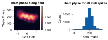
(2) In both experimental (e.g., Hafting et al., 2008; Chaudhuri-Vayalambrone et al., 2023; Ouchi & Fujisawa, 2024) and theoretical studies (e.g., Kang & DeWeese, 2019), predictive coding in grid cells has been closely associated with the phase profile of theta oscillations, including phenomena such as phase precession and phase locking. The authors' model successfully reproduces theta-range membrane potential oscillations, consistent with physiological observations. Given this, it would be valuable to know whether the model allows for an integrated analysis of the relationship between predictive coding and spike timing relative to theta phase. If such a simulation could be included in the present study, it would likely strengthen the contribution of the manuscript. However, if this is beyond the scope of the current work, a brief discussion of the potential relevance of theta-phase dynamics to predictive coding, and how this might be addressed in future studies, would still help broaden the perspective and impact of the paper.
The predictive grid cells [9,10] are phase-locked to theta oscillations. In line with this observation, our model demonstrates that for sufficiently high theta amplitude stellate cells can be entrained to the input oscillations. We have added a paragraph to the Discussion (page 12, lines 365–370) describing this entrainment finding in the text. However, we have not included the analysis figure (see Author response image 1) itself in the manuscript.
Manuscript change: Discussion (page 12, lines 365–370): added a new paragraph noting that the model reproduces theta-entrainment of stellate cell spiking consistent with phase-locked predictive grid cells, without including the underlying analysis figure in the manuscript.
Author response image 1.
(3) The Discussion section currently ends somewhat abruptly. Adding a concluding paragraph that summarizes the key findings and their broader implications would help reinforce the significance of the work for readers.
We have now modified the discussion section (page 15, lines 450–463) to summarize key findings and the broader implications of the study.
Manuscript change: Discussion (page 15, lines 450–463): a concluding paragraph has been added at the end of the Discussion section.
(4) "These properties are due to the presence of a hyperpolarization-activated depolarizing current (Ih) and a persistent sodium current (INaP) in addition to leak and spiking currents (IL, IK, INa). An interplay between Ih and INaP is needed to generate the membrane potential oscillations observed here. In addition, these oscillations fall squarely within the range of theta oscillations (4-12 Hz)." (Page 3, Line 97)
Please provide supporting evidence for these claims, such as simulation results or relevant literature references.
We have added references supporting our claims.
Manuscript change: Results, “Biophysically detailed conductance-based grid cell attractor model” (page 3, lines 97–100): added reference to "An interplay between I_h and I_NaP is needed to generate the membrane potential oscillations observed here. In addition, these oscillations fall squarely within the range of theta oscillations (4–12 Hz)."
(5) "How does HCN give rise to a predictive bias? The HCN channel depolarizes the cell and is activated by inhibitory input from interneurons in the model network. Since HCN increases the cell's excitability, it speeds up the onset of spiking, translating grid fields that are farther away to locations closer to the animal." (Page 7, Line 285).
Is this explanation derived directly from simulation data? If so, please indicate the relevant figures or results that support this interpretation.
Reference to support the effect of HCN on the cell’s excitability has been added (page 10, lines 296–299). A paragraph in the manuscript explains how this change in excitability can cause the grid field to be translated closer to the animal (page 10, lines 300-305).
Manuscript change: Results, “Cellular and network-based mechanisms for predictive and retrospective coding” (page 10, lines 296–299): added reference to "How does HCN give rise to a predictive bias? The HCN channel depolarizes the cell and is activated by inhibitory input from interneurons in the model Network. Since HCN increases the cell's excitability, it speeds up the onset of spiking, translating grid fields that are farther away to locations closer to the animal."
(6) Some elements in the main figures are not clearly explained or referenced in the main text. For example, Figures 1E and F, and Figure 3B appear to lack corresponding descriptions or interpretation. Please ensure that all figure panels are explicitly discussed in the results.
Missing explanations for Figure 1E and F have been added to the result section. Figure 3b is mentioned in the article (Page 8, line 235)
Manuscript change: Results, “Biophysically detailed conductance-based grid cell attractor model” (page 4, lines 153–154): added missing references to Figure 1e and Figure 1f in the manuscript.
(7) Typos
(a) Page 4, Line 140: "as shown in Figure 1A". It seems "Figure 1D" is intended.
(b) Figure 2A (color legend): "m.p. eamp (mV)" should be "m.p. ramp (mV)".
These typos have been fixed.
Manuscript change: fixed the Figure 1 panel-letter typo (Results, “Biophysically detailed conductance-based grid cell attractor model,” page 4, lines 146–147): fixed figure reference from Figure 1A (bottom) to Figure 1d (bottom) and the Figure 3 colour-legend typo (Figure 3a, page 9): fixed colour label typo from "m.p. eamp (mV)" to "m.p. ramp (mV)". and standardized all figure panel references to lowercase throughout the manuscript: Figure panel references were changed to lowercase throughout the manuscript.
Reviewer #2 (Recommendations for the authors):
(1) The model is based on five interconnected neuronal sheets organized in a toroidal topology, with four layers of stellate cells (71×71 neurons each) and one inhibitory interneuron layer. Key architectural features included: Stellate cells interacted exclusively via feedforward inhibition through interneurons, consistent with experimental observations (Couey et al., 2013). Interneurons projected symmetrically to other interneurons within their sheet, and each interneuron inhibited a concentric ring of stellate cells. This spatially structured inhibition, combined with toroidal boundary conditions, generated stable hexagonal activity patterns in both stellate and interneuron populations. In this model, it will be very likely that the interneurons also exhibited perspective coding, but this is not consistent with the physiology data. Can the authors comment on this?
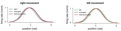
As stated in materials and methods, we used the interneuron activity as the “true” position of the animal. This was done because the errors accumulated during path integration were far greater than the predictive bias shown by the grid network. Therefore, it would not have been possible to discern the predictive bias from the path integration error. The interneuron field does show a skew in the opposite direction to the direction of travel. However, unlike stellate cells, this is not translation of the grid field, See Author response image 2. This is due to asymmetry in the firing rates of interneurons along its field. We have added a sentence to the Methods section (“Predictive and retrospective coding,” page 17, lines 521–525) making this distinction, between a firing-rate asymmetry and an actual translation of positional tuning, explicit.
Manuscript change: Methods and Materials, “Predictive and retrospective coding” (page 17, lines 521–525): added a sentence clarifying that interneuron firing-rate asymmetry does not reflect a translation of positional tuning, justifying its use as the position reference.
Author response image 2.
(2) In the original study (Ouchi et al., 2024), it reported predictive grid cells; it has been shown that theta-rhythmic population activity in the MEC supports dynamic spatial representation, with phase-organized firing patterns enabling continuous trajectory prediction. The current model did not examine this in much detail. Could the authors discuss this or test this in their model?
Our simulations have shown that the impact of theta oscillations on the temporal organisation of grid fields is more intricate than we initially anticipated, and further work is underway to elucidate these mechanisms. However, in line with experimental observations [9], our model demonstrates that stellate cells can be entrained to the input oscillations by increasing the amplitude of theta. See Author response image 1. As above, we have added a Discussion paragraph describing this entrainment result in the text, without including the corresponding figure in the manuscript.
Manuscript change: Discussion (page 12, lines 365–370): added a new paragraph noting that the model reproduces theta-entrainment of stellate cell spiking consistent with phase-locked predictive grid cells, without including the underlying analysis figure in the manuscript.
(3) In the data, the shift of the predictive grid is in a range of distribution from 22.5 {plus minus} 10.2 cm; the model did not clearly show the shift distance of the grid in the current model. It would be helpful to examine why the grid cells with different spacing could show a range of their shift in the current model.
The expansion of grid spacing is accompanied by a lengthening of HCN conductance time scales. As demonstrated in Figure 5e, the extent of predictive coding can be enhanced by increasing these intrinsic temporal constants. We have added a sentence to the Results section (“Cellular and network-based mechanisms for predictive and retrospective coding”, page 10, lines 315–320) relating this range of model-predicted shift distances explicitly to experiments.
Manuscript change: Results, “Cellular and network-based mechanisms for predictive and retrospective coding” (page 10, lines 315–320): added a sentence relating the range of model-predicted shift distances across grid scales to the experimentally reported range.
(4) In the non-linearity and ensure accurate path integration, the author mapped each value of speed𝑠 measured in (𝑟𝑎𝑑∕𝑠) to its corresponding depolarising drive 𝐼 = 𝑓(𝑠) (Figure 2a). It is necessary to discuss the biological meaning of the depolarizing drive. Is the value reasonable in the biological range?
The depolarizing drive I = f(s) is used to represent the population speed signal that is delivered to stellate cells as a somatic current, and across the simulated range of speeds its magnitude remains within the sub-nanoampere range used for current-clamp characterization of stellate cell and interneuron excitability in the original single-cell studies from which our conductance parameters were taken [2,3,11,12] We have added a sentence to the Results section (“Grid cell network can accurately path integrate 1D trajectories,” page 6, lines 184–188) stating this explicitly, so that the biological plausibility of the depolarizing drive is clear to the reader.
Manuscript change: Results, “Grid cell network can accurately path integrate 1D trajectories” (page 6, lines 184–188): added a sentence clarifying that the depolarizing drive I=f(s) remains within the sub-nanoampere range used experimentally to characterize stellate cell and interneuron excitability.
(5) In the study after HCN knockout, the spacing change of grid cells exhibited a linear drift to higher spacing (Giocomo et al., 2011a). This also shows grid cells with discrete modules (Stensola et al., 2012, Nature). Regarding this, how does the current model represent the discretized modules and their predictive coding?
In our model, all stellate cells share the same grid scale and can therefore be treated as belonging to a single module. Experimentally, however, grid scale varies along the dorsoventral axis of the MEC, in parallel with systematic changes in inhibitory connectivity and HCN channel conductance [13–15]. In the model, we can reproduce different grid spacings (as seen across modules) by increasing the range of lateral inhibition, and the grid expansion observed after HCN knockout would similarly apply to modules with different grid scales. Our current implementation does not include a mechanism for generating discrete modules, which would require a network with a graded change in lateral inhibition across its spatial extent. In networks with such graded organization, several mechanisms have been proposed for how modules emerge [16,17] and these would likely apply to our model as well. We have added a paragraph to the Discussion (page 15, lines 428–435) stating this limitation and the proposed extension explicitly, with supporting citations.
Manuscript change: Discussion (page 15, lines 428–435): added a new paragraph on the model's single-module limitation and possible extensions to discrete, dorsoventrally organized modules.
(6) The grid cells have been shown in both the Stella cells and the pyramidal cell group? Will the current model bias their conclusion toward different cell groups? Or can it not represent these cell groups? It could be necessary to discuss this.
Within Layer II of the MEC, stellate cells and pyramidal cells form separate, largely non-overlapping microcircuits. Pyramidal cells are commonly connected via 5− HT3a positive interneurons, while stellate cells are primarily connected via fast-spiking parvalbumin (PV+) cells and slower-spiking somatostatin-positive cells [5–7]. Further, stellate cells show a topographic organization with stellate cell clusters being separated by pyramidal neurons [1].
Given these observations we chose a model stellate cell and fast-spiking interneuron.
Based on our work it would be hard to say what, if any, the effect of pyramidal neurons would be to the stellate grid cell network. We have added a sentence to the Results section
(“Biophysically detailed conductance-based grid cell attractor model,” page 3, lines 110–117) stating explicitly that the model does not include pyramidal cells and that our conclusions are specific to the stellate-cell grid-cell population.
Manuscript change: Results, “Biophysically detailed conductance-based grid cell attractor model” (page 3, lines 110–117): added two sentences grounding the network topology (stellate-interneuron-only connectivity, exclusion of pyramidal cells) in anatomical data, which states the exclusion of pyramidal cells explicitly.
(7) In Kropff et al., 2015 Nature, speed cells have been shown also represent predictive shift in their coding, the author achieved accurate path integration with a linear relationship between the speed of the animal and the movement of attractor is necessary and for this by introducing an invertible mapping between speed and the amplitude of the depolarizing input used to drive the stellate cells. The current model did not address the possible contribution of speed coding to the retrospective coding in grid cells. Will the authors discuss this?
This is an interesting point. Kropff et al. (2015) [18] showed that a subset of speed-tuned cells in the MEC represent running speed with a temporal offset, effectively encoding a predictive or retrospective estimate of speed itself. Our model currently represents the population speed signal as a single, instantaneous depolarizing drive I = f(s) and does not incorporate this heterogeneity. We have added a passage to the Discussion (page 14, lines 393–402) noting that if even a fraction of the depolarizing drive to stellate cells were itself temporally offset in the way Kropff et al. describe, this could shift the magnitude, or potentially the sign, of the retrospective bias that we attribute to network asymmetry, providing an additional, speed-coding-dependent route to retrospective coding that could act alongside the intrinsic and network mechanisms we describe. Disentangling the relative contributions of speed-signal timing and network asymmetry to retrospective coding is a promising direction for future modeling work.
Manuscript change: Discussion (page 14, lines 393–402): added a discussion of how heterogeneity in speed-cell timing (Kropff et al., 2015) could interact with network-asymmetry-based retrospective coding.
References
(1) Huang, L.-W., Garden, D. L., McClure, C. & Nolan, M. F. Synaptic interactions between stellate cells and parvalbumin interneurons in layer 2 of the medial entorhinal cortex are organized at the scale of grid cell clusters. eLife 12, RP92854 (2024).
(2) Acker, C. D., Kopell, N. & White, J. A. Synchronization of Strongly Coupled Excitatory Neurons: Relating Network Behavior to Biophysics. J. Comput. Neurosci. 15, 71–90 (2003).
(3) Fransén, E., Alonso, A. A., Dickson, C. T., Magistretti, J. & Hasselmo, M. E. Ionic mechanisms in the generation of subthreshold oscillations and action potential clustering in entorhinal layer II stellate neurons. Hippocampus 14, 368–384 (2004).
(4) Gu, Y. et al. A Map-like Micro-Organization of Grid Cells in the Medial Entorhinal Cortex. Cell 175, 736-750.e30 (2018).
(5) Couey, J. J. et al. Recurrent inhibitory circuitry as a mechanism for grid formation. Nature neuroscience 16, 318–324 (2013).
(6) Nilssen, E. S., Doan, T. P., Ohara, S. & Witter, M. P. A reappraisal of the lateral and medial entorhinal subdivisions mediating parallel cortical pathways. Hippocampus in press, 1–17 (2019).
(7) Witter, M. P., Doan, T. P., Jacobsen, B., Nilssen, E. S. & Ohara, S. Architecture of the entorhinal cortex: a review of entorhinal anatomy in rodents with some comparative notes. Frontiers in systems neuroscience 11, 1–12 (2017).
(8) Nilssen, X. E. S. et al. Inhibitory Connectivity Dominates the Fan Cell Network in Layer II of Lateral Entorhinal Cortex. The Journal of neuroscience: the official journal of the Society for Neuroscience 38, 9712–9727 (2018).
(9) Ouchi, A. & Fujisawa, S. Predictive grid coding in the medial entorhinal cortex. Science 385, 776–784 (2024).
(10) Ouchi, A. Predictive grid cells: Future spatial representations in the hippocampal-entorhinal circuit. Neurosci. Res. 227, 105053 (2026).
(11) Dickson, C. T. et al. Properties and role of I(h) in the pacing of subthreshold oscillations in entorhinal cortex layer II neurons. Journal of neurophysiology 83, 2562–2579 (2000).
(12) Alonso, A. & Klink, R. Differential electroresponsiveness of stellate and pyramidal-like cells of medial entorhinal cortex layer II. J. Neurophysiol. 70, 128–143 (1993).
(13) Garden, D. L. F., Dodson, P. D., O’Donnell, C., White, M. D. & Nolan, M. F. Tuning of Synaptic Integration in the Medial Entorhinal Cortex to the Organization of Grid Cell Firing Fields. Neuron 60, 875–889 (2008).
(14) Giocomo, L. M. et al. Grid Cells Use HCN1 Channels for Spatial Scaling. Cell 147, 1159–1170 (2011).
(15) Yoshida, M., Giocomo, L. M., Boardman, I. & Hasselmo, M. E. Frequency of subthreshold oscillations at different membrane potential voltages in neurons at different anatomical positions on the dorsoventral axis in the rat medial entorhinal cortex. The Journal of neuroscience: the official journal of the Society for Neuroscience 31, 12683–12694 (2011).
(16) Kang, L. & Balasubramanian, V. A geometric attractor mechanism for self-organization of entorhinal grid modules. eLife 8, e46687 (2019).
(17) Khona, M., Chandra, S. & Fiete, I. Global modules robustly emerge from local interactions and smooth gradients. Nature 640, 155–164 (2025).
(18) Kropff, E., Carmichael, J. E., Moser, M.-B. & Moser, E. I. Speed cells in the medial entorhinal cortex. Nature 523, 419–424 (2015).
-

-
-
eLife Assessment
This valuable study presents a mechanistic model of predictive coding by medial entorhinal cortex grid cells, implemented with biologically detailed conductance-based neurons. The evidence supporting the emergence of this coding scheme from specific membrane currents and the anatomical connectivity among inhibitory neurons is solid. However, the justification for the choice of connectivity patterns and other network parameters remains somewhat incomplete. This work will be of interest to neuroscientists working on spatial navigation, circuit dynamics, and neuronal coding.
-
Reviewer #1 (Public review):
Summary:
In this manuscript, the authors aim to elucidate the mechanisms by which grid cells in the medial entorhinal cortex generate predictive representations of spatial location. To address this, they built a computational model integrating intrinsic neuronal dynamics with structured network connectivity. Specifically, they combine a conductance-based single-cell model incorporating biologically realistic HCN channels with a continuous attractor network that reflects known properties of grid cell circuitry. Their simulations show that HCN conductance can shift grid fields forward by approximately 5% of their diameter, consistent with experimental observations in layer II grid cells. Additionally, by introducing asymmetry in the connectivity of interneurons, the model produces larger forward shifts, which …
Reviewer #1 (Public review):
Summary:
In this manuscript, the authors aim to elucidate the mechanisms by which grid cells in the medial entorhinal cortex generate predictive representations of spatial location. To address this, they built a computational model integrating intrinsic neuronal dynamics with structured network connectivity. Specifically, they combine a conductance-based single-cell model incorporating biologically realistic HCN channels with a continuous attractor network that reflects known properties of grid cell circuitry. Their simulations show that HCN conductance can shift grid fields forward by approximately 5% of their diameter, consistent with experimental observations in layer II grid cells. Additionally, by introducing asymmetry in the connectivity of interneurons, the model produces larger forward shifts, which parallel properties observed in layer III grid cells. Together, these two mechanisms provide a unified framework for explaining layer-specific predictive coding in the entorhinal cortex.
Strengths:
A major strength of the study lies in its conceptual contribution. The authors propose two distinct mechanisms to generate forward-shifted grid fields for predictive coding. One mechanism is intrinsic and depends on the time constants associated with HCN channels. The other is network-based and results from asymmetries in interneuron connectivity. These two mechanisms correspond to different observed properties of grid cells in layer II and layer III, respectively. The modeling is based on previously validated frameworks of continuous attractor network models (e.g., Burak & Fiete; Kang & DeWeese), but it incorporates several novel features, including the incorporation of biophysically realistic HCN channels, a network architecture that excludes stellate-stellate connections and relies on interneurons, and asymmetric interneuron connectivity.
Weaknesses:
One of the proposed mechanisms for predictive coding, namely asymmetric interneuron connectivity, is a novel idea. However, this type of connectivity has not yet been demonstrated experimentally in the medial entorhinal cortex. Therefore, the biological plausibility of this mechanism remains uncertain and will need to be evaluated in future empirical studies.
-
Reviewer #2 (Public review):
Summary:
This study proposes that predictive spatial representations in medial entorhinal cortex (MEC) grid cells arise through two distinct biophysical mechanisms: (1) HCN conductance-dependent temporal dynamics, which generate modest forward shifts (~5% of grid field diameter) in Layer II cells, and (2) network asymmetry, enabling larger predictive shifts (~25% of grid field diameter) in Layer III cells. The model further predicts a dorsoventral gradient in predictive coding magnitude, correlating with observed HCN conductance variations. These results provide a mechanistic framework for understanding how intrinsic cellular properties and circuit architecture collectively enable prospective spatial coding in the MEC. This is an important study.
Strengths:
These findings reveal how cellular properties and …
Reviewer #2 (Public review):
Summary:
This study proposes that predictive spatial representations in medial entorhinal cortex (MEC) grid cells arise through two distinct biophysical mechanisms: (1) HCN conductance-dependent temporal dynamics, which generate modest forward shifts (~5% of grid field diameter) in Layer II cells, and (2) network asymmetry, enabling larger predictive shifts (~25% of grid field diameter) in Layer III cells. The model further predicts a dorsoventral gradient in predictive coding magnitude, correlating with observed HCN conductance variations. These results provide a mechanistic framework for understanding how intrinsic cellular properties and circuit architecture collectively enable prospective spatial coding in the MEC. This is an important study.
Strengths:
These findings reveal how cellular properties and circuit design enable prospective spatial coding. This novel, impactful study will be of interest to the field.
Weaknesses:
Some of the models are too mathematical and do not fit with the biological observation.
-
Reviewer #3 (Public review):
Summary:
The manuscript by Shaikh and Assisi addresses a timely and important question related to the neural circuit mechanisms underlying spatial representations during navigation. Concretely, they present a model of the medial entorhinal cortex (MEC) with biophysically detailed conductance-based stellate cells that can perform path integration and reveal two potential mechanisms underlying two forms of predictive coding by grid cells in the MEC. One mechanism uses HCN channels to explain predictive coding in MEC layer II grid cells equivalent to ~5% of the diameter of a grid field, and the other uses asymmetric connections between interneurons and stellate cells, resulting in a ~25% predictive bias of layer III grid cells. The methods and model are technically sound, and the model is expected to be useful …
Reviewer #3 (Public review):
Summary:
The manuscript by Shaikh and Assisi addresses a timely and important question related to the neural circuit mechanisms underlying spatial representations during navigation. Concretely, they present a model of the medial entorhinal cortex (MEC) with biophysically detailed conductance-based stellate cells that can perform path integration and reveal two potential mechanisms underlying two forms of predictive coding by grid cells in the MEC. One mechanism uses HCN channels to explain predictive coding in MEC layer II grid cells equivalent to ~5% of the diameter of a grid field, and the other uses asymmetric connections between interneurons and stellate cells, resulting in a ~25% predictive bias of layer III grid cells. The methods and model are technically sound, and the model is expected to be useful for computational neuroscientists studying the neural mechanisms of spatial navigation.
Strengths:
One strength of the model is its use of conductance-based neuron models of stellate cells and interneurons, adding important biophysical constraints and details to existing continuous attractor network models of grid cells. The model fills a gap in the literature by providing mechanisms for predictive coding constrained by biophysical properties of stellate cells and simplified network topology.
Weaknesses:
A weakness of the model is that the neural network is relatively small (five sheets with 71 × 71 neurons each), and the 2-D toroidal topology is further simplified to a 1-D ring attractor consisting of three rings with 192 neurons each. The model incorporates biophysical detail at the single-neuron level, but not at the network level. For example, it includes only stellate cells and a generic interneuron type, and does not implement data-driven connectivity patterns.
The restricted network size and the limited experimental knowledge about connectivity among stellate cells, principal cells, and different interneuron types in the MEC could be addressed in more detail. Moreover, the manuscript lacks a thorough discussion of assumptions common to most continuous attractor network (CAN) models of grid cells, such as the use of "hand-crafted" connections between direction-sensitive conjunctive grid cells and network cells to drive attractor shifts. Including such a discussion would strengthen the manuscript. This is especially relevant given the authors' explicit claim that they have revealed two mechanisms underlying the emergence of a predictive code in the MEC. In this reviewer's view, the work demonstrates a potential mechanism, but one that requires experimental verification. The significance of the model would thus be increased by providing more experimentally testable predictions of the model.
-
-
Intrinsic and circuit mechanisms of predictive coding in a grid cell network model https://www.biorxiv.org/content/10.1101/2025.04.11.648301v1
-
Intrinsic and circuit mechanisms of predictive coding in a grid cell network model https://www.biorxiv.org/content/10.1101/2025.04.11.648301v1
-