Prophase Chromosomes Relocalization to Nuclear Periphery in NPP-3/NUP205 Depletion Protects Genome Stability
Curation statements for this article:-
Curated by eLife

eLife Assessment
This study presents valuable findings on the functional consequences of nuclear envelope rupture caused by the depletion of nuclear pore complex subunits on the behaviour and organisation of chromosomes during mitosis in C. elegans embryos. The experiments are generally well-designed and executed; however, the evidence for some of the main conclusions is incomplete. This work is of potential interest to cell biologists working on cell division.
This article has been Reviewed by the following groups
Discuss this preprint
Start a discussion What are Sciety discussions?Listed in
- Evaluated articles (eLife)
Abstract
The nuclear envelope (NE) mediates transport between nucleus and cytoplasm in eukaryotic cells and protects genetic materials against cytoplasmic enzymes. Nuclear pore complexes (NPCs) regulate chromosome architecture, genome integrity, gene transcription, and cell division. In Caenorhabditis elegans embryos, depletion of NPP-3/NUP205 causes NE rupture, premature chromosome condensation, and relocalization of condensed chromosomes to the nuclear periphery, similar to responses during anoxia and quiescence. This chromosomal relocalization depends on the spindle assembly checkpoint (SAC), inner kinetochore proteins, and partially on the NE rupture repair proteins BAF-1 and LEM-2. NPP-3 depletion prolongs prophase and prometaphase, as mediated by SAC proteins MDF-1 and MDF-2. Additionally, NPP-3 depletion alters MDF-1 localization, removing it from NE and increasing its nuclear accumulation, while reducing import of kinetochore components such as KNL-1, BUB-1, and HCP-1. In 20-30 cell-stage embryos, MDF-1 foci are observed on peripheral chromosomes during prophase. Both MDF-1 and MDF-2 accumulate on chromosomes during prometaphase. The increased incidence of lagging chromosomes, DNA damage, and micronuclei upon NPP-3 and MDF-1 depletion, suggesting that peripheral chromosome localization may serve as a protective mechanism against DNA damage. These findings shed light into cellular responses to NE rupture, with potential implications for laminopathies and cancers involving nuclear envelope defects.
Article activity feed
-

eLife Assessment
This study presents valuable findings on the functional consequences of nuclear envelope rupture caused by the depletion of nuclear pore complex subunits on the behaviour and organisation of chromosomes during mitosis in C. elegans embryos. The experiments are generally well-designed and executed; however, the evidence for some of the main conclusions is incomplete. This work is of potential interest to cell biologists working on cell division.
-
Reviewer #1 (Public review):
In this manuscript, the authors investigate the functional consequences of nuclear envelope rupture caused by the depletion of the nucleoporin NPP-3.
They observe that loss of NPP-3 causes condensed chromosomes to localize to the nuclear periphery. This anchoring is independent of the pathway required to anchor heterochromatin and telomeres, but it depends on spindle assembly checkpoint proteins as well as centromere and kinetochore proteins. While the authors propose that relocalization of chromosomes to the nuclear periphery protects genome stability, they do not demonstrate this.
Overall, some of the observations are interesting, but several points should be addressed. Furthermore, the manuscript could be much clearer if certain sections were shortened, simplified, or removed.
Major points:
(1) The title …
Reviewer #1 (Public review):
In this manuscript, the authors investigate the functional consequences of nuclear envelope rupture caused by the depletion of the nucleoporin NPP-3.
They observe that loss of NPP-3 causes condensed chromosomes to localize to the nuclear periphery. This anchoring is independent of the pathway required to anchor heterochromatin and telomeres, but it depends on spindle assembly checkpoint proteins as well as centromere and kinetochore proteins. While the authors propose that relocalization of chromosomes to the nuclear periphery protects genome stability, they do not demonstrate this.
Overall, some of the observations are interesting, but several points should be addressed. Furthermore, the manuscript could be much clearer if certain sections were shortened, simplified, or removed.
Major points:
(1) The title is misleading because the authors provide no experimental evidence that chromosome relocalisation protects genome stability. They are more cautious in the abstract, where they state that it 'may serve a protective role'. If they could provide stronger experimental evidence that chromosome relocalization protects genome stability, this would significantly strengthen the manuscript.
(2) Here, the authors use acute inactivation of npp-3. Do chromosomes also localize to the periphery upon partial npp-3 inactivation? What are the minimal levels of nuclear envelope rupture that cause chromosomes to localize to the periphery? Given that NPP-3 and NPCs have pleiotropic functions, it would be important to analyze conditions where only a few nuclear envelope ruptures are induced. In such conditions, they might be able to explore the link between chromosome localization and genome stability.
(3) The authors primarily examined P1 cells. Is the behaviour of the chromosome different between cells of different lineages?
(4) The authors mentioned that defective chromosomal localisation does not occur upon npp-2 or npp-4 depletion. How do they explain this? Did they attempt to inactivate other NPPs in the Y complexes, and can they be certain that NPP-2 depletion is complete?
(5) The section on AIR-1 (line 147) is confusing and could be removed. To my knowledge, air-1 depletion does not cause the appearance of multiple centrosomes, except maybe in a very few embryos. air-1 depletion causes major defects, so it is difficult to draw a parallel with npp-3 depletion.
(6) The authors show that condensed chromosomes tend to localize to the nuclear envelope upon NPP-3 depletion. Do they condense at the nuclear envelope (NE), or do they condense first and then move to the periphery? This is unclear from the data presented in Figure 1D. Also, why do chromosomes condense earlier? This point could be discussed.
(7) The authors evaluated the consequences of NPP-3 depletion on transcription using RNA sequencing. The relevance of this experiment is questionable, however, as npp-3(RNAi) embryos have significant general defects and not only mislocalised chromosomes.
(8) In the co-depletion experiment npp-3(RNAi), X(RNAi) presented in Figure 3B, the levels of NPP-3 depletion seem highly variable. All the images shown are not similarly exposed, so it is difficult to evaluate these data.
(9) Inactivation of mdf-1/2 suppresses the mislocalization of the chromosomes observed upon npp-3 inactivation. Does it also suppress the premature chromosome condensation phenotype?
(10) Figure 5B: The delay induced by npp-3 depletion is not severe, based on the micrographs presented. The authors should show more representative images. The graph shows the elapsed time between NEBD and NER, and not NER to NEBD, as indicated.
(11) The authors observed that depleting mdf-1 slightly enhanced the lethality associated with npp-3 inactivation. Based on this observation, they conclude that loss of chromosome anchoring exacerbates genomic instability and severely impairs embryonic survival. However, the genetic interaction is not strong, as npp-3(RNAi) embryos already present more than 95% embryonic lethality and have defects other than just mislocalized chromosomes (e.g., defects in kinetochore and spindle assembly).
-
Reviewer #2 (Public review):
Summary:
The authors aimed to determine the molecular mechanisms by which nuclear pore component NPP-3/NUP205 regulates chromosome localization in C. elegans embryos. Previous studies had shown that depletion of NPP-3 caused premature chromosome condensation and movement of chromosomes to the nuclear periphery. Peripheral location of chromosomes is also observed under respiratory stress conditions, suggesting that peripheral chromosome positioning could act as a protective response to stress conditions. How NPP-3 affects chromosome positioning was unknown. Here, the authors conduct a screen to identify factors that promote chromosome relocation to the periphery in npp-3-depleted embryos, identifying an important role for spindle assembly checkpoint components in this process.
Strengths:
Using cytological …
Reviewer #2 (Public review):
Summary:
The authors aimed to determine the molecular mechanisms by which nuclear pore component NPP-3/NUP205 regulates chromosome localization in C. elegans embryos. Previous studies had shown that depletion of NPP-3 caused premature chromosome condensation and movement of chromosomes to the nuclear periphery. Peripheral location of chromosomes is also observed under respiratory stress conditions, suggesting that peripheral chromosome positioning could act as a protective response to stress conditions. How NPP-3 affects chromosome positioning was unknown. Here, the authors conduct a screen to identify factors that promote chromosome relocation to the periphery in npp-3-depleted embryos, identifying an important role for spindle assembly checkpoint components in this process.
Strengths:
Using cytological tools to visualise chromosomes and nuclear envelope markers, the authors show that, in addition to the peripheral location of chromosomes, NPP-3 depletion causes partial rupture of the nuclear envelope and premature chromosome condensation. By systematically co-depleting NPP-3 and factors required for heterochromatin association with nuclear lamina (CEC-4), telomere binding to nuclear envelope (SUN-1 and POT-1), proteins required for the nuclear rupture repair machinery (BAF-1 and LEM-2), kinetochore proteins and components of the spindle assembly checkpoint (SAC) (MDF-1 and MDF-2), the authors convincingly show that SAC components are required for peripheral relocation of chromosomes in absence of NPP-3. The study also provides convincing evidence that peripheral relocation of chromosomes in the absence of NPP-3 has functional implications as it causes transcriptional deregulation and premature relocation of SAC components from the nuclear envelope to chromosomes. Co-depletion of NPP-3 and SAC components accelerates progression through miotic prophase and increases the incidence of defects in chromosome segregation during mitosis. These findings demonstrate that SAC proteins play an important role in regulating chromosome positioning during prophase (at least in the absence of NPP-3) and that they can regulate cell cycle progression at earlier stages than previously thought.
Weaknesses:
The authors also propose that NPP-3 depletion causes DNA damage; however, the evidence presented to support this claim is not as strong as that presented for the effects mentioned above. Also, the premature condensation of chromosomes appears as a clear consequence of NPP-3 depletion, but this intriguing phenotype remains unexplored.
-
Reviewer #3 (Public review):
Summary:
This manuscript reports that RNAi depletion of the inner-ring nucleoporin NPP-3/NUP205 in Caenorhabditis elegans embryos causes nuclear envelope rupture, premature chromatin condensation, and relocalization of condensed prophase chromosomes to the nuclear periphery. Through a candidate epistasis screen, the authors argue that this relocalization requires spindle assembly checkpoint (SAC) components (MDF-1, MDF-2, SAN-1), inner kinetochore proteins (HCP-3, HCP-4, and partially KNL-1), and NE rupture-repair factors (BAF-1, LEM-2), but not the CEC-4 heterochromatin- or SUN-1/POT-1 telomere-anchoring pathways. They further show that NPP-3 loss extends prophase and the NEBD-to-anaphase interval in a SAC-dependent manner, redistributes MDF-1/MDF-2, and reduces import of KNL-1/BUB-1/HCP-1. Co-depletion of …
Reviewer #3 (Public review):
Summary:
This manuscript reports that RNAi depletion of the inner-ring nucleoporin NPP-3/NUP205 in Caenorhabditis elegans embryos causes nuclear envelope rupture, premature chromatin condensation, and relocalization of condensed prophase chromosomes to the nuclear periphery. Through a candidate epistasis screen, the authors argue that this relocalization requires spindle assembly checkpoint (SAC) components (MDF-1, MDF-2, SAN-1), inner kinetochore proteins (HCP-3, HCP-4, and partially KNL-1), and NE rupture-repair factors (BAF-1, LEM-2), but not the CEC-4 heterochromatin- or SUN-1/POT-1 telomere-anchoring pathways. They further show that NPP-3 loss extends prophase and the NEBD-to-anaphase interval in a SAC-dependent manner, redistributes MDF-1/MDF-2, and reduces import of KNL-1/BUB-1/HCP-1. Co-depletion of NPP-3 with MDF-1 abolishes both the arrest and the peripheral localization while increasing lagging chromosomes, HUS-1 foci, micronuclei, and lethality, which the authors interpret as evidence that peripheral positioning is protective.
Weaknesses:
(1) The "protective" conclusion is largely correlative. The protective claim rests on the observation that co-depleting MDF-1 (or MDF-2) with NPP-3 removes the peripheral localization and simultaneously increases DNA damage, micronuclei, and lethality. However, depleting a SAC component removes at least three things at once: the peripheral localization, the prophase extension, and the NEBD-to-anaphase arrest. Because loss of the SAC independently causes premature anaphase and genomic instability through well-established mechanisms unrelated to chromosome positioning, the current design cannot separate damage caused by loss of a protective peripheral location from damage caused by checkpoint bypass. As presented, the increased damage is at least as consistent with simple SAC bypass. To support the protective model, the authors should provide a manipulation that disrupts peripheral positioning without abrogating the SAC-dependent arrest (for example, via the BAF-1/LEM-2 or kinetochore depletion) and show that damage still increases. The LEM-2 co-depletion, which partially suppresses positioning, is a natural place to test whether micronuclei and HUS-1 foci also rise.
(2) Knockdown efficiency of the partner gene in double RNAi is not verified. The double depletions are performed by cloning both gene fragments into a single vector. This risks reducing the effective dose of each dsRNA, so an apparent suppression in an npp-3; gene X (RNAi) condition could reflect weaker NPP-3 knockdown rather than a true epistatic relationship. The authors partially address this by showing that NPP-3::mCherry is still reduced in npp-3;mdf-1 (Figure S4A/B), which is helpful, but they do not demonstrate efficient knockdown of the partner genes in any double condition. For the key epistasis conclusions (MDF-1, MDF-2, HCP-3, HCP-4 suppressions), the knockdown of the second gene should be independently validated with a reporter strain for the second protein.
(3) Alternative explanations for the transcriptomic and H3K9me3 data are not excluded. NPP-3 depletion blocks nuclear import of molecules smaller than ~70 kDa and arrests development at early gastrulation. Both the RNA-seq changes (30% of genes downregulated) and the increased H3K9me3 signal could therefore be secondary consequences of nucleocytoplasmic transport failure and developmental arrest rather than evidence of position-dependent transcriptional repression. Notably, the authors' own finding that up- and down-regulated genes show no chromosomal positional bias (Figure S2C/D) argues against a model in which peripheral repositioning drives silencing of specific chromatin domains. This section should be reframed more cautiously, with the transport/arrest confound explicitly discussed, and RNA-seq replicate number and differential-expression thresholds reported.
(4) Evidence for SAC "activation in prophase" is indirect, and the effect is small. The claim of a novel prophase role for the SAC rests on MDF-1/MDF-2 intensity changes that are repeatedly described as "modest," "slight," or "mild," measured with small n and Student's t-tests, together with phenotypic suppression of prophase extension. There is no direct readout of SAC catalytic activity (for example, MCC assembly). The prophase-extension suppression by MDF-1 is the strongest evidence; the intensity data are weak support. I recommend tempering "the SAC is activated in prophase" to a hypothesis, and strengthening it with a more direct assay if feasible.
(5) The BAF-1 arm of the model is inferred rather than demonstrated. The authors state that baf-1(RNAi) and npp-3;baf-1 produced clustering too severe for epistasis, so BAF-1's requirement for peripheral localization is not actually established genetically; it rests on increased BAF-1 accumulation (correlative) plus the LEM-2 partial suppression. The proposed BAF-1/CENP-C bridge is extrapolated from Drosophila (ref. 71). This is reasonable as a discussion hypothesis but should not be presented in the abstract or summary model as an established dependency.
-
Author response:
Public Reviews:
Reviewer #1 (Public review):
In this manuscript, the authors investigate the functional consequences of nuclear envelope rupture caused by the depletion of the nucleoporin NPP-3.
They observe that loss of NPP-3 causes condensed chromosomes to localize to the nuclear periphery. This anchoring is independent of the pathway required to anchor heterochromatin and telomeres, but it depends on spindle assembly checkpoint proteins as well as centromere and kinetochore proteins. While the authors propose that relocalization of chromosomes to the nuclear periphery protects genome stability, they do not demonstrate this.
Overall, some of the observations are interesting, but several points should be addressed. Furthermore, the manuscript could be much clearer if certain sections were shortened, …
Author response:
Public Reviews:
Reviewer #1 (Public review):
In this manuscript, the authors investigate the functional consequences of nuclear envelope rupture caused by the depletion of the nucleoporin NPP-3.
They observe that loss of NPP-3 causes condensed chromosomes to localize to the nuclear periphery. This anchoring is independent of the pathway required to anchor heterochromatin and telomeres, but it depends on spindle assembly checkpoint proteins as well as centromere and kinetochore proteins. While the authors propose that relocalization of chromosomes to the nuclear periphery protects genome stability, they do not demonstrate this.
Overall, some of the observations are interesting, but several points should be addressed. Furthermore, the manuscript could be much clearer if certain sections were shortened, simplified, or removed.
Major points:
(1) The title is misleading because the authors provide no experimental evidence that chromosome relocalisation protects genome stability. They are more cautious in the abstract, where they state that it 'may serve a protective role'. If they could provide stronger experimental evidence that chromosome relocalization protects genome stability, this would significantly strengthen the manuscript.
We understand the more severe chromosome missegregation and DNA damage phenotypes in the npp-3 mdf-1 or npp-3 mdf-2 double RNAi may suggest that npp-3 RNAi is more sensitized to the loss of spindle assembly checkpoint (SAC) component MDF-1 or MDF-2 for chromosome protection, but may not clearly show that the localization of chromosomes to the nuclear periphery serves a chromosome protective function. Based on our current phenotypic analyses, we will tone down the title to “Prophase Chromosomes Relocalization to Nuclear Periphery in NPP-3/NUP205 Depletion Depends on the Spindle Assembly Checkpoint and Inner Kinetochore Proteins”.
We have thought about artificially tethering the chromosomes to the nuclear periphery. However, without the same stimulus/defect and activation of the response pathway, the effects could be different. In future, to further clarify the functions of chromosome relocalization, we could analyze the chromosomal missegregation and DNA damage phenotypes in npp-3 lem-2 RNAi, which has a partial chromosomal nuclear periphery phenotype, to see if there is any quantitative relationship between chromosome nuclear periphery location and DNA protection.
(2) Here, the authors use acute inactivation of npp-3. Do chromosomes also localize to the periphery upon partial npp-3 inactivation? What are the minimal levels of nuclear envelope rupture that cause chromosomes to localize to the periphery? Given that NPP-3 and NPCs have pleiotropic functions, it would be important to analyze conditions where only a few nuclear envelope ruptures are induced. In such conditions, they might be able to explore the link between chromosome localization and genome stability.
We have not performed partial npp-3 RNAi yet. To explore whether the level of nuclear envelope rupture correlates with chromosome localization to the periphery or if a minimal level of nuclear rupture is required, we have performed the lacI::GFP reporter assay in different NPP RNAi to indicate the nuclear permeability defects (in Fig. S1A and B). npp-2, npp4 or npp-5 RNAi also causes increased permeability, at a comparable level as npp-3, npp-7 and npp-13 RNAi, yet only npp-3, npp-7 and npp-13 RNAi causes chromosome relocalization. This may explain the peripheral chromosome relocalization response may be more closely linked to the specific NPC subcomplex’s (inner ring and nuclear basket) or individual NPP’s function, rather than the general permeability defect or rupture size.
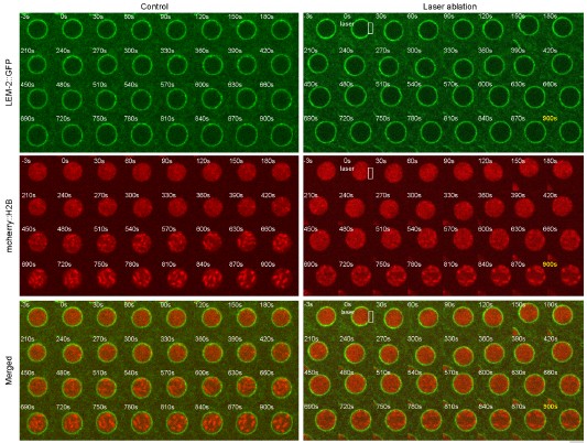
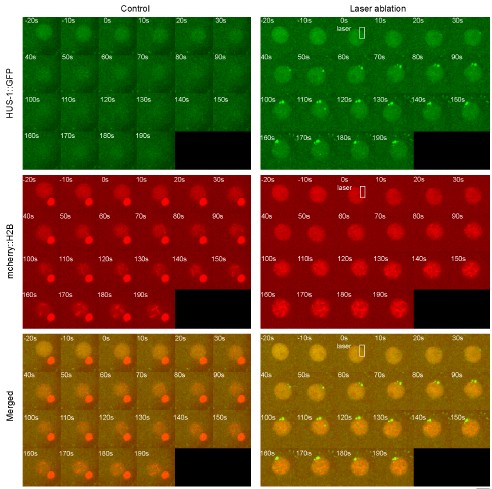
However, we have also attempted to use other methods to induce targeted nuclear rupture with specific rupture size by laser ablation (Author response image 1, 355 nm UV pulsed laser marked by the white rectangle), and we could occasionally observe all chromosomes relocalizing to all over the nuclear periphery after 660 s upon laser ablation in different strains. However, the chromosome relocalization phenotype is not very consistent (~33%, n =12). Importantly, the laser also introduces DNA damage at the rupture sites, as marked by HUS-1 (Author response image 2), complicating the interpretation, so we did not include this attempt in the manuscript.
Author response image 1.
Time-lapse imaging of embryos expressing LEM-2::GFP and mCherry::H2B, a 15*30pixel rectangle was laser-microirradiated to induce nuclear envelope rupture (white rectangle). 0s is the time applying laser ablation. Scale bar 5 μm.
Author response image 2.
Time-lapse imaging of embryos expressing HUS-1::GFP and mCherry::H2B, a 15*30pixel rectangle was laser-microirradiated to induce DNA damage (white rectangle). 0 s is the time applying laser ablation. Scale bar 5 μm.
Another attempt to achieve different nuclear rupture sizes is by observing nucleus in different stages of embryos. By npp-3 feeding RNAi approach, we observed an increase of the proportion of the nuclear circumference with nuclear rupture during embryonic development (Fig. S3D). Yet, the chromosome periphery phenotype is displayed in all embryo stages, suggesting that the localization of chromosomes to the nuclear periphery occurs across a range of rupture sizes.
Given these observations, it appears that the extent of nuclear rupture or permeability increase may not be the sole determinant of chromosome relocalization. Instead, disruption of certain NUP subcomplexes or NPPs might stimulate this process.
(3) The authors primarily examined P1 cells. Is the behaviour of the chromosome different between cells of different lineages?
We observed that chromosomes localize to the nuclear periphery in all cells during embryonic development until the embryo dies, as demonstrated by snapshots and time-lapse imaging (Fig. S1C and Fig. S1E).
We focused on the P1 blastomere for our analyses because its cell cycle timing and division orientation is well characterized, which facilitates precise examination of chromosome localization and cell cycle dynamics under different perturbation conditions. We will add a sentence at the beginning of that result section to clarify this choice: "The chromosome nuclear periphery localization in npp-3 RNAi is consistent in all cells at different embryonic stages (Fig. S1C and Fig. S1E). For analysis in distinct perturbation conditions, we specifically examined the P1 cells, whose cell cycle timing and division orientation is well characterized and suitable for such studies.”
(4) The authors mentioned that defective chromosomal localisation does not occur upon npp-2 or npp-4 depletion. How do they explain this? Did they attempt to inactivate other NPPs in the Y complexes, and can they be certain that NPP-2 depletion is complete?
(see response to point 2) We observed that npp-2 or npp-4 RNAi can cause nuclear rupture with certain permeability defect compared to wildtype (Fig. S1A and S1B), but not chromosomal nuclear periphery localization, suggesting that such chromosome localization to the nuclear periphery does not just depend on the nuclear rupture but could be a more specific phenotype related to individual NUP subcomplex’s (inner ring and nuclear basket) or NPP’s function.
For Y complex, we also now tested NPP-5 depletion, which shows permeability defect but did not show nuclear rupture marked by NPP-1:GFP or chromosomal periphery phenotype (Fig. S1A and S1B), which is consistent to the paper cited [1].
To confirm the efficiency of NPP-2 depletion, we have observed the presence of smaller nuclei (as described in phenobank) by NPP-1::GFP marker (Fig. S1A) and a marked reduction in NPP2::GFP signal in npp-2 RNAi-treated embryos (unpublished data). These evidences support that NPP-2 depletion was effective.
(5) The section on AIR-1 (line 147) is confusing and could be removed. To my knowledge, air-1 depletion does not cause the appearance of multiple centrosomes, except maybe in a very few embryos. air-1 depletion causes major defects, so it is difficult to draw a parallel with npp-3 depletion.
We agree with this suggestion and have decided to remove the section on AIR-1 in the manuscript text to avoid confusion. To clarify, air-1(RNAi) causes multiple centrosomes in only a small proportion of embryos (approximately 13%) [2] , primarily by affecting centrosome positioning [3]. and does not routinely lead to significant centrosome amplification.
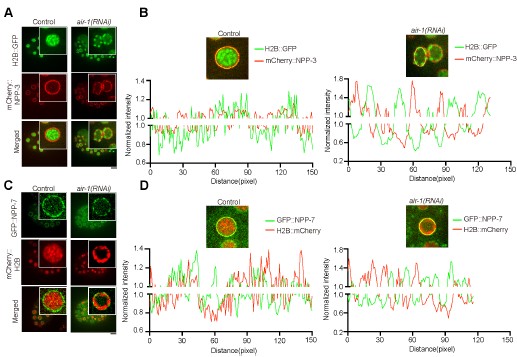
Our interest in AIR-1 was inspired by previous findings by Hachet et al., showing that AIR1/Aurora A localizes at sites without NPP-3 during mitotic entry—these sites are believed to correspond to centrosome locations [4]. This relationship prompted us to explore how AIR-1 depletion might influence NPP-3 localization at the nuclear envelope and its potential effects on chromosome positioning. Interestingly, we discovered that following air-1(RNAi) treatment, nuclei exhibit discontinuous NPP-3 localization on the nuclear envelope. Thereby, we could investigate the interplay between NPP-3 and chromosomal dynamics. Chromosomes localize to nuclear periphery specifically without NPP-3 (Author response image 3). This suggests a potential negative correlation of NPP-3 localization with the chromosomes. We did not imply any regulation by AIR-1.
Author response image 3.
Condensed chromosomes tend to localize at nuclear envelope sites without NPP-3 or NPP-7. (A) (C) Selected representative confocal images of all chromosomes localizing at the nuclear periphery in the control and air-1(RNAi) embryos expressing H2B::GFP and mCherry::NPP-3 (A) and GFP::NPP-7 and mCherry::H2B (C). The upper right image is the zoom-in view of the nucleus. Scale bar 10 μm. (B) (D) The intensity of GFP and mCherry is normalized to the average intensity along the nuclear envelope and plotted in the control and air- 1(RNAi) nucleus from (A) and (C).
(6) The authors show that condensed chromosomes tend to localize to the nuclear envelope upon NPP-3 depletion. Do they condense at the nuclear envelope (NE), or do they condense first and then move to the periphery? This is unclear from the data presented in Figure 1D. Also, why do chromosomes condense earlier? This point could be discussed.
Based on our time-lapse data in Figure 1D, most chromosomes appear to condense at or near the nuclear periphery, but there are still some chromosomes inside the nuclear space at approximately -600 seconds before NEBD, and then subsequently moving to the periphery during condensation (with full condensation at 100 s past NEBD). This suggests that initial condensation may occur both within the nucleus and at the periphery. Over time, chromosomes condense and cluster at the nuclear envelope. However, we did not separately measure the condensation level of individual chromosomes at the nuclear periphery versus in the middle of the nucleus, which could be challenging in live cells. So far, we cannot separate the chromosome nuclear periphery phenotype and the condensation.
To confirm whether chromosome condensation precedes or follows relocation to the nuclear periphery, we could perform depletion of condensin II component, e.g. hcp-6, and see if lack of condensation affects chromosome relocalization.
(7) The authors evaluated the consequences of NPP-3 depletion on transcription using RNA sequencing. The relevance of this experiment is questionable, however, as npp-3(RNAi) embryos have significant general defects and not only mislocalised chromosomes.
We recognize that npp-3 RNAi embryos exhibit broad developmental defects, and therefore global transcriptional changes. Our RNA-seq analysis revealed that differentially expressed genes did not display positional bias within the genome. NPP-3 depletion downregulates many pathways, including pathways related to RNA polymerase II activity and cell cycle regulation, consistent with a global transcriptional downregulation. Thus, while the transcriptomic data are broad, it is consistent with the chromosome condensation phenotype.
(8) In the co-depletion experiment npp-3(RNAi), X(RNAi) presented in Figure 3B, the levels of NPP-3 depletion seem highly variable. All the images shown are not similarly exposed, so it is difficult to evaluate these data.
In our co-depletion experiments, the images for npp-3(RNAi) and X(RNAi) (Fig. 4) are displayed side-by-side under the same exposure conditions and scaled the same way with the same intensity thresholds to facilitate comparison. Despite that, some differences in the background intensity can be observed across samples. Thus, the normalised mCherry::NPP-3 signal intensity (subtracting the background) the single and double RNAi samples will be quantified and added to supplementary figure S4.
To ensure consistency of double RNAi, we used ligation-based RNAi constructs designed to simultaneously target both NPP-3 and X, aiming to achieve comparable knockdown efficiencies. Nonetheless, variability in RNAi efficiency is a recognized limitation, and we interpret our data within this context. To further validate the knockdown, we will also assess the efficiency of the other gene X using the corresponding fluorescent reporter (see response to Reviewer 3, point 2).
(9) Inactivation of mdf-1/2 suppresses the mislocalization of the chromosomes observed upon npp-3 inactivation. Does it also suppress the premature chromosome condensation phenotype?
Inactivation of MDF-1 or MDF-2 suppresses both the chromosome nuclear periphery localization and extended duration of interphase to prophase and prometaphase in npp-3 inactivation. We have performed the analysis to assess the timing and extent of chromosome condensation in the single and double depletion conditions (Author response image 4). The chromosome condensation dynamics is similar in the mdf-1 RNAi and npp-3 mdf-1 double RNAi, as well as the control group, indicating that MDF-1 is also involved the premature chromosome condensation phenotype caused by NPP-3 depletion.
Author response image 4.
The dynamic changes of chromosome condensation parameter, in which 30% of pixels in the ROI analyzed is below the threshold scaled intensity (<77), in the different groups. The sample size is 5. Error bars show mean ± SEM.
(10) Figure 5B: The delay induced by npp-3 depletion is not severe, based on the micrographs presented. The authors should show more representative images. The graph shows the elapsed time between NEBD and NER, and not NER to NEBD, as indicated.
We have aligned the nuclear envelope reassembly (NER) time and highlighted the time point in the images (Fig. 5A). Our data show that in control embryos, this duration is approximately 780 seconds, while in npp-3(RNAi) embryos, it extends to about 930 seconds. This difference is statistically significant, as determined by one-way ANOVA (or appropriate nonparametric/mixed tests). The representative image is consistent with the quantification presented in Fig. 5B. We could add the corresponding videos to the supplementary information.
(11) The authors observed that depleting mdf-1 slightly enhanced the lethality associated with npp-3 inactivation. Based on this observation, they conclude that loss of chromosome anchoring exacerbates genomic instability and severely impairs embryonic survival. However, the genetic interaction is not strong, as npp-3(RNAi) embryos already present more than 95% embryonic lethality and have defects other than just mislocalized chromosomes (e.g., defects in kinetochore and spindle assembly).
It is correct that the average embryonic lethality observed in npp-3(RNAi) embryos reachs 95% (Fig. S6). Given the broad developmental defects and such high baseline lethality, the genetic interaction with mdf-1 is modest.
Nevertheless, our findings highlight that in the npp-3 mdf-1 double RNAi condition, we observe significantly increased rates of lagging chromosomes (Fig. 5D), micronuclei formation (Fig. 7A), and elevated DNA damage (Fig. 7B). These effects support the idea that MDF-1-, MDF-2dependent chromosome anchoring to the nuclear periphery (and condensation) plays a positive role in NPP-3 depleted cells.
Reviewer #2 (Public review):
Summary:
The authors aimed to determine the molecular mechanisms by which nuclear pore component NPP- 3/NUP205 regulates chromosome localization in C. elegans embryos. Previous studies had shown that depletion of NPP-3 caused premature chromosome condensation and movement of chromosomes to the nuclear periphery. Peripheral location of chromosomes is also observed under respiratory stress conditions, suggesting that peripheral chromosome positioning could act as a protective response to stress conditions. How NPP-3 affects chromosome positioning was unknown. Here, the authors conduct a screen to identify factors that promote chromosome relocation to the periphery in npp-3-depleted embryos, identifying an important role for spindle assembly checkpoint components in this process.
Strengths:
Using cytological tools to visualise chromosomes and nuclear envelope markers, the authors show that, in addition to the peripheral location of chromosomes, NPP-3 depletion causes partial rupture of the nuclear envelope and premature chromosome condensation. By systematically codepleting NPP-3 and factors required for heterochromatin association with nuclear lamina (CEC4), telomere binding to nuclear envelope (SUN-1 and POT-1), proteins required for the nuclear rupture repair machinery (BAF-1 and LEM-2), kinetochore proteins and components of the spindle assembly checkpoint (SAC) (MDF-1 and MDF-2), the authors convincingly show that SAC components are required for peripheral relocation of chromosomes in absence of NPP-3. The study also provides convincing evidence that peripheral relocation of chromosomes in the absence of NPP-3 has functional implications as it causes transcriptional deregulation and premature relocation of SAC components from the nuclear envelope to chromosomes. Codepletion of NPP-3 and SAC components accelerates progression through miotic prophase and increases the incidence of defects in chromosome segregation during mitosis. These findings demonstrate that SAC proteins play an important role in regulating chromosome positioning during prophase (at least in the absence of NPP-3) and that they can regulate cell cycle progression at earlier stages than previously thought.
Weaknesses:
The authors also propose that NPP-3 depletion causes DNA damage; however, the evidence presented to support this claim is not as strong as that presented for the effects mentioned above. Also, the premature condensation of chromosomes appears as a clear consequence of NPP-3 depletion, but this intriguing phenotype remains unexplored.
The DNA damage evidence is based on lagging chromosomes in 2-cell stages, the HUS-1 reporter, and micronuclei in embryos at the 20-30 cell stage. It is noted that npp-3 RNAi is pleiotropic and also causes DNA damage. There is additional DNA damage caused by loss of MDF-1 and MDF-2 in npp-3 RNAi, but we agree that it is difficult to say whether the effect is additive or not, complicating the interpretation. Thus, we will tone down in our title to describe the dependency of the chromosomal nuclear periphery phenotype (see response to Reviewer 1 point 1).
As for the premature chromosome condensation phenotype in NPP-3 depletion, we hypothesize it may result from accumulation of factors such as BAF-1 at the nuclear periphery, which could facilitate chromatin condensation. To confirm whether chromosome condensation precedes or follows relocation to the nuclear periphery, we could perform depletion of condensin II component, e.g. hcp-6, and see if lack of condensation affects chromosome relocalization (also see response to Reviewer 1 point 6).
Reviewer #3 (Public review):
Summary:
This manuscript reports that RNAi depletion of the inner-ring nucleoporin NPP-3/NUP205 in Caenorhabditis elegans embryos causes nuclear envelope rupture, premature chromatin condensation, and relocalization of condensed prophase chromosomes to the nuclear periphery. Through a candidate epistasis screen, the authors argue that this relocalization requires spindle assembly checkpoint (SAC) components (MDF-1, MDF-2, SAN-1), inner kinetochore proteins (HCP- 3, HCP-4, and partially KNL-1), and NE rupture-repair factors (BAF-1, LEM-2), but not the CEC-4 heterochromatin- or SUN-1/POT-1 telomere-anchoring pathways. They further show that NPP-3 loss extends prophase and the NEBD-to-anaphase interval in a SAC-dependent manner, redistributes MDF-1/MDF-2, and reduces import of KNL-1/BUB-1/HCP-1. Codepletion of NPP-3 with MDF-1 abolishes both the arrest and the peripheral localization while increasing lagging chromosomes, HUS-1 foci, micronuclei, and lethality, which the authors interpret as evidence that peripheral positioning is protective.
Weaknesses:
(1) The "protective" conclusion is largely correlative. The protective claim rests on the observation that co-depleting MDF-1 (or MDF-2) with NPP-3 removes the peripheral localization and simultaneously increases DNA damage, micronuclei, and lethality. However, depleting a SAC component removes at least three things at once: the peripheral localization, the prophase extension, and the NEBD-to-anaphase arrest. , they unrelated to chromosome positioning, the current design cannot separate damage caused by loss of a protective peripheral location from damage caused by checkpoint bypass. As presented, the increased damage is at least as consistent with simple SAC bypass. To support the protective model, the authors should provide a manipulation that disrupts peripheral positioning without abrogating the SACdependent arrest (for example, via the BAF-1/LEM-2 or kinetochore depletion) and show that damage still increases. The LEM-2 co-depletion, which partially suppresses positioning, is a natural place to test whether micronuclei and HUS-1 foci also rise.
We agree that the current data are largely correlative. In the early C. elegans embryos, single depletion of SAC components MDF-1 or MDF-2 does not affect the mitosis duration or chromosome segregation. When spindles are defective, the functional SAC delays progression through mitosis [5]. Depleting SAC components such as MDF-1 in npp-3 RNAi indeed impacts multiple processes, including prophase and prometaphase duration, chromosome repositioning and condensation, making it challenging to disentangle effects specifically to related chromosome positioning.
To address this, future experiments involving NPP-3 LEM-2 co-depletion, which has been shown to partially impair peripheral chromosome positioning, will be utilized to assess whether disruption of partial peripheral localization results in increased DNA damage, micronuclei formation, or HUS-1 foci accumulation, independent of SAC function (see response to Reviewer 1 point 1).
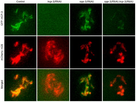
(2) Knockdown efficiency of the partner gene in double RNAi is not verified. The double depletions are performed by cloning both gene fragments into a single vector. This risks reducing the effective dose of each dsRNA, so an apparent suppression in an npp-3; gene X (RNAi) condition could reflect weaker NPP-3 knockdown rather than a true epistatic relationship. The authors partially address this by showing that NPP-3::mCherry is still reduced in npp-3;mdf-1 (Figure S4A/B), which is helpful, but they do not demonstrate efficient knockdown of the partner genes in any double condition. For the key epistasis conclusions (MDF-1, MDF-2, HCP-3, HCP4 suppressions), the knockdown of the second gene should be independently validated with a reporter strain for the second protein.
We acknowledge that in our double RNAi experiments, the knockdown efficiency of the npp-3 is checked by imaging (see response to Reviewer 1 point 8), whereas that of the second gene was not validated in each condition. To address this, we confirmed the effectiveness of certain partner gene depletions, e.g. HCP-3, by examining the levels of the respective proteins using available GFP-marked strains or immunofluorescence (Author response image 5). Additionally, for genes like HCP-3, KNL-1, and BUB-1, we also assessed the functional consequences on chromosome segregation, where severe defects observed (Fig. S4C) can support effective depletion. However, for some strains, we do not have GFP makers and will need to check the RNA levels.
Author response image 5.
Representative confocal image of GFP::HCP-3 and mCherry::H2B at the NEBD time point of P1 cell in the control, single and double RNAi. Scale bar, 5 μm.
(3) Alternative explanations for the transcriptomic and H3K9me3 data are not excluded. NPP-3 depletion blocks nuclear import of molecules smaller than ~70 kDa and arrests development at early gastrulation. Both the RNA-seq changes (30% of genes downregulated) and the increased H3K9me3 signal could therefore be secondary consequences of nucleocytoplasmic transport failure and developmental arrest rather than evidence of position-dependent transcriptional repression. Notably, the authors' own finding that up- and down-regulated genes show no chromosomal positional bias (Figure S2C/D) argues against a model in which peripheral repositioning drives silencing of specific chromatin domains. This section should be reframed more cautiously, with the transport/arrest confound explicitly discussed, and RNA-seq replicate number and differential-expression thresholds reported.
We agree that these chromatin modifications and transcriptional alterations could be related to the nuclear transport failure and developmental delay in npp-3 disruption. We will discuss this possibility in results and discussion.
(4) Evidence for SAC "activation in prophase" is indirect, and the effect is small. The claim of a novel prophase role for the SAC rests on MDF-1/MDF-2 intensity changes that are repeatedly described as "modest," "slight," or "mild," measured with small n and Student's t-tests, together with phenotypic suppression of prophase extension. There is no direct readout of SAC catalytic activity (for example, MCC assembly). The prophase-extension suppression by MDF-1 is the strongest evidence; the intensity data are weak support. I recommend tempering "the SAC is activated in prophase" to a hypothesis, and strengthening it with a more direct assay if feasible.
We agree that the evidence for SAC activation during prophase is indirect. The prophase extension (100 s) in npp-3 RNAi is a functional assay to support SAC activation, and the suppression in npp-3 mdf-1 double RNAi suggests dependency. The changes in MDF1/MDF-2 intensities are modest. Biochemical analyses of MCC assembly in C. elegans mixed cell cycle stage embryos is challenging to demonstrate SAC activity in prophase.
(5) The BAF-1 arm of the model is inferred rather than demonstrated. The authors state that baf- 1(RNAi) and npp-3;baf-1 produced clustering too severe for epistasis, so BAF-1's requirement for peripheral localization is not actually established genetically; it rests on increased BAF-1 accumulation (correlative) plus the LEM-2 partial suppression. The proposed BAF- 1/CENP-C bridge is extrapolated from Drosophila (ref. 71). This is reasonable as a discussion hypothesis but should not be presented in the abstract or summary model as an established dependency.
We did not include the BAF-1/CENP-C bridge hypothesis in the abstract or the model, and we will discuss this as a speculative mechanism rather than an established dependency.
References:
(1) Rodenas, E., Gonzalez-Aguilera, C., Ayuso, C. & Askjaer, P. Dissection of the NUP107 nuclear pore subcomplex reveals a novel interaction with spindle assembly checkpoint protein MAD1 in Caenorhabditis elegans. Mol Biol Cell 23, 930-944 (2012).
(2) Schumacher, J.M., Ashcroft, N., Donovan, P.J. & Golden, A. A highly conserved centrosomal kinase, AIR-1, is required for accurate cell cycle progression and segregation of developmental factors in Caenorhabditis elegans embryos. Development 125, 4391-4402 (1998).
(3) Kotak, S., Afshar, K., Busso, C. & Gonczy, P. Aurora A kinase regulates proper spindle positioning in C. elegans and in human cells. J Cell Sci 129, 3015-3025 (2016).
(4) Hachet, V. et al. The nucleoporin Nup205/NPP-3 is lost near centrosomes at mitotic onset and can modulate the timing of this process in Caenorhabditis elegans embryos. Molecular Biology of the Cell 23, 3111-3121 (2012).
(5) Encalada, S.E., Willis, J., Lyczak, R. & Bowerman, B. A spindle checkpoint functions during mitosis in the early Caenorhabditis elegans embryo. Molecular Biology of the Cell 16, 1056-1070 (2005).
-
