Sperm motility in mice with oligo-astheno-teratozoospermia restored by in vivo injection and electroporation of naked mRNA
Curation statements for this article:-
Curated by eLife

eLife Assessment
This study reports an mRNA-based strategy for restoring sperm motility in a mouse model of monogenic male infertility. The work is technically innovative and potentially valuable, as it demonstrates feasibility of in vivo testicular mRNA delivery without genomic integration of foreign DNA. However, although partial recovery of sperm motility is supported, the evidence for meaningful restoration of fertility remains incomplete, with weak IVF outcomes and difficult-to-interpret ICSI results. In addition, mechanistic questions regarding the persistence of mRNA and the specificity of germ-cell targeting remain insufficiently resolved, limiting the strength of the authors' conclusions.
This article has been Reviewed by the following groups
Discuss this preprint
Start a discussion What are Sciety discussions?Listed in
- Evaluated articles (eLife)
Abstract
Oligo-astheno-teratozoospermia (OAT), a recurrent cause of male infertility, is the most frequent disorder of spermatogenesis with a predominantly genetic origin. Patients and mice bearing mutations in the ARMC2 gene exhibit reduced sperm concentration, multiple morphological defects, and impaired motility, defining a canonical OAT phenotype. Intracytoplasmic sperm injection (ICSI) is required to treat this condition; however, it is associated with a slightly increased risk of birth defects compared with natural conception, highlighting the need for novel targeted therapies. Here, in vivo testicular injection followed by electroporation of capped, polyadenylated naked messenger RNA (mRNA) was evaluated as a strategy to treat ARMC2 -related infertility in mice. mRNAs encoding reporter proteins were used to assess expression efficiency and kinetics using in vivo and in vitro 2D and 3D imaging. Reporter proteins were detected in germ cells for up to three weeks, demonstrating the feasibility of mRNA-based approaches. These results were compared with a non-integrative plasmid Enhanced Episomal Vector, which induced weak and transient expression in spermatogenic cells. Delivery of Armc2 mRNA restored morphologically normal and motile sperm in deficient males, capable of producing embryos via in vitro fertilization and ICSI. These findings provide proof-of-concept that mRNA electroporation can restore sperm motility and fertilizing potential, offering a novel strategy to correct monogenic male infertility.
Article activity feed
-

-
-
-

eLife Assessment
This study reports an mRNA-based strategy for restoring sperm motility in a mouse model of monogenic male infertility. The work is technically innovative and potentially valuable, as it demonstrates feasibility of in vivo testicular mRNA delivery without genomic integration of foreign DNA. However, although partial recovery of sperm motility is supported, the evidence for meaningful restoration of fertility remains incomplete, with weak IVF outcomes and difficult-to-interpret ICSI results. In addition, mechanistic questions regarding the persistence of mRNA and the specificity of germ-cell targeting remain insufficiently resolved, limiting the strength of the authors' conclusions.
-
Reviewer #4 (Public review):
I maintain that the images in Figure 12 (new Figure 14) do not support the authors' interpretation that 2-cell embryos resulted from in vitro fertilization (IVF) of Amrc-/- rescued sperm. They are clearly not normal 2-cell embryos and instead look very much like fragmented eggs that can be seen occasionally following in vitro fertilization procedures even when that is done with wild type eggs and sperm. The only portion of current Figure 14 that has normal looking 2-cell embryos is in panel 14A4, where wild type B6D2 sperm were used. Even in that panel, there are some fragmented eggs that the authors identify as 2-cell embryos.
The authors offer the explanation that CD1 eggs fertilized by B6D2F1 hybrid male sperm do not develop beyond the 2-cell stage, citing a 2008 paper published in Biology of Reproduction …
Reviewer #4 (Public review):
I maintain that the images in Figure 12 (new Figure 14) do not support the authors' interpretation that 2-cell embryos resulted from in vitro fertilization (IVF) of Amrc-/- rescued sperm. They are clearly not normal 2-cell embryos and instead look very much like fragmented eggs that can be seen occasionally following in vitro fertilization procedures even when that is done with wild type eggs and sperm. The only portion of current Figure 14 that has normal looking 2-cell embryos is in panel 14A4, where wild type B6D2 sperm were used. Even in that panel, there are some fragmented eggs that the authors identify as 2-cell embryos.
The authors offer the explanation that CD1 eggs fertilized by B6D2F1 hybrid male sperm do not develop beyond the 2-cell stage, citing a 2008 paper published in Biology of Reproduction by Fernandez-Goonzalez et al. I read through that paper very carefully and even had a colleague read through it in case I missed something, but that paper says nothing at all about strain incompatibilities, much less 2-cell arrest due to them. The only crosses done in that paper are CD1 eggs x CD1 sperm and B6D2 eggs x B6D2 sperm, all by intracytoplasmic sperm injection and not standard in vitro fertilization. [Note that the paper does mention performing in vitro fertilization but says nothing about how it was done or what mouse strains were used.] I even searched the literature for information regarding incompatibility between these strains and could find nothing relevant. But even if the authors are correct and there happens to be a strain incompatibility and 2-cell arrest is expected, what the authors are calling 2-cell embryos are clearly not.
A second explanation offered by the authors is that they used collagenase to remove the cumulus cells and that this may have affected the appearance of the embryos. This technique is actually used to remove both the cumulus cells and the zona pellucida and has been described as a gentler way to do so than other standard methods (hyaluronidase treatment followed by acid Tyrodes to remove the zona pellucida) (Yamatoya et al., Reprod Med Biol 2011, DOI 10.1007/s12522-011-0075-8). I think it is highly relevant to the current study that the method they used to remove cumulus cells also dissolves the zona, either partially or completely. Given that many of the eggs, fragmented eggs, and 2-cell embryos (from the WT sperm) in Figure 14A are lacking a zona pellucida, it seems very likely that many of the eggs were either zona-free or had partial zona dissolution from the start. In fact, the authors state in the Methods section that "Cumulus-free and zona-free eggs were collected..." for how IVF was done. Partial zona dissolution is standard in some protocols for performing IVF using frozen mouse sperm, which usually have much lower motility and overall efficacy than fresh sperm. In any case, it would improve transparency if the manuscript made clear somewhere other than buried in the Methods that the IVF procedure was done on eggs with partially or fully removed zonas, to allow proper interpretation.
In the rebuttal, the authors go on to state: "To provide additional functional evidence, we complemented the IVF experiments with ICSI using rescued Armc2-/- sperm and B6D2 oocytes, which allowed embryos to develop to the blastocyst stage. In these experiments, 25% of injected oocytes reached the blastocyst stage with rescued sperm compared to 13% for untreated Armc2-/- sperm (Supplementary Fig. 9) These results support the functional competence of rescued sperm and demonstrate partial recovery of fertilization ability following Armc2 mRNA electroporation."
Their conclusion that the data support partial recovery of fertilization ability following Armc2 mRNA electroporation in my opinion has no basis. This experiment was done only once, and no information is provided regarding how many eggs underwent ICSI or how many reached the blastocyst stage. The authors claim that the rescued sperm were better than the Armc2-/- sperm in producing blastocysts, but this is based on a simple percentage report of 25% vs 13% without any statistical analysis, even on the results from the single experiment presented.
Overall, the paper shows rescue of some sperm motility by the new method they use, and the new title is therefore appropriate. The authors have also dealt reasonably with many of the original concerns regarding documenting that their methodology was effective in producing protein (at least the GFP marker) in spermatogenic cells. In my view the authors have, however, not shown any indication of functional recovery over what is already known for the knockout sperm, that ICSI can support blastocyst stage embryo development. They also have not, in my view, justified the claims at the end of the abstract "These motile sperm were able to produce embryos by IVF..." and that "...mRNA electroporation can restore...partially fertilizing ability..."
-
Reviewer #5 (Public review):
While the study presents an innovative and potentially impactful mRNA-based approach for addressing monogenic causes of male infertility, several significant weaknesses limit the strength of the authors' central conclusions.
First, the functional evidence for true fertility restoration remains incomplete. Although the authors convincingly demonstrate partial recovery of sperm motility, the downstream reproductive outcomes, particularly for IVF, are weak. Importantly, these concerns are shared by all three reviewers and the former Reviewing Editor, and to my eye they are both thoughtfully articulated and well warranted. The ICSI data show modest improvement, but this rescue is difficult to interpret.
In parallel, significant mechanistic questions persist regarding the unusually prolonged persistence of naked …
Reviewer #5 (Public review):
While the study presents an innovative and potentially impactful mRNA-based approach for addressing monogenic causes of male infertility, several significant weaknesses limit the strength of the authors' central conclusions.
First, the functional evidence for true fertility restoration remains incomplete. Although the authors convincingly demonstrate partial recovery of sperm motility, the downstream reproductive outcomes, particularly for IVF, are weak. Importantly, these concerns are shared by all three reviewers and the former Reviewing Editor, and to my eye they are both thoughtfully articulated and well warranted. The ICSI data show modest improvement, but this rescue is difficult to interpret.
In parallel, significant mechanistic questions persist regarding the unusually prolonged persistence of naked mRNA and reporter protein expression in germ cells, which is not fully reconciled with established mRNA and protein half-life biology and is supported largely by inference rather than by direct decay measurements.
Finally, although the authors have conducted additional cellular analyses, concerns about the extent and specificity of germ-cell targeting versus Sertoli-cell expression remain unresolved. Together, these issues do not negate the technical novelty of the work, but they do constrain the confidence with which the current dataset can support the authors' strongest therapeutic claims.
-
Author response:
The following is the authors’ response to the previous reviews
Public Reviews:
Reviewer #1 (Public review):
The authors assess the effectiveness of electroporating mRNA into male germ cells to rescue the expression of proteins required for spermatogenesis progression in individuals where these proteins are mutated or depleted. To set up the methodology, they first evaluated the expression of reporter proteins in wild-type mice, which showed expression in germ cells for over two weeks. Then, they attempted to recover fertility in a model of late spermatogenesis arrest that produces immotile sperm. By electroporating the mutated protein, the authors recovered the motility of ~5% of the sperm; although the sperm regenerated was not able to produce offspring using IVF, the embryos reached the 2-cell state (in contrast to …
Author response:
The following is the authors’ response to the previous reviews
Public Reviews:
Reviewer #1 (Public review):
The authors assess the effectiveness of electroporating mRNA into male germ cells to rescue the expression of proteins required for spermatogenesis progression in individuals where these proteins are mutated or depleted. To set up the methodology, they first evaluated the expression of reporter proteins in wild-type mice, which showed expression in germ cells for over two weeks. Then, they attempted to recover fertility in a model of late spermatogenesis arrest that produces immotile sperm. By electroporating the mutated protein, the authors recovered the motility of ~5% of the sperm; although the sperm regenerated was not able to produce offspring using IVF, the embryos reached the 2-cell state (in contrast to controls that did not progress past the zygote state).
This is a comprehensive evaluation of the mRNA methodology with multiple strengths. First, the authors show that naked synthetic RNA, purchased from a commercial source or generated in the laboratory with simple methods, is enough to express exogenous proteins in testicular germ cells. The authors compared RNA to DNA electroporation and found that germ cells are efficiently electroporated with RNA, but not DNA. The differences between these constructs were evaluated using in vivo imaging to track the reporter signal in individual animals through time. To understand how the reporter proteins affect the results of the experiments, the authors used different reporters: two fluorescent (eGFP and mCherry) and one bioluminescent (Luciferase). Although they observed differences among reporters, in every case expression lasted for at least two weeks. The authors used a relevant system to study the therapeutic potential of RNA electroporation. The ARMC2-deficient animals have impaired sperm motility phenotype that affects only the later stages of spermatogenesis. The authors showed that sperm motility was recovered to ~5%, which is remarkable due to the small fraction of germ cells electroporated with RNA with the current protocol. The sperm motility parameters were thoroughly assessed by CASA. The 3D reconstruction of an electroporated testis using state-of-the-art methods to show the electroporated regions is compelling.
The main weakness of the manuscript is that although the authors manage to recover motility in a small fraction of the sperm population, it is unclear whether the increased sperm quality is substantial to improve assisted reproduction outcomes. The authors found that the rescued sperm could be used to obtain 2-cell embryos via IVF, but no evidence for more advanced stages of embryo differentiation was provided. The motile rescued sperm was also successfully used to generate blastocyst by ICSI, but the statistical significance of the rate of blastocyst production compared to non-rescued sperm remains unclear. The title is thus an overstatement since fertility was never restored for IVF, and the mutant sperm was already able to produce blastocysts without the electroporation intervention.
Overall, the authors clearly show that electroporating mRNA can improve spermatogenesis as demonstrated by the generation of motile sperm in the ARMC2 KO mouse model.
We thank the reviewer for this thoughtful and constructive comment. We agree that our study demonstrates a partial functional recovery of spermatogenesis rather than a complete restoration of fertility. Our main objective was to establish and validate a proof-of-concept approach showing that mRNA electroporation can rescue the expression of a missing or mutated protein in post-meiotic germ cells and result in the production of motile sperm.
To address the reviewer’s concern, we have the title and discussion to more accurately reflect the scope of our findings. The new title reads:
“Sperm motility in mice with oligo-astheno-teratozoospermia restored by in vivo injection and electroporation of naked mRNA”
In the manuscript, we now emphasize that while motility recovery was significant, complete fertility restoration was not achieved. We have also clarified that:
The 5% recovery in motile sperm represents a substantial improvement considering the small population of germ cells reached by the current electroporation method.
The 2-cell embryo formation observed after IVF serves as a strong indication of partial functional recovery
Finally, we now explicitly state in the Discussion that this approach should be considered a therapeutic proof-of-concept, demonstrating feasibility and potential, rather than a fully curative intervention.
Reviewer #2 (Public review):
The authors inject, into the rete testes, mRNA and plasmids encoding mRNAs for GFP and then ARMC2 (into infertile Armc2 KO mice) in a gene therapy approach to express exogenous proteins in male germ cells. They do show GFP epifluorescence and ARMC2 protein in KO tissues, although the evidence presented is weak. Overall, the data do not necessarily make sense given the biology of spermatogenesis and more rigorous testing of this model is required to fully support the conclusions, that gene therapy can be used to rescue male infertility.
In this revision, the authors attempt to respond to the critiques from the first round of reviews. While they did address many of the minor concerns, there are still a number to be addressed. With that said, the data still do not support the conclusions of the manuscript.
We thank the reviewer for their careful and detailed assessment of our manuscript. We appreciate the concerns raised regarding mRNA stability, GFP localization, and the interpretation of spermatogenesis stages, and we have addressed these points in the manuscript and in the responses below.
(1) The authors have not satisfactorily provided an explanation for how a naked mRNA can persist and direct expression of GFP or luciferase for ~3 weeks. The most stable mRNAs in mammalian cells have half-lives of ~24-60 hours. The stability of the injected mRNAs should be evaluated and reported using cell lines. GFP protein's half-life is ~26 hours, and luciferase protein's half-life is ~2 hours.
We thank the reviewer for this important comment. The stability of mRNA-GFP was assessed by RT-QPCR in HEK cells and seminiferous tubule cells (Fig. 5). mRNA-GFP was detected for up to 60 hours in HEK cells and for up to two weeks in seminiferous tubule cells (Fig. 5A). Together, these results suggest that the long-lasting fluorescence observed in our experiments reflects a combination of transcript stability, efficient translation within germ cells and the slow protein turnover that is typical of the spermatogenic lineage.
(2) There is no convincing data shown in Figs. 1-8 that the GFP is even expressed in germ cells, which is obviously a prerequisite for the Armc2 KO rescue experiment shown in the later figures! In fact, to this reviewer the GFP appears to be in Sertoli cell cytoplasm, which spans the epithelium and surrounds germ cells - thus, it can be oft-confused with germ cells. In addition, if it is in germ cells, then the authors should be able to show, on subsequent days, that it is present in clones of germ cells that are maturing. Due to intracellular bridges, a molecule like GFP has been shown to diffuse readily and rapidly (in a matter of minutes) between adjacent germ cells. To clarify, the authors must generate single cell suspensions and immunostain for GFP using any of a number of excellent commercially-available antibodies to verify it is present in germ cells. It should also be present in sperm, if it is indeed in the germline.
We thank the reviewer for this insightful comment. To directly address the concern, we performed additional experiments to assess GFP expression in germ cells following in vivo mRNA delivery. GFP-encoding mRNA was injected and electroporated into the testes on day 0. On day 1, testes were collected, enzymatically dissociated, and the resulting seminiferous tubule cell suspensions were cultured for 12 hours. Live cells were then analyzed by fluorescence microscopy (Fig. 10).
We observed GFP expression in various germ cell types, including pachytene spermatocytes (53,4 %) (Fig 10 A-), round spermatids (25 %) (Fig 10B-E) and in elongated spermatids (11,4%) (Fig 10 C-E). The identification of these cell types was based on DAPI nuclear staining patterns, cell size fig 10 F, non-adherent characteristics, and the use of an enzymatic dissociation protocol.
Fluorescence imaging revealed strong cytoplasmic GFP signals in each of these populations, confirming efficient transfection and translation of the delivered mRNA. These results demonstrate that the in vivo injection and electroporation protocol enables effective mRNA transfection across multiple stages of spermatogenesis. These results confirm that the injected mRNA is efficiently translated in germ cells at various stages of spermatogenesis. Together, these data validate the germ cell-specific nature of the GFP signal, supporting the Armc2 KO rescue experiments.
As mentioned previously, we assessed the stability of mRNA-GFP using RT-QPCR in HEK cells and seminiferous tubule cells (see Fig. 5). mRNA-GFP was detected for up to 60 hours in HEK cells and for up to two weeks in seminiferous tubule cells. Together, these results suggest that the long-lasting fluorescence observed in our experiments reflects a combination of transcript stability and local translation within germ cells, as well as the slow protein turnover typical of the spermatogenic lineage.
Other comments:
70-1 This is an incorrect interpretation of the findings from Ref 5 - that review stated there were ~2,000 testis-enriched genes, but that does not mean "the whole process involves around two thousand of genes"
We thank the reviewer for this helpful comment. We agree that our previous phrasing was imprecise. We have revised the sentence to clarify that approximately 2,000 genes show testis-enriched expression, rather than implying that the entire spermatogenic process is limited to these genes. The corrected sentence now reads:
“Spermatogenesis involves the coordinated expression of a large number of genes, with approximately 2,000 showing testis-enriched expression, about 60% of which are expressed exclusively in the testes”
74 would specify 'male':
we have now specified it as you suggested.
79-84 Are the concerns with ICSI due to the procedure itself, or the fact that it's often used when there is likely to be a genetic issue with the male whose sperm was used? This should be clarified if possible, using references from the literature, as this reviewer imagines this could be a rather contentious issue with clinicians who routinely use this procedure, even in cases where IVF would very likely have worked:
We thank the reviewer for this important comment. Concerns about ICSI outcomes indeed reflect two partly overlapping causes: the procedure itself (direct sperm injection and associated laboratory manipulations) and the clinical/genetic background of couples undergoing ICSI (especially men with severe male-factor infertility). Large reviews and meta-analyses report a small increase in some perinatal and congenital risks after ART/ICSI, but these studies conclude that it is difficult to fully disentangle procedural effects from parental factors. Importantly, genetic or epigenetic abnormalities in the male (which motivate use of ICSI) likely contribute to adverse outcomes in offspring, while some studies also suggest that ICSI-specific manipulations may alter epigenetic marks in embryos. For these reasons professional bodies recommend reserving ICSI for appropriate male-factor indications rather than as routine insemination for non-male-factor cases
We have revised the text accordingly to clarify this distinction:
“ICSI can efficiently overcome the problems faced. Nevertheless, concerns persist regarding the potential risks associated with this technique, including blastogenesis defect, cardiovascular defect, gastrointestinal defect, musculoskeletal defect, orofacial defect, leukemia, central nervous system tumors, and solid tumors [1-4]. Statistical analyses of birth records have demonstrated an elevated risk of birth defects, with a 30-40 % increased likelihood in cases involving ICSI [1-4], and a prevalence of birth defects between 1 % and 4 % [3]. It is important to note, however, that the origin of these risks remains debated. Several large epidemiological and mechanistic studies indicate that both the procedure itself (direct microinjection and in vitro manipulation) and the underlying genetic or epigenetic abnormalities often present in men requiring ICSI contribute to the observed outcomes [1, 3] [5, 6] . To overcome these drawbacks, a number of experimental strategies have been proposed to bypass ARTs and restore spermatogenesis and fertility, including gene therapy [7-10].”
199 Codon optimization improvement of mRNA stability needs a reference;
We have added the references accordingly: [11-15]
In one study using yeast transcripts, optimization improved RNA stability on the order of minutes (e.g., from ~5 minutes to ~17 minutes); is there some evidence that it could be increased dramatically to days or weeks?
We agree with the reviewer that codon optimization can enhance mRNA stability, but available evidence indicates that this effect is moderate. In Saccharomyces cerevisiae, Presnyak et al. (2015) [16] showed that codon optimization increased mRNA half-life from approximately 5 minutes to ~17 minutes, representing a several-fold improvement rather than a shift to days or weeks. Similar codon-dependent stabilization has been observed in mammalian systems, where transcripts enriched in optimal codons exhibit longer half-lives and enhanced translation efficiency [11]; [17]). However, these studies consistently report effects on the scale of minutes to hours. In mammalian cells, the prolonged stability of therapeutic or vaccine mRNAs—lasting for days—is primarily achieved through additional features such as optimized untranslated regions, chemical nucleotide modifications (e.g., N¹-methylpseudouridine), and protective delivery systems, rather than codon usage alone ([18]; [19]).
Other molecular optimizations that improve in vivo mRNA stability and translation include a poly(A) tail, which binds poly(A)-binding proteins to protect the transcript from 3′ exonuclease degradation and promotes ribosome recycling, and a CleanCap structure at the 5′ end, which mimics the natural Cap 1 configuration, protects against 5′ exonuclease attack, and enhances translational initiation [11-15]. Together, these modifications act synergistically to stabilize the transcript and support efficient translation.
472-3 The reported half-life of EGFP is ~36 hours - so, if the mRNA is unstable (and not measured, but certainly could be estimated by qRT-PCR detection of the transcript on subsequent days after injection) and EGFP is comparatively more stable (but still hours), how does EGFP persist for 21 days after injection of naked mRNA??
We thank the reviewer for this important comment. The stability of mRNA-GFP was assessed by RT-QPCR in HEK cells and seminiferous tubule cells (Fig. 5). mRNA-GFP was detected for up to 60 hours in HEK cells and for up to two weeks in seminiferous tubule cells (Fig. 5). Together, these results suggest that the long-lasting fluorescence observed in our experiments reflects a combination of transcript stability, efficient translation within germ cells and the slow protein turnover that is typical of the spermatogenic lineage.
Curious why the authors were unable to get anti-GFP to work in immunostaining?
We appreciate the reviewer’s question. We attempted to detect GFP using several commercially available anti-GFP antibodies under various standard immunostaining conditions. However, in our hands, these antibodies consistently produced either no signal or high background staining, making the results unreliable. We therefore relied on direct detection of GFP fluorescence, which provides a more accurate and specific readout of protein expression in our system.
In Fig. 3-4, the GFP signals are unremarkable, in that they cannot be fairly attributed to any structure or cell type - they just look like blobs; and why, in Fig. 4D-E, why does the GFP signal appear stronger at 21 days than 15 days? And why is it completely gone by 28 days? This data is unconvincing.
We would like to thank the reviewer for their comments. Figure 3–4 provides a global overview of GFP expression on the surface of the testis. The entire testis was imaged using an inverted epifluorescence microscope, and the GFP signal represents a composite of multiple seminiferous tubules across the tissue surface. Due to this whole-organ imaging approach, it is not possible to resolve individual structures such as the basement membrane or lumen, which is why the signals may appear as diffuse “blobs.”
Regarding the time-course in Figure 4D–E, the apparent increase in GFP signal at 21 days compared with 15 days likely reflects accumulation and translation of the delivered mRNA in germ cells over time, whereas the absence of signal at 28 days corresponds to the natural turnover and degradation of GFP protein and mRNA in the tissue. We hope this explanation clarifies the observed patterns of fluorescence.
If the authors did a single cell suspension, what types or percentage of cells would be GFP+? Since germ cells are not adherent in culture, a simple experiment could be done whereby a single cell suspension could be made, cultured for 4-6 hours, and non-adherent cells "shaken off" and imaged vs adherent cells. Cells could also be fixed and immunostained for GFP, which has worked in many other labs using anti-GFP.
We thank the reviewer for this insightful comment. To directly address the concern, we performed additional experiments to assess GFP expression in germ cells following in vivo mRNA delivery. GFP-encoding mRNA was injected and electroporated into the testes on day 0. On day 1, testes were collected, enzymatically dissociated, and the resulting seminiferous tubule cell suspensions were cultured for 12 hours. Live cells were then analyzed by fluorescence microscopy (Fig. 10).
We observed GFP expression in various germ cell types, including pachytene spermatocytes (53,4 %) (Fig 10 A-), round spermatids (25 %) (Fig 10B-E) and in elongated spermatids (11,4%) (Fig 10 C-E). The identification of these cell types was based on DAPI nuclear staining patterns, cell size fig 10 F, non-adherent characteristics, and the use of an enzymatic dissociation protocol.
Fluorescence imaging revealed strong cytoplasmic GFP signals in each of these populations, confirming efficient transfection and translation of the delivered mRNA. These results demonstrate that the in vivo injection and electroporation protocol enables effective mRNA transfection across multiple stages of spermatogenesis.
These results confirm that the injected mRNA is efficiently translated in germ cells at various stages of spermatogenesis. Together, these data validate the germ cell-specific nature of the GFP signal, supporting the Armc2 KO rescue experiments.
As mentioned previously, we assessed the stability of mRNA-GFP using RT-QPCR in HEK cells and seminiferous tubule cells (see Fig. 5). mRNA-GFP was detected for up to 60 hours in HEK cells and for up to two weeks in seminiferous tubule cells. Together, these results suggest that the long-lasting fluorescence observed in our experiments reflects a combination of transcript stability and local translation within germ cells, as well as the slow protein turnover typical of the spermatogenic lineage.
In Fig. 5, what is the half-life of luciferase? From this reviewer's search of the literature, it appears to be ~2-3 h in mammalian cells. With this said, how do the authors envision detectable protein for up to 20 days from a naked mRNA? The stability of the injected mRNAs should be shown in a mammalian cell line - perhaps this mRNA has an incredibly long half-life, which might help explain these results. However, even the most stable endogenous mRNAs (e.g., globin) are ~24-60 hrs.
We did not directly assess the stability of luciferase mRNA, but we evaluated the persistence of GFP mRNA, which was synthesized and optimized using the same sequence optimization and chemical modification strategy as the luciferase mRNA. In these experiments, mRNA-GFP was detectable in seminiferous tubule cells for up to two weeks after injection. We therefore expect a similar stability profile for the luciferase mRNA. These findings suggest that the prolonged fluorescence or bioluminescence observed in our study likely reflects a combination of factors, including enhanced transcript stability, local translation within germ cells, and the inherently slow protein turnover characteristic of the spermatogenic lineage.
527-8 The Sertoli cell cytoplasm is not just present along the basement membrane as stated, but also projects all the way to the lumina:
we clarified the sentence " Sertoli cells have an oval to elongated nucleus and the cytoplasm presents a complex shape (“tombstone” pattern) along the basement membrane, with long projections that extend toward the lumen."
529-30 This is incorrect, as round spermatids are never "localized between the spermatocytes and elongated spermatids" - if elongated spermatids are present, rounds are not - they are never coincident in the same testis section:
We thank the reviewer for this important comment and for drawing attention to the detailed staging of the seminiferous epithelium. We agree that the spatial organization of germ cells varies depending on the stage of spermatogenesis. While round spermatids (steps 1–8) and elongated spermatids (steps 9–16) are typically associated with distinct stages, transitional stages of the seminiferous epithelium can contain both cell types in close proximity, reflecting the continuous and overlapping nature of spermatid differentiation (Meistrich, 2013, Methods Mol. Biol. 927:299–307). We have revised the text to clarify this point, indicating that the relative positioning of germ cell types depends on the stage of the seminiferous cycle rather than implying their constant coexistence within the same tubule section.
Fig. 7. To this reviewer, all of the GFP appears to be in Sertoli cell cytoplasm In Figs 1-8 there is no convincing evidence presented that GFP is expressed in germ cells! In fact, it appears to be in Sertoli cells.
We thank the reviewer for their observation. As previously mentioned, we have included an additional experiment specifically demonstrating GFP expression in germ cells (fig 10). This new data provides clear evidence that the GFP signal is not restricted to Sertoli cells and confirms successful uptake and translation of GFP mRNA in germ cells.
Fig. 9 - alpha-tubuline?
We corrected the figure.
Fig. 11 - how was sperm morphology/motility not rescued on "days 3, 6, 10, 15, or 28 after surgery", but it was in some at 21 and 35? How does this make sense, given the known kinetics of male germ cell development??
We note the reviewer’s concern regarding the timing of motile sperm appearance. Variability among treated mice is expected because transfection efficiency differed between spermatogonia and spermatids. Full spermiogenesis requires ~15 days, and epididymal transit adds ~8 days, consistent with motile sperm appearing around 21 days post-injection in some mice.
And at least one of the sperm in the KO in Fig. B5 looks relatively normal, and the flagellum may be out-of-focus in the image? With only a few sperm for reviewers to see, how can we know these represent the population?
We thank the reviewer for their comment. Upon closer examination of the image, the flagellum of the spermatozoon in question is clearly abnormally short and this is not due to being out of focus. Furthermore, the supplementary figure shows that the KO consistently lacks normal spermatozoa. These defects are consistent with previous findings from our laboratory [22], confirming that the observed phenotype is representative of the KO population rather than an isolated occurrence.
Reviewer #3 (Public review):
Summary:
The authors used a novel technique to treat male infertility. In a proof-of-concept study, the authors were able to rescue the phenotype of a knockout mouse model with immotile sperm using this technique. This could also be a promising treatment option for infertile men.
Strengths:
In their proof-of-concept study, the authors were able to show that the novel technique rescues the infertility phenotype of Armc2 knockout spermatozoa. In the current version of the manuscript, the authors have added data on in vitro fertilisation experiments with Armc2 mRNA-rescued sperm. The authors show that Armc2 mRNA-rescued sperm can successfully fertilise oocytes that develop to the blastocyst stage. This adds another level of reliability to the data.
Weaknesses:
Some minor weaknesses identified in my previous report have already been fixed. The technique is new and may not yet be fully established for all issues. Nevertheless, the data presented in this manuscript opens the way for several approaches to immotile spermatozoa to ensure successful fertilisation of oocytes and subsequent appropriate embryo development.
[Editors' note: The images in Figure 12 do not support the authors' interpretation that 2-cell embryos resulted from in vitro fertilization. Instead, the cells shown appear to be fragmented, unfertilized eggs. Combined with the lack of further development, it seems highly unlikely that fertilization was successful.]
We thank the reviewer for their careful evaluation and constructive feedback. We appreciate the acknowledgment of the strengths of our study, particularly the proof-of-concept demonstration that Armc2-mRNA electroporation can rescue sperm motility in Armc2 knockout mice.
Regarding the concern raised by the editor about Figure 12, we would like to clarify two technical points. First, the IVF experiments were performed using CD1 oocytes and B6D2 sperm. Due to strain-specific incompatibilities, fertilization of CD1 oocytes by B6D2 sperm typically does not progress beyond the two-cell stage (Fernández-González [23] et al., 2008, Biology of Reproduction). Therefore, the observation of two-cell embryos represents the expected limit of development in this cross and serves as a strong indication of successful fertilization, even though further development is not possible. Second, the oocytes used in these experiments were treated with collagenase to remove cumulus cells. This enzymatic treatment can sometimes affect the morphology of early embryos, which may explain why the two-cell embryos in Figure 12 appear less regular or somewhat fragmented. We also included a control showing embryos from B6D2 sperm with the same collagenase treatment on CD1 oocytes, which yielded similar appearances (Fig14 A4).
To provide additional functional evidence, we complemented the IVF experiments with ICSI using rescued Armc2–/– sperm and B6D2 oocytes, which allowed embryos to develop to the blastocyst stage. In these experiments, 25% of injected oocytes reached the blastocyst stage with rescued sperm compared to 13% for untreated Armc2–/– sperm (Supplementary Fig. 9) These results support the functional competence of rescued sperm and demonstrate partial recovery of fertilization ability following Armc2 mRNA electroporation.
We have clarified these points in the revised Results and Discussion sections to emphasize that the IVF data indicate partial functional recovery of rescued sperm rather than full fertility restoration. These clarifications address the editor’s concern while accurately representing the technical limitations of the strain combination used in our experiments.
Recommendations for the authors:
Reviewer #1 (Recommendations for the authors):
Fig 12 and Supplementary Fig 9 are mislabeled in the text and rebuttal.
We thank the reviewer for pointing this out. We have carefully checked the manuscript and the rebuttal text, and corrected all references to Figure 12 and Supplementary Figure 9 to ensure they are accurately labeled and consistent throughout the text.
Reviewer #3 (Recommendations for the authors):
The contribution of the newly added authors should be clarified. All other aspects of inadequacy raised in my previous report have been adequately addressed.
No further comments.
We thank the reviewer for noting this. The contributions of the newly added authors have been clarified in the Author Contributions section of the revised manuscript. All other points raised in the previous review have been addressed as indicated.
References
(1) Hansen, M., et al., Assisted reproductive technologies and the risk of birth defects--a systematic review. Hum Reprod, 2005. 20(2): p. 328-38.
(2) Halliday, J.L., et al., Increased risk of blastogenesis birth defects, arising in the first 4 weeks of pregnancy, after assisted reproductive technologies. Hum Reprod, 2010. 25(1): p. 59-65.
(3) Davies, M.J., et al., Reproductive technologies and the risk of birth defects. N Engl J Med, 2012. 366(19): p. 1803-13.
(4) Kurinczuk, J.J., M. Hansen, and C. Bower, The risk of birth defects in children born after assisted reproductive technologies. Curr Opin Obstet Gynecol, 2004. 16(3): p. 201-9.
(5) Graham, M.E., et al., Assisted reproductive technology: Short- and long-term outcomes. Dev Med Child Neurol, 2023. 65(1): p. 38-49.
(6) Palermo, G.D., et al., Intracytoplasmic sperm injection: state of the art in humans. Reproduction, 2017. 154(6): p. F93-f110.
(7) Usmani, A., et al., A non-surgical approach for male germ cell mediated gene transmission through transgenesis. Sci Rep, 2013. 3: p. 3430.
(8) Raina, A., et al., Testis mediated gene transfer: in vitro transfection in goat testis by electroporation. Gene, 2015. 554(1): p. 96-100.
(9) Michaelis, M., A. Sobczak, and J.M. Weitzel, In vivo microinjection and electroporation of mouse testis. J Vis Exp, 2014(90).
(10) Wang, L., et al., Testis electroporation coupled with autophagy inhibitor to treat non-obstructive azoospermia. Mol Ther Nucleic Acids, 2022. 30: p. 451-464.
(11) Wu, Q., et al., Translation affects mRNA stability in a codon-dependent manner in human cells. eLife, 2019. 8: p. e45396.
(12) Gallie, D.R., The cap and poly(A) tail function synergistically to regulate mRNA translational efficiency. Genes & Development, 1991. 5(11): p. 2108-2116.
(13) Henderson, J.M., et al., Cap 1 messenger RNA synthesis with co-transcriptional CleanCap® analog improves protein expression in mammalian cells. Nucleic Acids Research, 2021. 49(8): p. e42.
(14) Stepinski, J., et al., Synthesis and properties of mRNAs containing novel “anti-reverse” cap analogs. RNA, 2001. 7(10): p. 1486-1495.
(15) Sachs, A.B., P. Sarnow, and M.W. Hentze, Starting at the beginning, middle, and end: translation initiation in eukaryotes. Cell, 1997. 89(6): p. 831-838.
(16) Presnyak, V., et al., Codon optimality is a major determinant of mRNA stability. Cell, 2015. 160(6): p. 1111-24.
(17) Cao, D., et al., Unlock the sustained therapeutic efficacy of mRNA. J Control Release, 2025. 383: p. 113837.
(18) Karikó, K., et al., Incorporation of pseudouridine into mRNA yields superior nonimmunogenic vector with increased translational capacity and biological stability. Mol Ther, 2008. 16(11): p. 1833-40.
(19) Pardi, N., et al., mRNA vaccines — a new era in vaccinology. Nature Reviews Drug Discovery, 2018. 17(4): p. 261-279.
(20) Meistrich, M.L. and R.A. Hess, Assessment of Spermatogenesis Through Staging of Seminiferous Tubules, in Spermatogenesis: Methods and Protocols, D.T. Carrell and K.I. Aston, Editors. 2013, Humana Press: Totowa, NJ. p. 299-307.
(21) Au - Mäkelä, J.-A., et al., JoVE, 2020(164): p. e61800.
(22) Coutton, C., et al., Bi-allelic Mutations in ARMC2 Lead to Severe Astheno-Teratozoospermia Due to Sperm Flagellum Malformations in Humans and Mice. Am J Hum Genet, 2019. 104(2): p. 331-340.
(23) Fernández-Gonzalez, R., et al., Long-term effects of mouse intracytoplasmic sperm injection with DNA-fragmented sperm on health and behavior of adult offspring. Biol Reprod, 2008. 78(4): p. 761-72.
-
eLife Assessment
This study reports an approach for restoring sperm motility in mice. The strength lies in in the novelty of the methodology being developed, but the evidence for the success of the method or its mechanism is inadequate. Additional experimental support would be required to support the conclusions of the authors.
-
Reviewer #1 (Public review):
The authors assess the effectiveness of electroporating mRNA into male germ cells to rescue the expression of proteins required for spermatogenesis progression in individuals where these proteins are mutated or depleted. To set up the methodology, they first evaluated the expression of reporter proteins in wild-type mice, which showed expression in germ cells for over two weeks. Then, they attempted to recover fertility in a model of late spermatogenesis arrest that produces immotile sperm. By electroporating the mutated protein, the authors recovered the motility of ~5% of the sperm; although the sperm regenerated was not able to produce offspring using IVF, the embryos reached the 2-cell state (in contrast to controls that did not progress past the zygote state).
This is a comprehensive evaluation of the …
Reviewer #1 (Public review):
The authors assess the effectiveness of electroporating mRNA into male germ cells to rescue the expression of proteins required for spermatogenesis progression in individuals where these proteins are mutated or depleted. To set up the methodology, they first evaluated the expression of reporter proteins in wild-type mice, which showed expression in germ cells for over two weeks. Then, they attempted to recover fertility in a model of late spermatogenesis arrest that produces immotile sperm. By electroporating the mutated protein, the authors recovered the motility of ~5% of the sperm; although the sperm regenerated was not able to produce offspring using IVF, the embryos reached the 2-cell state (in contrast to controls that did not progress past the zygote state).
This is a comprehensive evaluation of the mRNA methodology with multiple strengths. First, the authors show that naked synthetic RNA, purchased from a commercial source or generated in the laboratory with simple methods, is enough to express exogenous proteins in testicular germ cells. The authors compared RNA to DNA electroporation and found that germ cells are efficiently electroporated with RNA, but not DNA. The differences between these constructs were evaluated using in vivo imaging to track the reporter signal in individual animals through time. To understand how the reporter proteins affect the results of the experiments, the authors used different reporters: two fluorescent (eGFP and mCherry) and one bioluminescent (Luciferase). Although they observed differences among reporters, in every case expression lasted for at least two weeks.
The authors used a relevant system to study the therapeutic potential of RNA electroporation. The ARMC2-deficient animals have impaired sperm motility phenotype that affects only the later stages of spermatogenesis. The authors showed that sperm motility was recovered to ~5%, which is remarkable due to the small fraction of germ cells electroporated with RNA with the current protocol. The sperm motility parameters were thoroughly assessed by CASA. The 3D reconstruction of an electroporated testis using state-of-the-art methods to show the electroporated regions is compelling.
The main weakness of the manuscript is that although the authors manage to recover motility in a small fraction of the sperm population, it is unclear whether the increased sperm quality is substantial to improve assisted reproduction outcomes. The authors found that the rescued sperm could be used to obtain 2-cell embryos via IVF, but no evidence for more advanced stages of embryo differentiation was provided. The motile rescued sperm was also successfully used to generate blastocyst by ICSI, but the statistical significance of the rate of blastocyst production compared to non-rescued sperm remains unclear. The title is thus an overstatement since fertility was never restored for IVF, and the mutant sperm was already able to produce blastocysts without the electroporation intervention.
Overall, the authors clearly show that electroporating mRNA can improve spermatogenesis as demonstrated by the generation of motile sperm in the ARMC2 KO mouse model.
-
Reviewer #2 (Public review):
The authors inject, into the rete testes, mRNA and plasmids encoding mRNAs for GFP and then ARMC2 (into infertile Armc2 KO mice) in a gene therapy approach to express exogenous proteins in male germ cells. They do show GFP epifluorescence and ARMC2 protein in KO tissues, although the evidence presented is weak. Overall, the data do not necessarily make sense given the biology of spermatogenesis and more rigorous testing of this model is required to fully support the conclusions, that gene therapy can be used to rescue male infertility.
In this revision, the authors attempt to respond to the critiques from the first round of reviews. While they did address many of the minor concerns, there are still a number to be addressed. With that said, the data still do not support the conclusions of the manuscript.
(1) …
Reviewer #2 (Public review):
The authors inject, into the rete testes, mRNA and plasmids encoding mRNAs for GFP and then ARMC2 (into infertile Armc2 KO mice) in a gene therapy approach to express exogenous proteins in male germ cells. They do show GFP epifluorescence and ARMC2 protein in KO tissues, although the evidence presented is weak. Overall, the data do not necessarily make sense given the biology of spermatogenesis and more rigorous testing of this model is required to fully support the conclusions, that gene therapy can be used to rescue male infertility.
In this revision, the authors attempt to respond to the critiques from the first round of reviews. While they did address many of the minor concerns, there are still a number to be addressed. With that said, the data still do not support the conclusions of the manuscript.
(1) The authors have not satisfactorily provided an explanation for how a naked mRNA can persist and direct expression of GFP or luciferase for ~3 weeks. The most stable mRNAs in mammalian cells have half-lives of ~24-60 hours. The stability of the injected mRNAs should be evaluated and reported using cell lines. GFP protein's half-life is ~26 hours, and luciferase protein's half-life is ~2 hours.
(2) There is no convincing data shown in Figs. 1-8 that the GFP is even expressed in germ cells, which is obviously a prerequisite for the Armc2 KO rescue experiment shown in the later figures! In fact, to this reviewer the GFP appears to be in Sertoli cell cytoplasm, which spans the epithelium and surrounds germ cells - thus, it can be oft-confused with germ cells. In addition, if it is in germ cells, then the authors should be able to show, on subsequent days, that it is present in clones of germ cells that are maturing. Due to intracellular bridges, a molecule like GFP has been shown to diffuse readily and rapidly (in a matter of minutes) between adjacent germ cells. To clarify, the authors must generate single cell suspensions and immunostain for GFP using any of a number of excellent commercially-available antibodies to verify it is present in germ cells. It should also be present in sperm, if it is indeed in the germline.
Other comments:
70-1 This is an incorrect interpretation of the findings from Ref 5 - that review stated there were ~2,000 testis-enriched genes, but that does not mean "the whole process involves around two thousand of genes"
74 would specify 'male'
79-84 Are the concerns with ICSI due to the procedure itself, or the fact that it's often used when there is likely to be a genetic issue with the male whose sperm was used? This should be clarified if possible using references from the literature, as this reviewer imagines this could be a rather contentious issue with clinicians who routinely use this procedure, even in cases where IVF would very likely have worked
199 Codon optimization improvement of mRNA stability needs a reference; in one study using yeast transcripts, optimization improved RNA stability on the order of minutes (e.g., from ~5 minutes to ~17 minutes); is there some evidence that it could be increased dramatically to days or weeks?
472-3 The reported half-life of EGFP is ~36 hours - so, if the mRNA is unstable (and not measured, but certainly could be estimated by qRT-PCR detection of the transcript on subsequent days after injection) and EGFP is comparatively more stable (but still hours), how does EGFP persist for 21 days after injection of naked mRNA??
Curious why the authors were unable to get anti-GFP to work in immunostaining?
In Fig. 3-4, the GFP signals are unremarkable, in that they cannot be fairly attributed to any structure or cell type - they just look like blobs; and why, in Fig. 4D-E, why does the GFP signal appear stronger at 21 days than 15 days? And why is it completely gone by 28 days? This data is unconvincing. If the authors did a single cell suspension, what types or percentage of cells would be GFP+? Since germ cells are not adherent in culture, a simple experiment could be done whereby a single cell suspension could be made, cultured for 4-6 hours, and non-adherent cells "shaken off" and imaged vs adherent cells. Cells could also be fixed and immunostained for GFP, which has worked in many other labs using anti-GFP.
In Fig. 5, what is the half-life of luciferase? From this reviewer's search of the literature, it appears to be ~2-3 h in mammalian cells. With this said, how do the authors envision detectable protein for up to 20 days from a naked mRNA? The stability of the injected mRNAs should be shown in a mammalian cell line - perhaps this mRNA has an incredibly long half-life, which might help explain these results. However, even the most stable endogenous mRNAs (e.g., globin) are ~24-60 hrs.
527-8 The Sertoli cell cytoplasm is not just present along the basement membrane as stated, but also projects all the way to the lumina
529-30 This is incorrect, as round spermatids are never "localized between the spermatocytes and elongated spermatids" - if elongated spermatids are present, rounds are not - they are never coincident in the same testis section
Fig. 7 To this reviewer, all of the GFP appears to be in Sertoli cell cytoplasm
In Figs 1-8 there is no convincing evidence presented that GFP is expressed in germ cells! In fact, it appears to be in Sertoli cells
Fig. 9 - alpha-tubuline?
Fig. 11 - how was sperm morphology/motility not rescued on "days 3, 6, 10, 15, or 28 after surgery", but it was in some at 21 and 35? How does this make sense, given the known kinetics of male germ cell development?? And at least one of the sperm in the KO in Fig. B5 looks relatively normal, and the flagellum may be out-of-focus in the image? With only a few sperm for reviewers to see, how can we know these represent the population?
-
Reviewer #3 (Public review):
Summary:
The authors used a novel technique to treat male infertility. In a proof-of-concept study, the authors were able to rescue the phenotype of a knockout mouse model with immotile sperm using this technique. This could also be a promising treatment option for infertile men.
Strengths:
In their proof-of-concept study, the authors were able to show that the novel technique rescues the infertility phenotype of Armc2 knockout spermatozoa. In the revised version of the manuscript, the authors have added data on in vitro fertilisation experiments with Armc2 mRNA-rescued sperm. The authors show that Armc2 mRNA-rescued sperm can successfully fertilise oocytes that develop to the blastocyst stage. This adds another level of reliability to the data.
Weaknesses:
Some minor weaknesses identified in my previous …
Reviewer #3 (Public review):
Summary:
The authors used a novel technique to treat male infertility. In a proof-of-concept study, the authors were able to rescue the phenotype of a knockout mouse model with immotile sperm using this technique. This could also be a promising treatment option for infertile men.
Strengths:
In their proof-of-concept study, the authors were able to show that the novel technique rescues the infertility phenotype of Armc2 knockout spermatozoa. In the revised version of the manuscript, the authors have added data on in vitro fertilisation experiments with Armc2 mRNA-rescued sperm. The authors show that Armc2 mRNA-rescued sperm can successfully fertilise oocytes that develop to the blastocyst stage. This adds another level of reliability to the data.
Weaknesses:
Some minor weaknesses identified in my previous report have already been fixed. The technique is new and may not yet be fully established for all issues. Nevertheless, the data presented in this manuscript opens the way for several approaches to immotile spermatozoa to ensure successful fertilisation of oocytes and subsequent appropriate embryo development.
[Editors' note: The images in Figure 12 do not support the authors' interpretation that 2-cell embryos resulted from in vitro fertilization. Instead, the cells shown appear to be fragmented, unfertilized eggs. Combined with the lack of further development, it seems highly unlikely that fertilization was successful.]
-
Author response:
The following is the authors’ response to the original reviews.
Public Reviews:
Reviewer #1 (Public Review):
The authors assess the effectiveness of electroporating mRNA into male germ cells to rescue the expression of proteins required for spermatogenesis progression in individuals where these proteins are mutated or depleted. To set up the methodology, they first evaluated the expression of reporter proteins in wild-type mice, which showed expression in germ cells for over two weeks. Then, they attempted to recover fertility in a model of late spermatogenesis arrest that produces immotile sperm. By electroporating the mutated protein, the authors recovered the motility of ~5% of the sperm, although the sperm regenerated was not able to produce offspring using IVF.
We actually did not write that “sperm regenerated …
Author response:
The following is the authors’ response to the original reviews.
Public Reviews:
Reviewer #1 (Public Review):
The authors assess the effectiveness of electroporating mRNA into male germ cells to rescue the expression of proteins required for spermatogenesis progression in individuals where these proteins are mutated or depleted. To set up the methodology, they first evaluated the expression of reporter proteins in wild-type mice, which showed expression in germ cells for over two weeks. Then, they attempted to recover fertility in a model of late spermatogenesis arrest that produces immotile sperm. By electroporating the mutated protein, the authors recovered the motility of ~5% of the sperm, although the sperm regenerated was not able to produce offspring using IVF.
We actually did not write that “sperm regenerated was not able to produce offspring using IVF” but rather that IVF was not attempted because the number of rescued sperm was too low. To address this important point, the ability of sperm to produce embryos was therefore challenged by two different assisted reproduction technologies, that are IVF and ICSI. To increase the number of motile sperm for IVF experiments, we have injected both testes from one male. We also conducted intracytoplasmic sperm injection (ICSI) experiments, using only rescued sperm, identified as motile sperm with a normal flagellum. The results of these new experiments have demonstrated that the rescued ARMC2 sperm successfully fertilized eggs and produced embryos at the two-cell stage by IVF and blastocysts by ICSI. These outcomes are presented in Figure 12.
This is a comprehensive evaluation of the mRNA methodology with multiple strengths. First, the authors show that naked synthetic RNA, purchased from a commercial source or generated in the laboratory with simple methods, is enough to express exogenous proteins in testicular germ cells. The authors compared RNA to DNA electroporation and found that germ cells are efficiently electroporated with RNA, but not DNA. The differences between these constructs were evaluated using in vivo imaging to track the reporter signal in individual animals through time. To understand how the reporter proteins affect the results of the experiments, the authors used different reporters: two fluorescent (eGFP and mCherry) and one bioluminescent (Luciferase). Although they observed differences among reporters, in every case expression lasted for at least two weeks.
The authors used a relevant system to study the therapeutic potential of RNA electroporation. The ARMC2-deficient animals have impaired sperm motility phenotype that affects only the later stages of spermatogenesis. The authors showed that sperm motility was recovered to ~5%, which is remarkable due to the small fraction of germ cells electroporated with RNA with the current protocol. The 3D reconstruction of an electroporated testis using state-of-the-art methods to show the electroporated regions is compelling.
The main weakness of the manuscript is that although the authors manage to recover motility in a small fraction of the sperm population, it is unclear whether the increased sperm quality is substantial to improve assisted reproduction outcomes. The quality of the sperm was not systematically evaluated in the manuscript, with the endpoints being sperm morphology and sperm mobility.
We would like to thank the reviewers for their comments. As previously stated above, we produced additional rescue experiments and performed CASA, morphology observation, IVF and ICSI with the rescued sperm. The rescued ARMC2 sperm exhibited normal morphology (new figure 11 and Supp Fig 8), motility (figure 11), and fecundity (figure 12). Whereas sperm from untreated KO males were unable to fertilize egg by IVF, the rescued sperm fertilized eggs in vitro at a significant level (mean 62%, n=5), demonstrating that our strategy improves the sperm quality and assisted reproduction outcome (from 0 to 62%).
Some key results, such as the 3D reconstruction of the testis and the recovery of sperm motility, are qualitative given the low replicate numbers or the small magnitude of the effects. The presentation of the sperm motility data could have been clearer as well. For example, on day 21 after Armc2-mRNA electroporation, only one animal out of the three tested showed increased sperm motility. However, it is unclear from Figure 11A what the percentage of sperm motility for this animal is since the graph shows a value of >5% and the reported aggregate motility is 4.5%. It would have been helpful to show all individual data points in Figure 11A.
We provide now in figure 11A, a graph showing the percentage of rescued sperm for all animals. (scatter dot plot). Moreover, we performed additional CASA experiments to analyze in detail sperm motility (Figure 11A2-A3). Individual CASA parameters for motile sperm cells were extracted as requested by reviewer 3 and represented in a new graph (Fig 11 A2).
The expression of the reporter genes is unambiguous; however, better figures could have been presented to show cell type specificity. The DAPI staining is diffused, and it is challenging to understand where the basement membranes of the tubules are. For example, in Figures 7B3 and 7E3, the spermatogonia seems to be in the middle of the seminiferous tubule. The imaging was better for Figure 8. Suboptimal staining appears to lead to mislabeling of some germ cell populations. For example, in Supplementary Figure 4A3, the round spermatid label appears to be labeling spermatocytes. Also, in some instances, the authors seem to be confusing, elongating spermatids with spermatozoa, such as in the case of Supplementary Figures 4D3 and D4.
Thanks for the comments, some spermatogenic cells were indeed mislabeled as you mentioned. We have therefore readjusted the labeling accordingly. We also changed spermatozoa to mature spermatids. The new sentence is now: “At the cellular level, fluorescence was detectable in germ cells (B1-B3) including Spermatogonia (Sg), Spermatocytes (Scytes),round Spermatids (RStids), mature spermatids (m-Sptids) and Sertoli cells (SC)”. Moreover, to indicate the localization of the basal membrane, we have also labelled myoid cells.
The characterization of Armc2 expression could have been improved as well. The authors show a convincing expression of ARMC2 in a few spermatids/sperm using a combination of an anti-ARMC2 antibody and tubules derived from ARMC2 KO animals. At the minimum, one would have liked to see at least one whole tubule of a relevant stage.
Thanks for the remark.
We present now new images showing transversal section of seminiferous tubules as requested (see supp fig 6). In this new figure, it is clear that Armc2 is only expressed in spermatids. We have also added in this figure an analysis of the RNA-seq database produced by Gan's team (Gan, Wen et al. 2013), confirming that ArmC2 expression is predominantly expressed at the elongated spermatid stage. This point is now clearly indicated in the text.
Overall, the authors show that electroporating mRNA can improve spermatogenesis as demonstrated by the generation of motile sperm in the ARMC2 KO mouse model.
Thank you
Reviewer #2 (Public Review):
Summary:
Here, the authors inject naked mRNAs and plasmids into the rete testes of mice to express exogenous proteins - GFP and later ARMC2. This approach has been taken before, as noted in the Discussion to rescue Dmc1 KO infertility. While the concept is exciting, multiple concerns reduce reviewer enthusiasm.
Strengths:
The approach, while not necessarily novel, is timely and interesting. Weaknesses:
Overall, the writing and text can be improved and standardized - as an example, in some places in vivo is italicized, in others it's not; gene names are italicized in some places, others not; some places have spaces between a number and the units, others not. This lack of attention to detail in the preparation of the manuscript is a significant concern to this reviewer - the presentation of the experimental details does cast some reasonable concern with how the experiments might have been done. While this may be unfair, it is all the reviewers have to judge. Multiple typographical and grammatical errors are present, and vague or misleading statements.
Thanks for the comment, we have revised the whole manuscript to remove all the mistakes. We have also added new experiments/figures to strengthen the message. Finally, we have substantially modified the discussion.
Reviewer #3 (Public Review):
Summary:
The authors used a novel technique to treat male infertility. In a proof-of-concept study, the authors were able to rescue the phenotype of a knockout mouse model with immotile sperm using this technique. This could also be a promising treatment option for infertile men.
Strengths:
In their proof-of-concept study, the authors were able to show that the novel technique rescues the infertility phenotype in vivo.
Weaknesses:
Some minor weaknesses, especially in the discussion section, could be addressed to further improve the quality of the manuscript.
We have substantially modified the discussion, following the remarks of the reviewers.
It is very convincing that the phenotype of Armc2 KO mice could (at least in part) be rescued by injection of Armc2 RNA. However, a central question remains about which testicular cell types have been targeted by the constructs. From the pictures presented in Figures 7 and 8, this issue is hard to assess. Given the more punctate staining of the DNA construct a targeting of Sertoli cells is more likely, whereas the more broader staining of seminiferous tubules using RNA constructs is talking toward germ cells. Further, the staining for up to 119 days (Figure 5) would point toward an integration of the DNA construct into the genome of early germ cells such as spermatogonia and/or possibly to Sertoli cells.
Thanks for the comment. We would like to recall the peculiar properties of the non-insertional Enhanced Episomes Vector (EEV) plasmid, which is a non-viral episome based on the Epstein-Barr virus (EBV: Epstein-Barr Virus). It allows the persistence of the plasmid for long period of time without integration. Its maintenance within the cell is made possible by its ability to replicate in a synchronous manner with the host genome and to segregate into daughter cells. This is due to the fact that EEV is composed of two distinct elements derived from EBV: an origin of replication (oriP) and an EpsteinBarr Nuclear Antigen 1 (EBNA1) expression cassette (Gil, Gallaher, and Berk, 2010). The oriP is a locus comprising two EBNA1-binding domains, designated as the Family of Repeats (FR) and Dyad Symmetry (DS). The FR is an array of approximately 20 EBNA1-binding sites (20 repeats of 30 bp) with high affinity, while the DS comprises four lower-affinity sites operating in tandem (Ehrhardt et al., 2008).
The 641-amino-acid EBNA1 protein contains numerous domains. The N-terminal domains are rich in glycines and alanines, which enable interaction with host chromosomes. The C-terminal region is responsible for binding to oriP (Hodin, Najrana, and Yates, 2013). The binding of EBNA1 to the DS element results in the recruitment of the origin of replication. This results in the synchronous initiation of extra-chromosomal EEV replication with host DNA at each S phase of the cell cycle (Düzgüneş, Cheung, and Konopka 2018). Furthermore, EBNA1 binding to the FR domain induces the formation of a bridge between metaphase chromosomes and the vector during mitosis. This binding is responsible for the segregation of the EEV episome in daughter cells (Düzgüneş, Cheung, and Konopka 2018). It is notable that EEV is maintained at a rate of 90-95% per cell division.
Because of the intrinsic properties of EEV described above, the presence of the reporter protein at 119 day after injection was likely due to the maintenance of the plasmid, mostly in Sertoli cells, and not to the DNA integration of the plasmid.
Of note, the specificity of EEV was already indicated in the introduction (lines 124-128 clean copy). Nevertheless, we have added more information about EEV to help the readers.
Given the expression after RNA transfection for up to 21 days (Figure 4) and the detection of motile sperm after 21 days (Figure 11), this would point to either round spermatids or spermatocytes. These aspects need to be discussed more carefully (discussion section: lines 549-574).
We added a sentence to highlight that spermatids are transfected and protein synthetized at this stage and this question is discussed in details (see lines 677-684 clean copy).
It would also be very interesting to know in which testicular cell type Armc2 is endogenously expressed (lines 575-591)
Thanks for the remarks. We present now new images showing the full seminiferous tubules as requested by reviewer 1 (see supp fig 6). In this new figure, it is clear that Armc2 is only expressed in spermatids. We have also added in this figure an analysis of the RNA-seq database produced by Gan's team (Gan, Wen et al. 2013), confirming that Armc2 is predominantly expressed at the elongated spermatid stage. This point is now clearly indicated in the text. (lines 570-579 clean copy).
Recommendations for the authors:
Reviewer #1 (Recommendations For The Authors):
The article is well-structured and easy to read. Nonetheless, there are typos and mistakes in some places that are distracting to the reader, such as the capitalization of the word "Oligo-" in the title of the manuscript, the use of the word "Materiel" in the title of the Materials and methods and the presence of space holders "Schorr staining was obtained from Merck (XXX)". Thank you, we corrected the misspelling of "Materials and Methods" and corrected our error: "obtained from Merck (Darmstadt, Germany)". We also carefully corrected the manuscript to remove typos and mistakes.
The discussion is too lengthy, with much repetition regarding the methods used and the results obtained. For example, these are two sentences from the discussion. "The vector was injected via the rete testis into the adult Armc2 KO mice. The testes were then electroporated." I would recommend shortening these passages.
Thanks for your comments, we removed the sentences and we have substantially modified the discussion, following the remarks of the reviewers.
The work is extensive, and many experiments have been done to prove the points made. However, a more in-depth analysis of critical experiments would have benefited the manuscript significantly. A more thorough analysis of sperm mobility and morphology using the CASA system would have been an initial step.
In response to the observations made, additional CASA experiments and sperm motility analysis were conducted, as illustrated in Figure 11 (A2-A3). Individual CASA parameters for motile sperm cells were extracted as suggested and represented in a new graph (Fig 11 A2). We have observed significant differences between WT and rescued sperm. In particular, the VSL and LIN parameters were lower for rescued sperm. Nevertheless, these differences were not sufficient to prevent IVF, maybe because the curvilinear velocity (VCL) was not modified.
In the case of ARMC2 localization, an analysis of the different stages of spermatogenesis to show when ARMC2 starts to be expressed.
Thanks for the remarks. This is an important remark pointed out by all reviewers. As explained above, we have performed more experiments. We present now new images showing transversal section of seminiferous tubules as requested (see supp fig 6). In this new figure, it is clear that Armc2 is only expressed in spermatid layers. We have also added in this figure an analysis of the RNA-seq database produced by Gan's team (Gan, Wen et al. 2013), confirming that ArmC2 expression is predominantly expressed at the elongated spermatid stage. This point is now clearly indicated in the text. (lines 575579 clean copy).
Finally, exploring additional endpoints to understand the quality of the sperm generated, such as the efficiency of ICSI or sperm damage, could have helped understand the degree of the recovery.
This point was underlined in public review. We paste here our answer: “To address this important point, the ability of sperm to produce embryos was therefore challenged by two different assisted reproduction technologies, that are IVF and ICSI. To increase the number of motile sperm for IVF experiments, we have injected both testes from one male. We also conducted intracytoplasmic sperm injection (ICSI) experiments, using only rescued sperm, identified as motile sperm with a normal flagellum. The results of these new experiments have demonstrated that the rescued ARMC2 sperm successfully fertilized eggs and produced embryos at the two-cell stage by IVF and blastocysts by ICSI. These outcomes are presented in Figure 12.”
Reviewer #2 (Recommendations For The Authors):
38,74 intracellular
Thanks, we changed it accordingly: "Intracytoplasmic sperm injection (ICSI) is required to treat such a condition, but it has limited efficacy and has been associated with a small increase in birth defects" and "such as intracytoplasmic sperm injection (ICSI)".
39 "limited efficacy" Versus what? And for what reason? "small increase in birth defects" - compared to what?
We changed to “… but it is associated with a small increase in birth defect with comparison to pregnancies not involving assisted conception.”
40 Just thinking through the logic of the argument thus far - the authors lay out that there are people with OAT (true), ICSI must be used (true), ICSI is bad (not convincing), and therefore a new strategy is needed... so is this an alternative to ICSI? And this is to restore fertility, not "restore spermatogenesis"
- because ICSI doesn't restore spermatogenesis. This logic flow needs to be cleaned up some
Thanks we changed it accordingly: “restore fertility.”
45 "mostly"?
Thank you, we removed the word: “We show that mRNA-coded reporter proteins are detected for up to 3 weeks in germ cells, making the use of mRNA possible to treat infertility.”
65 Reference missing.
We added the following reference Kumar, N. and A. K. Singh (2015). "Trends of male factor infertility, an important cause of infertility: A review of literature." J Hum Reprod Sci 8(4): 191-196.
68 Would argue meiosis is not a reduction of the number of chromosomes - that happens at the ends of meiosis I and II - but the bulk of meiosis is doubling DNA and recombination; would re-word; replace "differentiation" with morphogenesis, which is much more commonly used:
Thank you, we have changed the sentence accordingly: "proliferation (mitosis of spermatogonia), reduction of the number of chromosomes (meiosis of spermatocytes), and morphogenesis of sperm (spermiogenesis)".
70 "almost exclusively" is an odd term, and a bit of an oxymoron - if not exclusively, then where else are they expressed? Can you provide some sense of scale rather than using vague words like "large", "almost", "several", "strongly" and "most...likely" - need some support for these claims by being more specific:
Thanks for the comment, we changed the sentence: "The whole process involves around two thousand genes, 60% of which are expressed exclusively in the testes."
73 "severe infertility" is redundant - if they are infertile, is there really any more or less about it? I think what is meant is patients with immotile sperm can be helped by ICSI - so just be more specific...
We changed the transition : “Among infertility disorders, oligo-astheno-teratozoospermia (OAT) is the most frequent (50 % (Thonneau, Marchand et al. 1991); it is likely to be of genetic origin. Spermatocytograms of OAT patients show a decrease in sperm concentration, multiple morphological defects and defective motility. Because of these combined defects, patients are infertile and can only conceive by IntraCytoplasmic Sperm Injection (ICSI). IntraCytoplasmic Sperm Injection (ICSI) can efficiently overcome the problems faced. However, there are …”
75 "some" is vague - how many concerns, and who has them? Be specific!
Thanks for the comment, we removed the word.
76-7 Again, be specific - "real" has little meaning - what is the increased risk, in % or fold? This is likely a controversial point, so make sure you absolutely support your contention with data .
77 "these"? There was only one concern listed - increased birth defects; and "a number" is vague - what number, 1 or 1,000,000? A few (2-3), dozens, hundreds?
Thanks for the comment, we have reworded the sentence: “Nevertheless, concerns persist regarding the potential risks associated with this technique, including blastogenesis defect, cardiovascular defect, gastrointestinal defect, musculoskeletal defect, orofacial defect, leukemia, central nervous system tumors, and solid tumors. Statistical analyses of birth records have demonstrated an elevated risk of birth defects, with a 30–40% increased likelihood in cases involving ICSI, and a prevalence of birth defects between 1% and 4%.” We have added a list of references to support these claims.
79-81 So, basically transgenesis? Again, vague terms "widely" - I don't think it's all that widely used yet... and references are missing to support the statement that integration of DNA into patient genomes is widely used. Give specific numbers, and provide a reference to support the contention.
Thanks for the comment, we removed the word widely and add references.
81-5 Just finished talking about humans, but now it appears the authors have switched to talking about mice - got to let the readers know that! Unless you're talking about the Chinese group that deleted CCR5 in making transgenic humans?
Your feedback is greatly appreciated. In response to your comments, the sentence in question has been amended to provide a more comprehensive understanding. Indeed, the text refers to experiences carried in mice. The revised wording is as follows: “Given the genetic basis of male infertility, the first strategy, tested in mice, was to overcome spermatogenic failure associated with monogenic diseases by delivery of an intact gene to deficient germ cells (Usmani, Ganguli et al. 2013).
84-5 "efficiently" and "high" - provide context so the reader can understand what is meant - do the authors mean the experiments work efficiently, or that a high percentage of cells are transfected? And give some numbers or range of numbers - you're asking the readers to take your word for things when you choose adjectives - instead, provide values and let the readers decide for themselves.
Thanks for the comment, we have reworded the sentence: Gene therapy is effective in germ cells, as numerous publications have shown that conventional plasmids can be transferred into spermatogonia in several species with success, allowing their transcription in all cells of the germinal lineage (Usmani, Ganguli et al. 2013, Michaelis, Sobczak et al. 2014, Raina, Kumar et al. 2015, Wang, Liu et al. 2022).
93 Reference at the end of the sentence "most countries"
Thanks, we changed the sentence and added the reference: the new sentence is "… to avoid any eugenic deviations, transmissible changes in humans are illegal in 39 countries (Liu 2020)” (Liu, S. (2020). "Legal reflections on the case of genomeedited babies." Glob Health Res Policy 5: 24
93-4 Odd to say "multiple" and then list only one.
Thanks for the comment, we have reworded the sentence: “Furthermore, the genetic modification of germ cell lines poses biological risks, including the induction of cancer, off-target effects, and cell mosaicism. Errors in editing may have adverse effects on future generations. It is exceedingly challenging to anticipate the consequences of genetic mosaicism, for instance, in a single individual. (Sadelain, Papapetrou et al. 2011, Ishii 2017).”
97 Is this really a "small" change? Again, would use adjectives carefully - to this reviewer, this is not a small change, but a significant one! And "should be" is not altogether convincing
Thanks for the comment, we have reworded the sentence: “Thanks to this change, the risk of genomic insertion is avoided, and thus there is no question of heritable alterations.”
What chance is there of retrotransposition? Is there any data in the literature for that, after injecting millions of copies of RNA one or more might be reverse transcribed and inserted into the genome?
This is certainly possible and is the putative origin for multiple intronless spermatid-expressed genes:
The expert poses an interesting question, but one that unfortunately remains unanswered at present. Most papers on mRNA therapy state that there is no risk concerning genomic integration, but no reference is given (for instance see mRNA-based therapeutics: looking beyond COVID-19 vaccines. Lancet. 2024 doi: 10.1016/S0140-6736(23)02444-3). This is an important question, which deserves to be evaluated, but is beyond the scope of this manuscript. Nevertheless is remaining very debating (Igyarto and Qin 2024).
98 Odd to say "should be no risk" and then conclude with "there is no question" - so start the sentence with 'hedging', and then end with certainty - got to pick one or the other.
Thanks for the comment, we have reworded the sentence
99 "Complete" - probably not, would delete:
We removed the word: “The first part of this study presents a characterization of the protein expression patterns obtained following transfection of naked mRNA coding for reporter genes into the testes of mice”
101-2 Reference missing, as are numbers - what % of cases?
Thank you, we changed the sentence and added the reference: “Among infertility disorders, oligoastheno-teratozoospermia (OAT) is the most frequent (50 % (Thonneau, Marchand et al. 1991)” Thonneau, P., S. Marchand, A. Tallec, M. L. Ferial, B. Ducot, J. Lansac, P. Lopes, J. M. Tabaste and A. Spira (1991). "Incidence and main causes of infertility in a resident population (1,850,000) of three French regions (1988-1989)." Hum Reprod 6(6): 811-816.
103 Once again, the reference is missing:
We have added these references: (Colpi, Francavilla et al. 2018) (Cavallini 2006)
104-5 Awkward transition.
Thanks, we changed the transition: “The first part of this study presents a characterization of the protein expression patterns obtained following transfection of naked mRNA coding for reporter genes into the testes of mice. The second part is to apply the protocol to a preclinical mouse model of OAT.”
105 Backslash is odd - never seen it used in that way before
Removed
108 "completely infertile" is redundant;
Thank you, we changed it accordingly: “Patients and mice carrying mutations in the ARMC2 gene present a canonical OAT phenotype and are infertile”.
and is a KO mouse really "preclinical"?
The definition of preclinical research, is research involving the use of animals to ascertain the potential efficacy of a drug, procedure, or treatment. Preclinical studies are conducted prior to any testing in humans. Our KO mouse model has been shown to mimic human infertility. Indeed Armc2-/-mice exhibit a phenotype that is identical to that observed in humans. Our study is in line with this definition. For this reason, we have decided to maintain our current position and to use the term "preclinical" in the article.
110 Delete "sperm".
Thank you, we changed it accordingly: “The preclinical Armc2 deficient (Armc2 KO) mouse model is therefore a valuable model to assess whether in vivo injection of naked mRNA combined with electroporation can restore spermatogenesis”
111 "Easy"? Really?
We changed it accordingly: “We chose this model for several reasons: first, Armc2 KO mice are sterile and all sperm exhibit short, thick or coiled flagella [13].”
112-3 "completely immobile" is redundant - either they are immobile or not.
Thank you, we changed it accordingly: “As a result, 100 % of sperm are immobile, thus it should be easy to determine the efficacy of the technique by measuring sperm motility with a CASA system.”
108-33 Condense this lengthy text into a coherent few sentences to give readers a sense of what you sought to accomplish, broadly how it was done, and what you found. This reads more like a Results section
Thanks for the comment, we shortened the text.
Materials and Methods
The sections appear to have been written by different scientists - the authors should standardize so that similar detail and formatting are used - e.g., in some parts the source is in parentheses with catalog number, in others not, some have city, state, country, others do not... the authors should check eLife mandates for this type of information and provide.
We are grateful for your feedback. We standardized the text, and if we had missed some, as outlined on the E-Life website, we can finish to format the article once it has been accepted for publication in the journal before sending the VOR.
134 Misspelling
We corrected the misspelling
142 Just reference, don't need to spell it out.
Thanks, we changed it accordingly: “and the Armc2 KO mouse strain obtained by CRISPR-Cas9 (Coutton, Martinez et al. 2019). Experiments”
150 What is XXX?
We would like to express our gratitude for bringing this error to our attention. We have duly rectified the issue: “obtained from Merck (Darmstadt, Germany).”
157-60 Are enough details provided for readers to repeat this if necessary? Doesn't seem so to this reviewer; if kits were followed, then can say "using manufacturer's protocol", or refer to another manuscript - but this is too vague.
Thanks, we change it accordingly: After expansion, plasmids were purified with a NucleoBond Xtra Midi kit (740410-50; Macherey-Nagel, Düren, Germany) using manufacturer's protocol.”
165 Again, too few details - how was it purified? What liquid was it in?
Thanks for the comment, the EEV plasmids were purified like all other plasmids. We change the text: “All plasmids,EEV CAGs-GFP-T2A-Luciferase,((EEV604A-2), System Bioscience, Palo Alto, CA, USA), mCherry plasmid ( given by Dr. Conti MD at UCSF, San Francisco, CA, USA) and EEV-Armc2-GFP plasmid (CUSTOM-S017188-R2-3,Trilink,San Diego, USA) were amplified by bacterial transformation”
170 Seems some words are missing - and will everyone know Dr. Conti by last name alone? Would spell out, and the details of the plasmid must either be provided or a reference given; how was amplification done? Purification? What was it resuspended in?
Thank for the remark, the mcherry plasmids were purified like all other plasmids. We change the text: “All plasmids,EEV CAGs-GFP-T2A-Luciferase,((EEV604A-2), System Bioscience, Palo Alto, CA, USA), mCherry plasmid ( given by Dr. Conti MD, UCSF, San Francisco, CA, USA) and EEV-Armc2-GFP plasmid (CUSTOM-S017188-R2-3,Trilink,San Diego, USA) were amplified by bacterial transformation”
175 Again, for this plasmid provide more information - catalog number, reference, etc; how amplified and purified, what resuspension buffer?
Thank you for the remark, as We mentioned, we add this sentence for the preparation: “All plasmids, EEV CAGs-GFP-T2A-Luciferase,((EEV604A-2), System Bioscience, Palo Alto, CA, USA), mCherry plasmid (given by Dr. Conti MD at UCSF, San Francisco, CA, USA) and EEV-Armc2-GFP plasmid (CUSTOMS017188-R2-3,Trilink,San Diego, USA) were amplified by bacterial transformation” and we add these sentence “The EEV-Armc2-GFP plasmid used for in vivo testes microinjection and electroporation was synthesized and customized by Trilink (CUSTOM-S017188-R2-3,San Diego, USA).”
183 What sequence, or isoform was used? Mouse or human?
Thanks, we changed accordingly: “This non-integrative episome contains the mice cDNA sequences of Armc2 (ENSMUST00000095729.11)”
186-7 Provide sequence or catalog number; what was it resolubilized in?
Thanks we changed accordingly “the final plasmid concentration was adjusted to 9 μg μL-1 in water.” We provided the sequence of EEV-Armc2-GFP in supp data 6.
207-219 Much better, this is how the entire section needs to be written!
237-240 Font
Thanks for the comment, we changed it accordingly
246 Cauda, and sperm, not sperm cells
Thanks for the comment, we changed it accordingly
255-6 Which was done first? Would indicate clearly.
Thanks for the comment, we changed the sentence: “Adult mice were euthanized by cervical dislocation and then transcardiac perfused with 1X PBS”
281-2 Provide source for software - company, location, etc:
We changed it accordingly: FIJI software (Opened source software) was used to process and analyze images and Imaris software (Oxford Instruments Tubney Woods, Abingdon, Oxon OX13 5QX, UK) for the 3D reconstructions.
323 um, not uM.
Thanks for the comment, we changed our mistake: “After filtration (100 µm filter)”
Results
369 Weighed.
Thanks for the comment, we changed our mistake: “the testes were measured and weighed”
371 No difference in what, specifically?
Thanks for the comment, we changed the sentence to: “No statistical differences in length and weight were observed between control and treated testes”
375 "was respected"? What does this mean?
Thanks for the comment, we changed the sentence to “The layered structure of germ cells were identical in all conditions”
378 This is highly unlikely to be true, as even epididymal sperm from WT animals are often defective - the authors are saying there were ZERO morphological defects? Or that there was no difference between control and treated? Only showing 2-3 sperm for control vs treatment is not sufficient.
Your observation that the epididymal spermatozoa from wild-type animals exhibited defective morphology is indeed true. The prevalence of these defects varies by strain, with an average incidence of 20% to 40% (Kawai, Hata et al., 2006; Fan, Liu et al., 2015). To provide a more comprehensive representation, we conducted a Harris-Shorr staining procedure and included a histogram of the percentage of normal sperm in each condition (new figure 2F4). Furthermore, Harris-Shorr staining of the epididymal sperm cells revealed that there were no discernible increases in morphological defects when mRNA and EEV were utilized, in comparison with the control. We add the sentence “At last, Harris-Shorr staining of the epididymal sperm cells demonstrated that there were no increases in morphological defects when mRNA and EEV were used in comparison with the control”.
379 "safe" is not the right word - better to say "did not perturb spermatogenesis".
Thanks, we changed it accordingly: “these results suggest that in vivo microinjection and electroporation of EEV or mRNA did not perturb spermatogenesis”
382-3 This sentence needs attention, doesn't make sense as written:
Thanks for the remark, we changed the sentence to: “No testicular lesions were observed on the testes at any post injection time”
389 How long after injection?
Thanks for the comment, we changed the sentence to: “It is worth noting that both vectors induced GFP expression at one day post-injection”
390 Given the duration of mouse spermatogenesis (~35 days), for GFP to persist past that time suggests that it was maintained in SSCs? How can the authors explain how such a strong signal was maintained after such a long period of time? How stable are the episomally-maintained plasmids, are they maintained 100% for months? And if they are inherited by progeny of SSCs, shouldn't they be successively diluted over time? And if they are inherited by daughter cells such that they would still be expressed 49 days after injection, shouldn't all the cells originating from that SSC also be positive, instead of what appear to be small subsets as shown in Fig. 3H2? Overall, this reviewer is struggling to understand how a plasmid would be inherited and passed through spermatogenesis in the manner seen in these results.
Thanks for the comment.
This point was already underlined in public review. We paste here our answer: “The non-insertional Enhanced Episomes Vector (EEV) plasmid is a non-viral episome based on the Epstein-Barr virus (EBV: Epstein-Barr Virus). Its maintenance within the cell is made possible by its ability to replicate in a synchronous manner with the host genome and to segregate into daughter cells. This is due to the fact that EEV is composed of two distinct elements derived from EBV: an origin of replication (oriP) and an Epstein-Barr Nuclear Antigen 1 (EBNA1) expression cassette (Gil, Gallaher, and Berk, 2010). The oriP is a locus comprising two EBNA1-binding domains, designated as the Family of Repeats (FR) and Dyad Symmetry (DS). The FR is an array of approximately 20 EBNA1-binding sites (20 repeats of 30 bp) with high affinity, while the DS comprises four lower-affinity sites operating in tandem (Ehrhardt et al., 2008).
The 641-amino-acid EBNA1 protein contains numerous domains.The N-terminal domains are rich in glycines and alanines, which enable interaction with host chromosomes. The C-terminal region is responsible for binding to oriP (Hodin, Najrana, and Yates, 2013a). The binding of EBNA1 to the DS element results in the recruitment of the origin of replication. This results in the synchronous initiation of extra-chromosomal EEV replication with host DNA at each S phase of the cell cycle (Düzgüneş, Cheung, and Konopka 2018a). Furthermore, EBNA1 binding to the FR domain induces the formation of a bridge between metaphase chromosomes and the vector during mitosis. This binding is responsible for the segregation of the EEV episome in daughter cells (Düzgüneş, Cheung, and Konopka 2018b). It is notable that EEV is maintained at a rate of 90-95% per cell division.”
Because of the intrinsic properties of EEV described above, the presence of the reporter protein at 119 day after injection was likely due to the maintenance of the plasmid, mostly in Sertoli cells, and not to the DNA integration of the plasmid.
Of note, the specificity of EEV was already indicated in the introduction. Nevertheless, we have added more information about it to help the readers (lines 124-128 clean copy)
398 Which "cell types"?
Your feedback is greatly appreciated, and the sentence in question has been amended to provide a more comprehensive understanding. The revised wording is as follows: These results suggest that GFPmRNA and EEV-GFP targeted different seminiferous cell types, such as Sertoli cells and all germline cells, or that there were differences in terms of transfection efficiency.
409 Why is it important to inject similar copies of EEV and mRNA? Wouldn't the EEV be expected to generate many, many more copies of RNA per molecule than the mRNAs when injected directly??
We removed the word importantly.
415 How is an injected naked mRNA stably maintained for 3 weeks? What is the stability of this mRNA?? Wouldn't its residence in germ cells for 21 days make it more stable than even the most stable endogenous mRNAs? Even mRNAs for housekeeping genes such as actin, which are incredibly stable, have half-lives of 9-10 hours.
We appreciate your inquiry and concur with your assessment that mRNA stability is limited. It is our hypothesis that the source of the confusion lies in the fact that we injected mRNA coding for the GFP protein, rather than mRNA tagged with GFP. After a three-week observation period, we did not observe the mRNA, but we observed the expression of the GFP protein induced by the mRNA. To draw the reader's attention to this point, we have added the following sentence to the text “It is important to underline that the signal measured is the fluorescence emitted by the GFP. This signal is dependent of both the half-lives of the plasmid/mRNA and the GFP. Therefore, the kinetic of the signal persistence (which is called here expression) is a combination of the persistence of the vector and the synthetized protein. See lines 469-472 clean copy.
This being said, it is difficult to compare the lifespan of a cellular mRNA with that of a mRNA that has been modified at different levels, including 5’Cap, mRNA body, poly(A)tail modifications, which both increase mRNA stability and translation (see The Pivotal Role of Chemical Modifications in mRNA Therapeutics (2022) https://doi.org/10.3389/fcell.2022.901510). This question is discussed lines 687698 clean copy
467 "safely" should be deleted
Thanks, we removed the word: “To validate and confirm the capacity of naked mRNA to express proteins in the testes after injection and electroporation”
470 Except that apoptotic cells were clearly seen in Figure 2:
We would like to thank the reviewer for their comment. We agree that the staining of the provided sections were of heterogenous quality. To address the remark, we carried out additional HE staining for all conditions, and we now present testis sections correctly stained obtained in the different condition in Fig. 2 and Supp. 7. Our observations revealed that the number of apoptotic cells remained consistent across all conditions.
471 "remanence"?
We appreciate your feedback and have amended the sentence to provide clear meaning. The revised wording is as follows: “The assessment of the temporal persistence of testicular mCherry fluorescent protein expression revealed a robust red fluorescence from day 1 post-injection, which remained detectable for at least 15 days (Fig. Supp. 3 B2, C2, and D2).”
489 IF measures steady-state protein levels, not translation; should say you determined when ARMC2 was detectable.
Thanks for the remark, we changed the sentence to: “ By IF, we determined when ARMC2 protein was detectable during spermatogenesis.”
491 Flagella
Thanks for the comment, we changed our mistake: “in the flagella of the elongated spermatids (Fig 9A)”
Discussion
The Discussion is largely a re-hashing of the Methods and Results, with additional background.
Message stability must be addressed - how is a naked mRNA maintained for 21 days?
As previously stated, it is our hypothesis that the source of the confusion lies in the fact that we injected mRNA coding for the GFP protein, rather than mRNA tagged with GFP. After a three-week observation period, we did not observe the mRNA, but we observed the synthetized GFP protein. This point and the stability of protein in the testis is now discussed lines 677-684 (clean copy).
556 How do the authors define "safe"?
Thanks for the comment, we changed the sentence to be clearer: “Our results also showed that the combination of injection and electroporation did not perturb spermatogenesis when electric pulses are carefully controlled”
563 Synthesized
Thanks, we changed it accordingly
602 Again, this was not apparent, as there were more apoptotic cells in Fig. 2 - data must be provided to show "no effect".
As previously stated, we carried out additional HE staining for all conditions, as can be observed in Fig. 2 . Our observations revealed that the number of apoptotic cells remained consistent across all conditions.
629-30 This directly contradicts the authors' contention in the Introduction that ICSI was unsafe - how is this procedure going to be an advancement over ICSI as proposed, if ICSI needs to be used?? Why not just skip all this and do ICSI then?? Perhaps if this technique was used to 'repair' defects in spermatogonia or spermatocytes, then that makes more sense. But if ICSI is required, then this is not an advancement when trying to rescue a sperm morphology/motility defect.
In light of the latest findings (Fig 12), we have revised this part of the discussion and this paragraph no longer exist.
Nevertheless, to address specifically the reviewer’s remark, we would like to underline that ICSI with sperm from fertile donor is always more efficient than ICSI with sperm from patient suffering of OAT condition. Our strategy, by improving sperm quality, will improve the efficiency of ICSI and at the end will increase the live birth rate resulting from the first fresh IVF cycle.
640-2 What is meant by "sperm organelles" And what examples are provided for sperm proteins being required at or after fertilization?
This paragraph was also strongly modified and the notion of protein persistence during spermatogenesis was discussed in the paragraph on fluorescent signal duration. See lines 698-705.
651 "Dong team"??
Thanks for the comment, we added the references.
Figure 2D2 - tubule treated with EEV-GFP appears to have considerably more apoptotic cells - this reviewer counted ~10 vs 0 in control; also, many of the spermatocytes appear abnormal in terms of their chromatin morphology - the authors must address this by staining for markers of apoptosis - not fair to conclude there was no difference when there's a very obvious difference!
We would like to thank the reviewer for their comment. This point was already addressed. As previously stated, we provide now new testis sections for all condition (see Fig. 2). Our observations revealed that the number of apoptotic cells remained consistent across all conditions.
Figure 2D3 staining is quite different than D1-2, likely a technical issue - looks like no hematoxylin was added? Need to re-stain so results can be compared to the other 2 figures
As previously stated, we carried out additional HE staining for all conditions, and new images are provided, with similar staining.
Figure 3 - the fluorescent images lack any context of tubule structure so it is nearly impossible to get a sense of what cells express GFP, or whether they're in the basal vs adluminal compartment - can the authors outline them? Indicate where the BM and lumen are.
We would like to thank the reviewer for their comment. This figure provides actually a global view of the green fluorescent protein (GFP) expression at the surface of the testis. The entire testis was placed under an inverted epifluorescence microscope, and a picture of the GFP signal was recorded. For this reason, it is impossible to delineate the BM and the lumen. It should be noted that the fluorescence likely originates from different seminiferous tubules.
Author response image 1.
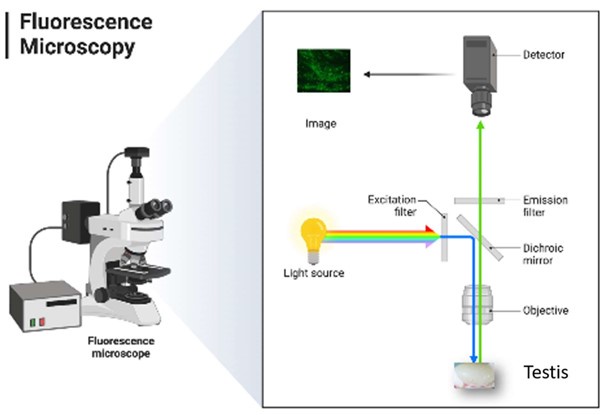
So, for Figure 3 if the plasmid is being uptaken by cells and maintained as an episome, is it able to replicate? Likely not.
Yes! it is the intrinsic property of the episome, see the detailed explanation provided above about the EEV plasmid
So, initially, it could be in spermatogonia, spermatocytes, and spermatids. As time progressed those initially positive spermatids and then spermatocytes would be lost - and finally, the only cells that should be positive would be the progeny of spermatogonia that were positive - but, as they proliferate shouldn't the GFP signal decline?
Because EEV is able to replicate in a synchronous manner with the host genome and to segregate into daughter cells at a level of 90% of the mother cell, the expected decline is very slow.
And, since clones of germ cells are connected throughout their development, shouldn't the GFP diffuse through the intercellular bridges so entire clones are positive? Was this observed?
We did not perform IF experiments further than 7 days after injection, a time too short to observe what the reviewer suggested. Moreover, if at 1 day after injection, GFP synthesized from injected EEV was found in both germ cells and Sertoli cells (Fig 7), after one week, the reporter proteins were only observable in Sertoli cells. This result suggests that EEV is maintained only in Sertoli cells, thus preventing the observation of stained clones.
Can these sections be stained for the ICB TEX14 so that clonality can be distinguished? Based on the apparent distance between cells, it appears some are clones, but many are not...
We thank the reviewer for this suggestion but we are not able to perform testis sectioning and costaining experiments because the PFA treatment bleaches the GFP signal. We also tested several GFP antibodies, but all failed.
Nevertheless, we were able to localize and identify transfected cells thank to the whole testis optical clearing, combined with a measure of GFP fluorescence and three-dimensional image reconstructions.
For Figure 4, with the mRNA-GFP, why does the 1-day image (which looks similar to the plasmidtransfected) look so different from days 7-21?
And why do days 7-21 look so different from those days in Fig 3?
Thank you for your feedback. It is an excellent question. Because of the low resolution of the whole testis epifluorescences imaging and light penetration issue, we decided to carry-out whole testis optical clearing and three-dimensional image reconstructions experiments, in order to get insights on the transfection process. At day 1, GFP synthesized from EEV injection was found in spermatogonia, spermatocytes and Sertoli cells (Fig 7). After one week, the reporter protein synthesized from injected EEV was only observable in Sertoli cells.
In contrast, for mRNA, on day 1 and day 7 post-injection, GFP fluorescent signal was associated with both Sertoli cells and germ cells. This explains why patterns between mRNA-GFP and EEV-GFP are similar at day 1 and different at day 7 between both conditions.
Why do the authors think the signal went from so strong at 21 to undetectable at 28? What changed so drastically over those 7 days?
What is the half-life of this mRNA supposed to be? It seems that 21 days is an unreasonably long time, but then to go to zero at 28 seems also odd... Please provide some explanation, and context for whether the residence of an exogenous mRNA for 21 days is expected.
As previously stated, it is our hypothesis that the source of the confusion lies in the fact that we injected mRNA coding for the GFP protein, rather than mRNA tagged with GFP. After a three-week observation period, we did not observe the mRNA, but we observed the GFP protein produced by the mRNA. The time of observation of the reporter proteins expressed by the respective mRNA molecules (mCherry, luciferase, or GFP) ranged from 15 to 21 days. Proteins have very different turnover rates, with half-lives ranging from minutes to days. Half-lives depend on proteins but also on tissues. As explained in the discussion, it has been demonstrated that proteins involved in spermatogenesis exhibit a markedly low turnover rate and this explains the duration of the fluorescent signal.
The authors should immunostain testis sections from controls and those with mRNA and plasmid and immunostain with established germ cell protein fate markers to show what specific germ cell types are GFP+
Thank you for your feedback. As previously mentioned, we were unable to perform testis sectioning and co-staining because the PFA treatment bleaches the GFP signal and because we were unable to reveal GFP with an GFP antibody, for unknown reasons.
For the GFP signal to be maintained past 35 days, the plasmid must have integrated into SSCs - and for that to happen, the plasmid would have to cross the blood-testis-barrier... is this expected?
We are grateful for your observation.
First, as explained above, we do not think that the plasmid has been integrated.
Concerning the blood-testing barrier. It bears noting that electroporation is a technique that is widely utilized in biotechnology and medicine for the delivery of drugs and the transfer of genes into living cells (Boussetta, Lebovka et al. 2009). This process entails the application of an electric current, which induces the formation of hydrophilic pores in the lipid bilayer of the plasma membrane (Kanduser, Miklavcic et al. 2009). The pores remain stable throughout the electroporation process and then close again once it is complete. Consequently, as electroporation destabilizes the cell membrane, it can also destabilize the gap junctions responsible of the blood-testis barrier. This was actually confirmed by several studies, which have observed plasmid transfection beyond the blood-testis barrier with injection into rete testis following electroporation (Muramatsu, Shibata et al. 1997, Kubota, Hayashi et al. 2005, Danner, Kirchhoff et al. 2009, Kanduser, Miklavcic et al. 2009, Michaelis, Sobczak et al. 2014).
Figure 9 - authors should show >1 cell - this is insufficient; also, it's stated it's only in the flagella, but it also appears to be in the head as well. And is this just the principal piece?? And are the authors sure those are elongating vs condensing spermatids? Need to show multiple tubules, at different stages, to make these claims
We have partly answered to this question in the public review; We pastehere our answer
“We present now new images showing the full seminiferous tubules as requested (see supp fig 6). In this new figure, it is clear that Armc2 is only expressed in spermatids. We have also added in this figure an analysis of the RNA-seq database produced by Gan's team (Gan, Wen et al. 2013), confirming that ArmC2 expression is predominantly expressed at the elongated spermatid stage. This point is now clearly indicated in the text.”
Concerning the localization of the protein in the head, we confirm that the base of the manchette is stained but we have no explanation so far. This point is now indicated in the manuscript.
Figure 10B2 image - a better resolution is necessary
We are grateful for your feedback. We concede that the quality of the image was not optimal. Consequently, We have replaced it with an alternative.
Figure 11 - in control, need to show >1 sperm; and lower-mag images should be provided for all samples to show population-wide effects; showing 1 "normal" sperm per group (white arrows) is insufficient:
We are grateful for your feedback. We conducted further experiments and provide now additional images in Supp. figure 8.
Reviewer #3 (Recommendations For The Authors):
In this study, Vilpreux et al. developed a microinjection/electroporation method in order to transfect RNA into testicular cells. The authors studied several parameters of treated testis and compared the injection of DNA versus RNA. Using the injection of Armc2 RNA into mice with an Armc2 knockout the authors were able to (partly) rescue the fertility phenotype.
Minor points.
Figure 6 + lines 553+554: might it be that the staining pattern primarily on one side of the testis is due to the orientation of the scissor electrode during the electroporation procedure and the migration direction of negatively charged RNA molecules (Figure 6)?
Your input is greatly appreciated. We concur that the observed peripheral expression is due to both the electroporation and injection. Accordingly, we have amended the sentence as follows: "The peripheral expression observed was due to the close vicinity of cells to the electrodes, and to a peripheral dispersal of the injected solution, as shown by the distribution of the fluorescent i-particles NIRFiP-180."
Discussion of the safety aspect (lines 601-608): The authors state several times that there are no visible tissue changes after the electroporation procedure. However, in order to claim that this procedure is "safe", it is necessary to examine the offspring born after microinjection/electroporation.
Your input is greatly appreciated. Consequently, the term "safe" has been replaced with "did not perturb spermatogenesis" in accordance with the provided feedback. Your assertion is correct; an examination of the offspring born would be necessary to ascertain the safety of the procedure. Due to the quantity of motile sperm obtained, it was not possible to produce offspring through natural mating. However, novel Armc2-/--rescued sperm samples have been produced and in vitro fertilization (IVF) and intracytoplasmic sperm injection (ICSI) experiments have been conducted. The results demonstrate that the Armc2-/--rescued sperm can successfully fertilize eggs and produce two-cell embryos by IVF and blastocysts by ICSI. These outcomes are visually represented in Figure 12. The development of embryos up to the blastocyst stage is a step in the right direction.
The discussion section could be shortened. Lines 632-646 are largely a repetition of the introductory section. In addition, the Dong paper (ref. 25) may be interesting; however, this part could also be shortened (lines 647-676). This reviewer would prefer the authors to focus on the technique (different application sites and applied nucleotides) and proof of concept for (partial) phenotype rescue in the knockout mice.
Your contribution is highly valued. In light of your observations and the latest findings, we have substantially revised the discussion accordingly.
Line 63: oocytes rather than eggs.
We are grateful for your input, but we have decided to retain our current position and to use the term "eggs" rather than "oocytes" in our writing because the definition of an oocyte is a female gametocyte or germ cell involved in reproduction. In other words, oocyte corresponds to a germ cell inside the ovary and after ovulation become an egg.
Boussetta, N., N. Lebovka, E. Vorobiev, H. Adenier, C. Bedel-Cloutour and J. L. Lanoiselle (2009). "Electrically assisted extraction of soluble matter from chardonnay grape skins for polyphenol recovery." J Agric Food Chem 57(4): 1491-1497.
Cavallini, G. (2006). "Male idiopathic oligoasthenoteratozoospermia." Asian J Androl 8(2): 143-157.
Colpi, G. M., S. Francavilla, G. Haidl, K. Link, H. M. Behre, D. G. Goulis, C. Krausz and A. Giwercman (2018). "European Academy of Andrology guideline Management of oligo-asthenoteratozoospermia." Andrology 6(4): 513-524.
Coutton, C., G. Martinez, Z. E. Kherraf, A. Amiri-Yekta, M. Boguenet, A. Saut, X. He, F. Zhang, M. Cristou-Kent, J. Escoffier, M. Bidart, V. Satre, B. Conne, S. Fourati Ben Mustapha, L. Halouani, O. Marrakchi, M. Makni, H. Latrous, M. Kharouf, K. Pernet-Gallay, M. Bonhivers, S. Hennebicq, N. Rives, E. Dulioust, A. Toure, H. Gourabi, Y. Cao, R. Zouari, S. H. Hosseini, S. Nef, N. Thierry-Mieg, C. Arnoult and P. F. Ray (2019). "Bi-allelic Mutations in ARMC2 Lead to Severe Astheno-Teratozoospermia Due to Sperm Flagellum Malformations in Humans and Mice." Am J Hum Genet 104(2): 331-340.
Danner, S., C. Kirchhoff and R. Ivell (2009). "Seminiferous tubule transfection in vitro to define postmeiotic gene regulation." Reprod Biol Endocrinol 7: 67.
Gan, H., L. Wen, S. Liao, X. Lin, T. Ma, J. Liu, C. X. Song, M. Wang, C. He, C. Han and F. Tang (2013). "Dynamics of 5-hydroxymethylcytosine during mouse spermatogenesis." Nat Commun 4: 1995. Igyarto, B. Z. and Z. Qin (2024). "The mRNA-LNP vaccines - the good, the bad and the ugly?" Front Immunol 15: 1336906.
Ishii, T. (2017). "Germ line genome editing in clinics: the approaches, objectives and global society." Brief Funct Genomics 16(1): 46-56.
Kanduser, M., D. Miklavcic and M. Pavlin (2009). "Mechanisms involved in gene electrotransfer using high- and low-voltage pulses--an in vitro study." Bioelectrochemistry 74(2): 265-271.
Kubota, H., Y. Hayashi, Y. Kubota, K. Coward and J. Parrington (2005). "Comparison of two methods of in vivo gene transfer by electroporation." Fertil Steril 83 Suppl 1: 1310-1318.
Michaelis, M., A. Sobczak and J. M. Weitzel (2014). "In vivo microinjection and electroporation of mouse testis." J Vis Exp(90).
Muramatsu, T., O. Shibata, S. Ryoki, Y. Ohmori and J. Okumura (1997). "Foreign gene expression in the mouse testis by localized in vivo gene transfer." Biochem Biophys Res Commun 233(1): 45-49.
Raina, A., S. Kumar, R. Shrivastava and A. Mitra (2015). "Testis mediated gene transfer: in vitro transfection in goat testis by electroporation." Gene 554(1): 96-100.
Sadelain, M., E. P. Papapetrou and F. D. Bushman (2011). "Safe harbours for the integration of new DNA in the human genome." Nat Rev Cancer 12(1): 51-58.
Thonneau, P., S. Marchand, A. Tallec, M. L. Ferial, B. Ducot, J. Lansac, P. Lopes, J. M. Tabaste and A. Spira (1991). "Incidence and main causes of infertility in a resident population (1,850,000) of three French regions (1988-1989)." Hum Reprod 6(6): 811-816.
Usmani, A., N. Ganguli, H. Sarkar, S. Dhup, S. R. Batta, M. Vimal, N. Ganguli, S. Basu, P. Nagarajan and S. S. Majumdar (2013). "A non-surgical approach for male germ cell mediated gene transmission through transgenesis." Sci Rep 3: 3430.
Wang, L., C. Liu, H. Wei, Y. Ouyang, M. Dong, R. Zhang, L. Wang, Y. Chen, Y. Ma, M. Guo, Y. Yu, Q. Y. Sun and W. Li (2022). "Testis electroporation coupled with autophagy inhibitor to treat nonobstructive azoospermia." Mol Ther Nucleic Acids 30: 451-464.
-
-
-
eLife assessment
This potentially useful study reports a new method for restoring sperm motility. Strengths are in the methodology being developed, but the conclusions require additional experimental support. The authors provide inadequate evidence for the success of the method or its mechanism.
-
Reviewer #1 (Public Review):
The authors assess the effectiveness of electroporating mRNA into male germ cells to rescue the expression of proteins required for spermatogenesis progression in individuals where these proteins are mutated or depleted. To set up the methodology, they first evaluated the expression of reporter proteins in wild-type mice, which showed expression in germ cells for over two weeks. Then, they attempted to recover fertility in a model of late spermatogenesis arrest that produces immotile sperm. By electroporating the mutated protein, the authors recovered the motility of ~5% of the sperm, although the sperm regenerated was not able to produce offspring using IVF.
This is a comprehensive evaluation of the mRNA methodology with multiple strengths. First, the authors show that naked synthetic RNA, purchased from a …
Reviewer #1 (Public Review):
The authors assess the effectiveness of electroporating mRNA into male germ cells to rescue the expression of proteins required for spermatogenesis progression in individuals where these proteins are mutated or depleted. To set up the methodology, they first evaluated the expression of reporter proteins in wild-type mice, which showed expression in germ cells for over two weeks. Then, they attempted to recover fertility in a model of late spermatogenesis arrest that produces immotile sperm. By electroporating the mutated protein, the authors recovered the motility of ~5% of the sperm, although the sperm regenerated was not able to produce offspring using IVF.
This is a comprehensive evaluation of the mRNA methodology with multiple strengths. First, the authors show that naked synthetic RNA, purchased from a commercial source or generated in the laboratory with simple methods, is enough to express exogenous proteins in testicular germ cells. The authors compared RNA to DNA electroporation and found that germ cells are efficiently electroporated with RNA, but not DNA. The differences between these constructs were evaluated using in vivo imaging to track the reporter signal in individual animals through time. To understand how the reporter proteins affect the results of the experiments, the authors used different reporters: two fluorescent (eGFP and mCherry) and one bioluminescent (Luciferase). Although they observed differences among reporters, in every case expression lasted for at least two weeks.
The authors used a relevant system to study the therapeutic potential of RNA electroporation. The ARMC2-deficient animals have impaired sperm motility phenotype that affects only the later stages of spermatogenesis. The authors showed that sperm motility was recovered to ~5%, which is remarkable due to the small fraction of germ cells electroporated with RNA with the current protocol. The 3D reconstruction of an electroporated testis using state-of-the-art methods to show the electroporated regions is compelling.
The main weakness of the manuscript is that although the authors manage to recover motility in a small fraction of the sperm population, it is unclear whether the increased sperm quality is substantial to improve assisted reproduction outcomes. The quality of the sperm was not systematically evaluated in the manuscript, with the endpoints being sperm morphology and sperm mobility.
Some key results, such as the 3D reconstruction of the testis and the recovery of sperm motility, are qualitative given the low replicate numbers or the small magnitude of the effects. The presentation of the sperm motility data could have been clearer as well. For example, on day 21 after Armc2-mRNA electroporation, only one animal out of the three tested showed increased sperm motility. However, it is unclear from Figure 11A what the percentage of sperm motility for this animal is since the graph shows a value of >5% and the reported aggregate motility is 4.5%. It would have been helpful to show all individual data points in Figure 11A.
The expression of the reporter genes is unambiguous; however, better figures could have been presented to show cell type specificity. The DAPI staining is diffused, and it is challenging to understand where the basement membranes of the tubules are. For example, in Figures 7B3 and 7E3, the spermatogonia seems to be in the middle of the seminiferous tubule. The imaging was better for Figure 8. Suboptimal staining appears to lead to mislabeling of some germ cell populations. For example, in Supplementary Figure 4A3, the round spermatid label appears to be labeling spermatocytes. Also, in some instances, the authors seem to be confusing, elongating spermatids with spermatozoa, such as in the case of Supplementary Figures 4D3 and D4.
The characterization of Armc2 expression could have been improved as well. The authors show a convincing expression of ARMC2 in a few spermatids/sperm using a combination of an anti-ARMC2 antibody and tubules derived from ARMC2 KO animals. At the minimum, one would have liked to see at least one whole tubule of a relevant stage.
Overall, the authors show that electroporating mRNA can improve spermatogenesis as demonstrated by the generation of motile sperm in the ARMC2 KO mouse model.
-
Reviewer #2 (Public Review):
Summary:
Here, the authors inject naked mRNAs and plasmids into the rete testes of mice to express exogenous proteins - GFP and later ARMC2. This approach has been taken before, as noted in the Discussion to rescue Dmc1 KO infertility. While the concept is exciting, multiple concerns reduce reviewer enthusiasm.
Strengths:
The approach, while not necessarily novel, is timely and interesting.
Weaknesses:
Overall, the writing and text can be improved and standardized - as an example, in some places in vivo is italicized, in others it's not; gene names are italicized in some places, others not; some places have spaces between a number and the units, others not. This lack of attention to detail in the preparation of the manuscript is a significant concern to this reviewer - the presentation of the experimental …
Reviewer #2 (Public Review):
Summary:
Here, the authors inject naked mRNAs and plasmids into the rete testes of mice to express exogenous proteins - GFP and later ARMC2. This approach has been taken before, as noted in the Discussion to rescue Dmc1 KO infertility. While the concept is exciting, multiple concerns reduce reviewer enthusiasm.
Strengths:
The approach, while not necessarily novel, is timely and interesting.
Weaknesses:
Overall, the writing and text can be improved and standardized - as an example, in some places in vivo is italicized, in others it's not; gene names are italicized in some places, others not; some places have spaces between a number and the units, others not. This lack of attention to detail in the preparation of the manuscript is a significant concern to this reviewer - the presentation of the experimental details does cast some reasonable concern with how the experiments might have been done. While this may be unfair, it is all the reviewers have to judge. Multiple typographical and grammatical errors are present, and vague or misleading statements.
-
Reviewer #3 (Public Review):
Summary:
The authors used a novel technique to treat male infertility. In a proof-of-concept study, the authors were able to rescue the phenotype of a knockout mouse model with immotile sperm using this technique. This could also be a promising treatment option for infertile men.
Strengths:
In their proof-of-concept study, the authors were able to show that the novel technique rescues the infertility phenotype in vivo.
Weaknesses:
Some minor weaknesses, especially in the discussion section, could be addressed to further improve the quality of the manuscript.
It is very convincing that the phenotype of Armc2 KO mice could (at least in part) be rescued by injection of Armc2 RNA. However, a central question remains about which testicular cell types have been targeted by the constructs. From the pictures …
Reviewer #3 (Public Review):
Summary:
The authors used a novel technique to treat male infertility. In a proof-of-concept study, the authors were able to rescue the phenotype of a knockout mouse model with immotile sperm using this technique. This could also be a promising treatment option for infertile men.
Strengths:
In their proof-of-concept study, the authors were able to show that the novel technique rescues the infertility phenotype in vivo.
Weaknesses:
Some minor weaknesses, especially in the discussion section, could be addressed to further improve the quality of the manuscript.
It is very convincing that the phenotype of Armc2 KO mice could (at least in part) be rescued by injection of Armc2 RNA. However, a central question remains about which testicular cell types have been targeted by the constructs. From the pictures presented in Figures 7 and 8, this issue is hard to assess. Given the more punctate staining of the DNA construct a targeting of Sertoli cells is more likely, whereas the more broader staining of seminiferous tubules using RNA constructs is talking toward germ cells.
Further, the staining for up to 119 days (Figure 5) would point toward an integration of the DNA construct into the genome of early germ cells such as spermatogonia and/or possibly to Sertoli cells. Given the expression after RNA transfection for up to 21 days (Figure 4) and the detection of motile sperm after 21 days (Figure 11), this would point to either round spermatids or spermatocytes.
These aspects need to be discussed more carefully (discussion section: lines 549-574).
It would also be very interesting to know in which testicular cell type Armc2 is endogenously expressed (lines 575-591).
-
