State-dependent representations of mixtures by the olfactory bulb
Curation statements for this article:-
Curated by eLife

Evaluation Summary:
This study examined how mixture odors are represented in the mouse olfactory bulb. The authors found that compared to the responses in anesthetized mice, mixture responses are more linear in awake mice regardless whether the mice were engaged in a behavioral task or not. The results are potentially important as the results differ from previous studies which were done mostly in anesthetized animals, but the reviewers raised concerns for the validity and the strength of the conclusions.
(This preprint has been reviewed by eLife. We include the public reviews from the reviewers here; the authors also receive private feedback with suggested changes to the manuscript. Reviewer #1 agreed to share their name with the authors.)
This article has been Reviewed by the following groups
Discuss this preprint
Start a discussion What are Sciety discussions?Listed in
- Evaluated articles (eLife)
Abstract
Sensory systems are often tasked to analyse complex signals from the environment, separating relevant from irrelevant parts. This process of decomposing signals is challenging when a mixture of signals does not equal the sum of its parts, leading to an unpredictable corruption of signal patterns. In olfaction, nonlinear summation is prevalent at various stages of sensory processing. Here, we investigate how the olfactory system deals with binary mixtures of odours under different brain states by two-photon imaging of olfactory bulb (OB) output neurons. Unlike previous studies using anaesthetised animals, we found that mixture summation is more linear in the early phase of evoked responses in awake, head-fixed mice performing an odour detection task, due to dampened responses. Despite smaller and more variable responses, decoding analyses indicated that the data from behaving mice was well discriminable. Curiously, the time course of decoding accuracy did not correlate strictly with the linearity of summation. Further, a comparison with naïve mice indicated that learning to accurately perform the mixture detection task is not accompanied by more linear mixture summation. Finally, using a simulation, we demonstrate that, while saturating sublinearity tends to degrade the discriminability, the extent of the impairment may depend on other factors, including pattern decorrelation. Altogether, our results demonstrate that the mixture representation in the primary olfactory area is state-dependent, but the analytical perception may not strictly correlate with linearity in summation.
Article activity feed
-

Author Response
Reviewer #1 (Public Review):
Adefuin and colleagues examined the interaction between components of binary odor mixtures in odor responses in mice. The authors used two-photon calcium imaging from the soma and apical dendrites of mitral/tufted cells in the olfactory bulb. Odor responses were measured in various conditions: under anesthesia (ketamine/xylazine), while well-trained mice were engaged in an odor discrimination task, or disengaged. The authors first show that mixture components interacted sublinearly in a large fraction of mitral/tufted cells (46%; Fig. 6D) consistent with previous studies. However, when odor responses were measured in awake animals, very few mitral/tufted cells showed sublinear responses at soma (8-9%; Fig. 6D). Interestingly, sublinear interaction was evident in apical dendrites of …
Author Response
Reviewer #1 (Public Review):
Adefuin and colleagues examined the interaction between components of binary odor mixtures in odor responses in mice. The authors used two-photon calcium imaging from the soma and apical dendrites of mitral/tufted cells in the olfactory bulb. Odor responses were measured in various conditions: under anesthesia (ketamine/xylazine), while well-trained mice were engaged in an odor discrimination task, or disengaged. The authors first show that mixture components interacted sublinearly in a large fraction of mitral/tufted cells (46%; Fig. 6D) consistent with previous studies. However, when odor responses were measured in awake animals, very few mitral/tufted cells showed sublinear responses at soma (8-9%; Fig. 6D). Interestingly, sublinear interaction was evident in apical dendrites of mitral/tufted cells (45%). Whether mixture components are represented linearly or not in the olfactory system is an important question, related to the animal's ability to identify or segment mixture components. Somewhat contrary to previous studies, this study demonstrate largely linear interactions. Furthermore, this study compares various behavioral conditions. These results are important and of interest to those who study sensory systems. I have a few concerns regarding data analysis.
Thank you for your helpful review, and for recognising the relevance our work. We hope that the reviewer finds the our point-by-point responses satisfactory.
- Non-linear interactions are detected by the activity showing a deviation from linearity greater than 2 standard deviations. Using this criterion, non-linear interactions might decrease if the trial-by-trial activity becomes more variable. This is concerning because the activity might be less variable in the anesthetized condition, and the reduction in sublinear interactions in awake conditions may be due to a general increase in response variability during awake. Can the authors exclude the possibility that the decrease in sublinear interactions is merely due to an increase in response variability in the awake conditions. This issue also applies to the comparison between apical dendrites versus soma; are the signals in apical dendrite less variable (maybe due to some averaging across dendrites from multiple cells; see the following point 5)?
Thank you for raising this valid point and for suggesting alternative analyses. We agree that the index we used previously is susceptible to noise, and not appropriate for comparing two datasets with different trial-by-trial variability. To quantify the deviation from linear sum more robustly, we now use the “Median fractional deviation”, which expresses a deviation from the linear sum as a fraction of predicted, linear sum - not normalised by the standard deviation – and take the median of the distribution from each field of view. As we describe in the revised Figure 4, this measure is more robust to noise. Notably, our finding that mixture summation is generally less sublinear in awake mice still stands for the early phase.
In the revised manuscript, we use the median fractional deviation whenever we compare linearity of summation across different conditions, which includes the comparison of anaesthetised vs. awake, behaving conditions (revised Fig. 4), comparison of dendrites vs. somata (revised Fig. 4-figure supplement 1), and comparisons of awake states (revised Fig. 6). This has given us, too, more confidence about our interpretation, so we are grateful for the reviewer’s suggestions.
- Related to the above issue, it would be useful to analyze the difference between conditions using different metrics to fully understand what really are different between conditions. The scatter plots shown in various figures do not show drastic differences between awake and anesthetized conditions, as might be indicated by the percent of sublinear responses. It would be useful to characterize the magnitude of sublinear/supralinear effects. For example, one can calculate a fractional change in the mean response. Does this measure show consistent difference between awake and anesthetized conditions?
Thank you for suggesting this analysis. As described above, we now use the fractional deviation to quantify how mixture summations differ from linear sums, which turned out to be a very useful way to express the property of summation (N.B.: noise is amplified for small responses when fractional deviation is used, which is another reason we use the median now). We thank the reviewer for suggesting this analysis.
Reviewer #2 (Public Review):
This study addresses how complex stimuli are represented in neural responses. This is particularly relevant to olfaction because the vast majority of stimuli are complex mixtures that perceptually, are not easy to decompose into parts. Nonetheless, the ability to discern a relevant odor from background odors is essential. This process is easier when neural responses to mixtures reflect the linear sum of the responses to the individual components. The main conclusion of this study is that the linearity of olfactory bulb responses to two-component mixtures increases awake versus anesthetized states. The authors provide some evidence to support this claim. However, this could be better quantified and there is a temporal aspect of linearization that is not addressed. Perhaps the most interesting aspect of the study is the difference in linearity between the dendrites and the somata of the mitral/tufted cells. But a statistical analysis of this finding was not evident. Overall a mechanistic or functional approach to understanding these findings is lacking. The differences linearity between the anesthetized and awake are simply explained by response saturation anesthetized animals. There are hints at mechanism by which linearity is supported in the OB with comparisons between soma and dendrite but these are not well developed. There is a model that addresses the functional significance of linearity but this is only supplemental and not well described.
Thank you for appreciating the significance of our work, and for your constructive comments.
Reviewer #3 (Public Review):
Adefuin et al use multiphoton imaging of M/T cell responses to investigate whether neuronal representations of binary mixtures can be explained as a sum of the components. The current view in the field (built largely from studies in anesthetized animals), is that mixture summation is non-linear and increases with the degree in glomerular response overlap elicited by the components. The authors reproduce these results and ask whether the same phenomenon is observed in the awake state, in particular when the animals are engaged in an odor discrimination task. Unlike in the anesthetized state, the authors find that mixture representations are linear in the awake brain. They use a series of systematic behavioral paradigms to show that the observed linearity in the awake state (compared to anesthetized) is not dependent on task engagement (reward is given randomly, post-odor) or stimulus relevance (reward is given before odor). While the experiments are well done and the data is presented clearly, I have several major concerns about the interpretation of their results.
- Given the data the authors present, it is unclear if one can conclude that the olfactory system is more or less linear in the awake state compared to the anaesthetised one. What seems to change most across the awake vs. anesthetized state is the response amplitude. Responses appear to be ~3x smaller in the awake mice. In the anesthetized state, non-linearity seems most apparent for large response amplitudes (>5 dF/F) with mixture responses being sub-linear, most likely due to saturation effects. The authors themselves do an analysis in Figure 6 - supplement 1 to show that most of the observed non-linearity in the anesthetized animals can be explained away after accounting for amplitude normalisation. The authors use this analysis to comment that the level of linearity is the same across all the three awake states, but the same figure shows that it is in fact the same even for the anaesthetized state.
To put it differently, it is indeed true from the authors data that the OB response gain is significantly lower in the awake state, but it is unclear if the summation is more linear if measured at similar response amplitude regimes in both awake and anaesthetised mice.
Thank you for the valuable comments. We agree that many differences between the anaesthetised vs. awake states should have been taken into account when comparing the linearity of summation. We address the reviewer’s concern now by expressing the deviation as a fraction of the predicted, linear sum of component responses. Further, we also considered another factor that could influence the anaesthetised vs. awake comparison, namely, the trial-by-trial variability. This is reproduced below.
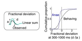
Figure R1: comparison of mixture summation for the early phase of responses, expressed as the fractional deviation.
- The authors argue that keeping response amplitudes small in the awake brain prevents sub-linear summation and therefore may lead to better mixture decomposition. They do a decoding analysis in anaesthetised mice to show that linear mixture representations (instead of using observed sub-linear representations) make odor classification easier. However, I find this analysis uninformative and misleading. It is no surprise that the decoders trained on single odor representations should perform better (or equivalent) when using linear sums as input instead of observed sub-linear representations. The authors use this observation to suggest that this mechanism aids discrimination ability in the awake state. However, given that even the single odor responses are much weaker and noisier in the awake state, it is likely that even the single odor discrimination ability is poorer in the awake state. By the same logic, mixture decomposition might be also much poorer in the awake brain than the anesthetized brain, even though summation is more linear, just because responses are weaker and noisier. In my opinion, the authors should compare decoding accuracy across awake vs. anesthetized responses if they want to assert that linearisation of responses in the awake brain leads to easier decomposition. Because otherwise, while linearisation in principle can aid decomposition, at least in the form that the authors observe here, it may come at a high cost on signal-to-noise ratio which would undo the gain that linearity provides, in principle, for discrimination.
Thank you very much for the insight and for the excellent suggestion to consider the discriminability of stimuli. In particular, we now include an analysis where a decoder trained on single responses is tested on observed mixture responses. Surprisingly, despite the substantial differences in the amplitudes of response and trial-by-trial variability, decoders using data from awake mice performed well, even better than anaesthetised data for the late phase of responses. This is now described in the revised figures (revised Fig. 5). We thank the reviewer for the excellent suggestion.
Interestingly, though, the time course of the decoder performance does not correlate well with the linearity of summation. This observation is now described in the abstract (lines 19-21): “…decoding analyses indicated that the data from behaving mice was able to encode mixture responses well, though the time course of decoding accuracy did not correlate with the linearity of summation“.
- At a more philosophical level, to this Reviewer, it is unclear if anesthesia vs. awake state difference in response should constitute the main focus of the manuscript. The authors explore summation properties under four different brain states, one of which is anaesthesia (also least behaviorally relevant). In three out of four states, they observe that summation is linear. In the fourth (anaesthesia), they observe that summation is sub-linear, but this happens at much larger response amplitude regimes compared to the three awake states sampled, presumably due to saturation. To me, it seems that the Authors here show that mixture summation in the OB, is largely independent of brain state since it is unaffected by whether the animal is task engaged or motivated etc.
Thank you for this thoughtful comment. This has made us reflect on the essence of our study. We believe we make three main observations. First, the anaesthesia vs. awake difference in the property of summation differ, and should be reported, because of the large volume of prior works reporting sublinear summations. However, as the reviewer recommends and as mentioned next, this is no longer the sole focus of our study. Our second observation is that the linearity of summation does not necessarily correlate with the ability to analyse mixtures, based on the decoder performance. We believe it is important to share this observation, since a number of previous studies speculated that nonlinear summation contributes to perceptual difficulty (Bell et al., 1987; Laing, 1994). Third, the decoder performance - especially one that is trained on single odour responses and tested on mixtures - shows differences depending on the awake states, where data from disengaged mice performed particularly poorly. This result is shown in the revised Figure 6. Further, we have edited the abstract and results to ensure that these are clearly communicated. We hope that this is more balanced and reflects the data better.
- It is unclear how to interpret the dendritic imaging comparison. First, the dendritic signal is pooled across many cells. If any of the cells that are being pooled shows sub-linearity, the pooled population response will look sub-linear, albeit less so than at the single cell level. Second, again like for the anesthetized vs. awake comparison, there is a discrepancy in response amplitudes - dendritic responses are ~2x stronger than the somatic responses and sub-linear summation would be more apparent as one approaches the saturation regime. Third, dendritic responses pool both mitral and tufted, while the somatic data the authors present is predominantly from tufted cells.
Thank you for commenting on ways to further understand the dendritic signal. Indeed, the early prevalence of sublinearity in the apical dendrites does seem to relate to the time course of responses. This is treated more directly in the revised Fig.4 – supplement 1.
To address the averaging effect, we tested how pulled signals may look like in terms of linearity of summation. To roughly approximate pooled responses, we reasoned that neighbouring TC/MC somata have higher chances of belonging to the same glomerulus. Thus, we averaged signals from somatic ROIs (TCs and MCs) from each field of view and calculated the fractional deviation from the linear sum (Fig. R2). While a simplistic averaging of neighbouring somata may not be perfectly accurate, but this analysis indicates that the difference between the apical dendrites vs. somata may not be simply explained by the averaging effect.
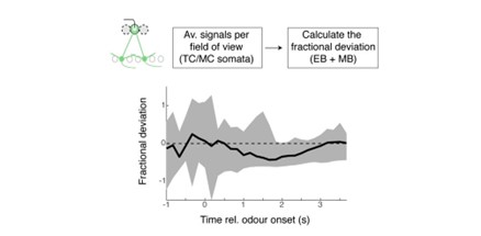
Figure R2: Analysis of pooled somatic signals
To approximate how dendritic signals might look like if they were simple averages of somatic responses, we pooled together signals from all TC/MC somata from each field of view, and treated it as “an approximate glomerular signal”. The plot above shows the fractional deviation from the linear sum. MC somata data comes from an additional set of experiments conducted for this rebuttal).
In terms of the unmatched amplitude distributions and trial-by-trial variability across conditions, as the reviewer points out, the issue is similar to the comparison of anaesthetised vs. awake data. To address this, all comparisons are now presented in terms of the median fractional deviations. Further, to explain if mitral cells contributed to the discrepancy in the linearity between the dendritic signal vs. somatic signal, we now provide additional data from 137 MCs (5 fields of view, 3 trained mice performing the mixture task). These changes are described in the revised manuscript (Figure 4- supplement 1).
-
Evaluation Summary:
This study examined how mixture odors are represented in the mouse olfactory bulb. The authors found that compared to the responses in anesthetized mice, mixture responses are more linear in awake mice regardless whether the mice were engaged in a behavioral task or not. The results are potentially important as the results differ from previous studies which were done mostly in anesthetized animals, but the reviewers raised concerns for the validity and the strength of the conclusions.
(This preprint has been reviewed by eLife. We include the public reviews from the reviewers here; the authors also receive private feedback with suggested changes to the manuscript. Reviewer #1 agreed to share their name with the authors.)
-
Reviewer #3 (Public Review):
Adefuin et al use multiphoton imaging of M/T cell responses to investigate whether neuronal representations of binary mixtures can be explained as a sum of the components. The current view in the field (built largely from studies in anesthetized animals), is that mixture summation is non-linear and increases with the degree in glomerular response overlap elicited by the components. The authors reproduce these results and ask whether the same phenomenon is observed in the awake state, in particular when the animals are engaged in an odor discrimination task. Unlike in the anesthetized state, the authors find that mixture representations are linear in the awake brain. They use a series of systematic behavioral paradigms to show that the observed linearity in the awake state (compared to anesthetized) is not …
Reviewer #3 (Public Review):
Adefuin et al use multiphoton imaging of M/T cell responses to investigate whether neuronal representations of binary mixtures can be explained as a sum of the components. The current view in the field (built largely from studies in anesthetized animals), is that mixture summation is non-linear and increases with the degree in glomerular response overlap elicited by the components. The authors reproduce these results and ask whether the same phenomenon is observed in the awake state, in particular when the animals are engaged in an odor discrimination task. Unlike in the anesthetized state, the authors find that mixture representations are linear in the awake brain. They use a series of systematic behavioral paradigms to show that the observed linearity in the awake state (compared to anesthetized) is not dependent on task engagement (reward is given randomly, post-odor) or stimulus relevance (reward is given before odor). While the experiments are well done and the data is presented clearly, I have several major concerns about the interpretation of their results.
- Given the data the authors present, it is unclear if one can conclude that the olfactory system is more or less linear in the awake state compared to the anaesthetised one. What seems to change most across the awake vs. anesthetized state is the response amplitude. Responses appear to be ~3x smaller in the awake mice. In the anesthetized state, non-linearity seems most apparent for large response amplitudes (>5 dF/F) with mixture responses being sub-linear, most likely due to saturation effects. The authors themselves do an analysis in Figure 6 - supplement 1 to show that most of the observed non-linearity in the anesthetized animals can be explained away after accounting for amplitude normalisation. The authors use this analysis to comment that the level of linearity is the same across all the three awake states, but the same figure shows that it is in fact the same even for the anaesthetized state.
To put it differently, it is indeed true from the authors data that the OB response gain is significantly lower in the awake state, but it is unclear if the summation is more linear if measured at similar response amplitude regimes in both awake and anaesthetised mice.
The authors argue that keeping response amplitudes small in the awake brain prevents sub-linear summation and therefore may lead to better mixture decomposition. They do a decoding analysis in anaesthetised mice to show that linear mixture representations (instead of using observed sub-linear representations) make odor classification easier. However, I find this analysis uninformative and misleading. It is no surprise that the decoders trained on single odor representations should perform better (or equivalent) when using linear sums as input instead of observed sub-linear representations. The authors use this observation to suggest that this mechanism aids discrimination ability in the awake state. However, given that even the single odor responses are much weaker and noisier in the awake state, it is likely that even the single odor discrimination ability is poorer in the awake state. By the same logic, mixture decomposition might be also much poorer in the awake brain than the anesthetized brain, even though summation is more linear, just because responses are weaker and noisier. In my opinion, the authors should compare decoding accuracy across awake vs. anesthetized responses if they want to assert that linearisation of responses in the awake brain leads to easier decomposition. Because otherwise, while linearisation in principle can aid decomposition, at least in the form that the authors observe here, it may come at a high cost on signal-to-noise ratio which would undo the gain that linearity provides, in principle, for discrimination.
At a more philosophical level, to this Reviewer, it is unclear if anesthesia vs. awake state difference in response should constitute the main focus of the manuscript. The authors explore summation properties under four different brain states, one of which is anaesthesia (also least behaviorally relevant). In three out of four states, they observe that summation is linear. In the fourth (anaesthesia), they observe that summation is sub-linear, but this happens at much larger response amplitude regimes compared to the three awake states sampled, presumably due to saturation. To me, it seems that the Authors here show that mixture summation in the OB, is largely independent of brain state since it is unaffected by whether the animal is task engaged or motivated etc.
It is unclear how to interpret the dendritic imaging comparison. First, the dendritic signal is pooled across many cells. If any of the cells that are being pooled shows sub-linearity, the pooled population response will look sub-linear, albeit less so than at the single cell level. Second, again like for the anesthetized vs. awake comparison, there is a discrepancy in response amplitudes - dendritic responses are ~2x stronger than the somatic responses and sub-linear summation would be more apparent as one approaches the saturation regime. Third, dendritic responses pool both mitral and tufted, while the somatic data the authors present is predominantly from tufted cells.
-
Reviewer #2 (Public Review):
This study addresses how complex stimuli are represented in neural responses. This is particularly relevant to olfaction because the vast majority of stimuli are complex mixtures that perceptually, are not easy to decompose into parts. Nonetheless, the ability to discern a relevant odor from background odors is essential. This process is easier when neural responses to mixtures reflect the linear sum of the responses to the individual components. The main conclusion of this study is that the linearity of olfactory bulb responses to two-component mixtures increases awake versus anesthetized states. The authors provide some evidence to support this claim. However, this could be better quantified and there is a temporal aspect of linearization that is not addressed. Perhaps the most interesting aspect of the …
Reviewer #2 (Public Review):
This study addresses how complex stimuli are represented in neural responses. This is particularly relevant to olfaction because the vast majority of stimuli are complex mixtures that perceptually, are not easy to decompose into parts. Nonetheless, the ability to discern a relevant odor from background odors is essential. This process is easier when neural responses to mixtures reflect the linear sum of the responses to the individual components. The main conclusion of this study is that the linearity of olfactory bulb responses to two-component mixtures increases awake versus anesthetized states. The authors provide some evidence to support this claim. However, this could be better quantified and there is a temporal aspect of linearization that is not addressed. Perhaps the most interesting aspect of the study is the difference in linearity between the dendrites and the somata of the mitral/tufted cells. But a statistical analysis of this finding was not evident. Overall a mechanistic or functional approach to understanding these findings is lacking. The differences linearity between the anesthetized and awake are simply explained by response saturation anesthetized animals. There are hints at mechanism by which linearity is supported in the OB with comparisons between soma and dendrite but these are not well developed. There is a model that addresses the functional significance of linearity but this is only supplemental and not well described.
-
Reviewer #1 (Public Review):
Adefuin and colleagues examined the interaction between components of binary odor mixtures in odor responses in mice. The authors used two-photon calcium imaging from the soma and apical dendrites of mitral/tufted cells in the olfactory bulb. Odor responses were measured in various conditions: under anesthesia (ketamine/xylazine), while well-trained mice were engaged in an odor discrimination task, or disengaged. The authors first show that mixture components interacted sublinearly in a large fraction of mitral/tufted cells (46%; Fig. 6D) consistent with previous studies. However, when odor responses were measured in awake animals, very few mitral/tufted cells showed sublinear responses at soma (8-9%; Fig. 6D). Interestingly, sublinear interaction was evident in apical dendrites of mitral/tufted cells (45%). …
Reviewer #1 (Public Review):
Adefuin and colleagues examined the interaction between components of binary odor mixtures in odor responses in mice. The authors used two-photon calcium imaging from the soma and apical dendrites of mitral/tufted cells in the olfactory bulb. Odor responses were measured in various conditions: under anesthesia (ketamine/xylazine), while well-trained mice were engaged in an odor discrimination task, or disengaged. The authors first show that mixture components interacted sublinearly in a large fraction of mitral/tufted cells (46%; Fig. 6D) consistent with previous studies. However, when odor responses were measured in awake animals, very few mitral/tufted cells showed sublinear responses at soma (8-9%; Fig. 6D). Interestingly, sublinear interaction was evident in apical dendrites of mitral/tufted cells (45%). Whether mixture components are represented linearly or not in the olfactory system is an important question, related to the animal's ability to identify or segment mixture components. Somewhat contrary to previous studies, this study demonstrate largely linear interactions. Furthermore, this study compares various behavioral conditions. These results are important and of interest to those who study sensory systems. I have a few concerns regarding data analysis.
1. Non-linear interactions are detected by the activity showing a deviation from linearity greater than 2 standard deviations. Using this criterion, non-linear interactions might decrease if the trial-by-trial activity becomes more variable. This is concerning because the activity might be less variable in the anesthetized condition, and the reduction in sublinear interactions in awake conditions may be due to a general increase in response variability during awake. Can the authors exclude the possibility that the decrease in sublinear interactions is merely due to an increase in response variability in the awake conditions. This issue also applies to the comparison between apical dendrites versus soma; are the signals in apical dendrite less variable (maybe due to some averaging across dendrites from multiple cells; see the following point 5)?
2. Related to the above issue, it would be useful to analyze the difference between conditions using different metrics to fully understand what really are different between conditions. The scatter plots shown in various figures do not show drastic differences between awake and anesthetized conditions, as might be indicated by the percent of sublinear responses. It would be useful to characterize the magnitude of sublinear/supralinear effects. For example, one can calculate a fractional change in the mean response. Does this measure show consistent difference between awake and anesthetized conditions?
-

-
