Kazrin promotes dynein/dynactin-dependent traffic from early to recycling endosomes
Curation statements for this article:-
Curated by eLife

eLife assessment
Hernandez-Perez et al. perform a detailed analysis of Kazrin, a widely expressed protein that appears to be involved in many diverse cellular processes, but whose exact function is unknown. The authors employ mouse embryonic fibroblasts and biochemistry to investigate the function of Kazrin and determine that Kazrin promotes the dynein/dynactin-dependent transport of early endosomes. These findings are valuable to those in the field of intracellular transport, but the story will benefit from additional experiments to prove the main claims, or from textual modifications.
This article has been Reviewed by the following groups
Discuss this preprint
Start a discussion What are Sciety discussions?Listed in
- Evaluated articles (eLife)
Abstract
Kazrin is a protein widely expressed in vertebrates whose depletion causes a myriad of developmental defects, in part derived from altered cell adhesion and migration, as well as failure to undergo epidermal to mesenchymal transition. However, the primary molecular role of kazrin, which might contribute to all these functions, has not been elucidated yet. We previously identified one of its isoforms, kazrin C, as a protein that potently inhibits clathrin-mediated endocytosis when overexpressed. We now generated kazrin knock-out mouse embryonic fibroblasts to investigate its endocytic function. We found that kazrin depletion delays juxtanuclear enrichment of internalized material, indicating a role in endocytic traffic from early to recycling endosomes. Consistently, we found that the C-terminal domain of kazrin C, predicted to be an intrinsically disordered region, directly interacts with several early endosome (EE) components, and that kazrin depletion impairs retrograde motility of these organelles. Further, we noticed that the N-terminus of kazrin C shares homology with dynein/dynactin adaptors and that it directly interacts with the dynactin complex and the dynein light intermediate chain 1. Altogether, the data indicate that one of the primary kazrin functions is to facilitate endocytic recycling by promoting dynein/dynactin-dependent transport of EEs or EE-derived transport intermediates to the recycling endosomes.
Article activity feed
-

-

Author Response
Reviewer #1 (Public Review):
Kazrin appears to be implicated in many diverse cellular functions, and accordingly, localizes to many subcellular sites. Exactly what it does is unclear. The authors perform a fairly detailed analysis of Kazrin in-cell function, and find that it is important for the perinuclear localization of TfN, and that it binds to members of the AP-1 complex (e.g., gamma-adaptin). The authors note that the C-terminus of Kazrin (which is predicted to be intrinsically disordered) forms punctate structures in the cytoplasm that colocalize with components of the endosomal machinery. Finally, the authors employ co-immunoprecipitation assays to show that both N and C-termini of Kazrin interacts with dynactin, and the dynein light-intermediate chain.
Much of the data presented in the manuscript are of …
Author Response
Reviewer #1 (Public Review):
Kazrin appears to be implicated in many diverse cellular functions, and accordingly, localizes to many subcellular sites. Exactly what it does is unclear. The authors perform a fairly detailed analysis of Kazrin in-cell function, and find that it is important for the perinuclear localization of TfN, and that it binds to members of the AP-1 complex (e.g., gamma-adaptin). The authors note that the C-terminus of Kazrin (which is predicted to be intrinsically disordered) forms punctate structures in the cytoplasm that colocalize with components of the endosomal machinery. Finally, the authors employ co-immunoprecipitation assays to show that both N and C-termini of Kazrin interacts with dynactin, and the dynein light-intermediate chain.
Much of the data presented in the manuscript are of fairly high quality and describe a potentially novel function for Kazrin C. However, I had a few issues with some of the language used throughout, the manner of data presentation, and some of their interpretations. Most notably, I think in its current form, the manuscript does not strongly support the authors' main conclusion: that Kazrin is a dynein-dynactin adaptor, as stated in their title. Without more direct support for this function, the authors need to soften their language. Specific points are listed below.
Major comments:
- I agree with the authors that the data provided in the manuscript suggest that Kazrin may indeed be an endosomal adaptor for dynein-dynactin. However, without more direct evidence to support this notion, the authors need to soften their language stating as much. For example, the title as stated would need to be changed, as would much of the language in the first paragraph of the discussion. Alternatively, the manuscript could be significantly strengthened if the authors performed a more direct assay to test this idea. For example, the authors could use methods employed previously (e.g., McKenney et al., Science 2014) to this end. In brief, the authors can simply use their recombinant Kazrin C (with a GFP) to pull out dynein-dynactin from cell extracts and perform single molecule assays as previously described.
While this is certainly an excellent suggestion, the in vitro dynein/dynactin motility assays are really not straight forward experiments for laboratories that do not use them as a routine protocol. That is why we asked Dr. Thomas Surrey (Centre for Genomic Regulation, Barcelona), an expert in the biochemistry and biophysics of microtubule dynamics, to help us with this kind of analysis. In their setting, TIRF microscopy is used to follow EGFPdynein/dynactin motility along microtubules immobilized on cover slides (Jha et al., 2017). As shown in figure R1, more binding of EGFP-dynein to the microtubules is observed when purified kazrin is added to the assay (from 20 to 400 nM), but there is no increase in the number or processivity of the EGFP-dynein motility events. These results are hard to interpret at this point. Kazrin might still be an activating adaptor but a component is missing in the assay (i. e. an activating posttranslational modification or a particular subunit of the dynein or dynactin complexes), or it could increase the processivity of dyneindynactin in complex with another bona fide activating adaptor, as it has been demonstrated for LIS1 (Baumbach et al., 2017; Gutierrez et al., 2017). Alternatively, kazrin could transport dynactin and/or dynein to the microtubule plus ends in a kinesin 1-dependent manner, in order to load the peripheral endosomes with the minus end directed motor (Yamada et al., 2008).
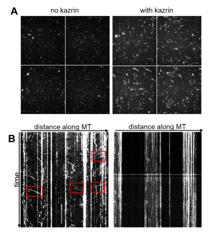
Figure R1. Kazrin C purified from E. coli increases binding of dynein to microtubules but does not increase the number or processivity of EGFP-dynein motility events. A. TIRF (Total Internal Reflexion Fluorescence) micrographs of microtubule-coated cover slides incubated in the presence of 10 nM EGFP-dynein and 20 nM dynactin in the presence or absence of 20 nM kazrin C, expressed and purified from E. coli. B. Kymographs of TIRF movies of microtubule-coated cover slides incubated in the presence of purified 10 nM EGFP-dynein, 20 nM dynactin and either 400 nM of the activating adaptor BICD2 (1:2:40 ratio) (left panel) or kazrin C (right panel). Red squares indicate processive dynein motility events induced by BICD2”.
Investigating the molecular activity of kazrin on the dynein/dynactin motility is a whole project in itself that we feel it is out of the scope of the present manuscript. Therefore, as suggested by the BRE, we have chosen to soften the conclusions and classify kazrin as a putative “candidate” dynein/dynactin adaptor based on its interactome, domain organization and subcellular localization, as well as on the defects installed in vivo on the endosome motility upon its depletion. We also discuss other possibilities as those outlined above.
- I'm not sure I agree with the use of the term 'condensates' used throughout the manuscript to describe the cytoplasmic Kazrin foci. 'Condensates' is a very specific term that is used to describe membraneless organelles. Given the presumed association of Kazrin with membrane-bound compartments, I think it's more reasonable to assume these foci are quite distinct from condensates.
We actually used condensates to avoid implying that the kazrin IDR generates membraneless compartments or induces liquid-liquid-phase separation, which is certainly not a conclusion from the manuscript. However, since all reviewers agreed that the word was misleading, we have substituted the term condensates for foci throughout the manuscript.
- The authors note the localization of Tfn as perinuclear. Although I agree the localization pattern in the kazKO cells is indeed distinct, it does not appear perinuclear to me. It might be useful to stain for a centrosomal marker (such as pericentrin, used in Figure 5B) to assess Tfn/EEA1 with respect to MT minus ends.
We have now changed the term perinuclear, which implies that endosomes surround the nucleus, by the term juxtanuclear, which more accurately define what we wanted to indicate (close to). We thank the reviewer for pointing out this lack of accuracy. We also more clearly describe in the text that in fibroblast, the Golgi apparatus and the Recycling Endosomes (REs) gather around the pericentriolar region ((Granger et al., 2014) and reference therein), which is usually close to the nucleus ((Tang and Marshall, 2012) and references therein). Nevertheless, as suggested by the reviewer, we have included pictures of the TxR-Tfn and EEA1-labelled endosomes accumulating around pericentrin in wild type mouse embryonic fibroblast (MEF) (Figure 1–supplement figure 3) to illustrate these points.
- "Treatment with the microtubule depolymerizing drug nocodazole disrupted the perinuclear localization of GFP-kazrin C, as well as the concomitant perinuclear accumulation of EE (Fig. 5C & D), indicating that EEs and GFP-kazrin C localization at the pericentrosomal region required minus end-directed microtubule-dependent transport, mostly affected by the dynactin/dynein complex (Flores-Rodriguez et al., 2011)."
- I don't agree that the nocodazole experiment indicates that minus end-directed motility is required for this perinuclear localization. In the absence of other experiments, it simply indicates that microtubules are required. It might, however, "suggest" the involvement of dynein. The same is true for the subsequent sentence ("Our observations indicated that kazrin C can be transported in and out of the pericentriolar region along microtubule tracks...").
We agree with the reviewer. To reinforce the point that GFP-kazrin C localization and the pericentriolar accumularion of EEA1 rely on dynein-dependent transport, we have now added an experiment in figure 5E and F, where we use ciliobrevin to inhibit dynein in cells expressing GFP-kazrin C. In the treated cells, we see that the GFP-kazrin C staining in the pericentrin foci is lost and that EEs have a more dispersed distribution, similar to kazKO MEF. We have also completed and rearranged the in vivo fluorescence microscopy data to more clearly show that small GFP-kazrin C foci can be observed moving towards the cell centre (Figure 5-S1 and movies 6 and 7). Taken all this data together, I think we can now suggest that kazrin might travel into the pericentriolar region, possibly along microtubules and powered by dynein.
- Although I see a few examples of directed motion of Tfn foci in the supplemental movies, it would be more useful to see the kymographs used for quantitation (and noted by the authors on line 272). Also related to this analysis, by "centripetal trajectories", I assume the authors are referring to those moving in a retrograde manner. If so, it would be more consistent with common vernacular (and thus more clear to readers) to use 'retrograde' transport.
We have now included some more examples of the time projections used in the analysis in figure 6-S1 and 2, where we have coloured in blue the fairly straight, longer trajectories, as opposed to the more confined movements that appeared as round dots in the time projections (coloured in red). We have also added more videos illustrating the differences observed in cells expressing endogenous or GFP-kazrin C versus kazKO cells or kazKO cells expressing GFP or GFP-kazrin C-Nt. Movies 8 and 11 show the endosome motility in representative WT and kazKO cells (movie 8) and kazKO cells expressing GFP, GFPkazrin C or GFP-kazrin C Nt (movie 11). Movies 9 and 10 show endosome motility in four magnified fields of different WT and kazKO cells, where longer and faster motility events can be observed when endogenous kazrin is expressed. Movies 12 to 14 show endosome motility in four magnified fields of different kazKO cells expressing, GFP-kazrin C (movie 12), GFP (movie 13) and GFP-kazrin C-Nt (movie 14). Longer and faster movements can be observed in the different insets of movie 12, as compared with movies 13 and 14. Finally, as suggested by the reviewer, we have re-worded centripetal movement to retrograde movement throughout the manuscript.
- The error bars on most of the plots appear to be extremely small, especially in light of the accompanying data used for quantitation. The authors state that they used SEM instead of SD, but their reasoning is not stated. All the former does is lead to an artificial reduction in the real deviation (by dividing SD by the square root of whatever they define as 'n', which isn't clear to me) of the data which I find to be misleading and very nonrepresentative of biological data. For example, the error bars for cell migration speed in Figure 2B suggest that the speeds for WT cells ranged from ~1.7-1.9 µm/sec, which I'm assuming is largely underrepresenting the range of values. Although I'm not a statistician, as someone that studies biochemical and biological processes, I strongly urge the authors to use plots and error bars that more accurately describe the data to your readers (e.g., scatter plots with standard deviation are the most transparent way to display data).
We have now changed all plots to scattered plots with standard deviations, as suggested.
-
eLife assessment
Hernandez-Perez et al. perform a detailed analysis of Kazrin, a widely expressed protein that appears to be involved in many diverse cellular processes, but whose exact function is unknown. The authors employ mouse embryonic fibroblasts and biochemistry to investigate the function of Kazrin and determine that Kazrin promotes the dynein/dynactin-dependent transport of early endosomes. These findings are valuable to those in the field of intracellular transport, but the story will benefit from additional experiments to prove the main claims, or from textual modifications.
-
Reviewer #1 (Public Review):
Kazrin appears to be implicated in many diverse cellular functions, and accordingly, localizes to many subcellular sites. Exactly what it does is unclear. The authors perform a fairly detailed analysis of Kazrin in-cell function, and find that it is important for the perinuclear localization of TfN, and that it binds to members of the AP-1 complex (e.g., gamma-adaptin). The authors note that the C-terminus of Kazrin (which is predicted to be intrinsically disordered) forms punctate structures in the cytoplasm that colocalize with components of the endosomal machinery. Finally, the authors employ co-immunoprecipitation assays to show that both N and C-termini of Kazrin interacts with dynactin, and the dynein light-intermediate chain.
Much of the data presented in the manuscript are of fairly high quality and …
Reviewer #1 (Public Review):
Kazrin appears to be implicated in many diverse cellular functions, and accordingly, localizes to many subcellular sites. Exactly what it does is unclear. The authors perform a fairly detailed analysis of Kazrin in-cell function, and find that it is important for the perinuclear localization of TfN, and that it binds to members of the AP-1 complex (e.g., gamma-adaptin). The authors note that the C-terminus of Kazrin (which is predicted to be intrinsically disordered) forms punctate structures in the cytoplasm that colocalize with components of the endosomal machinery. Finally, the authors employ co-immunoprecipitation assays to show that both N and C-termini of Kazrin interacts with dynactin, and the dynein light-intermediate chain.
Much of the data presented in the manuscript are of fairly high quality and describe a potentially novel function for Kazrin C. However, I had a few issues with some of the language used throughout, the manner of data presentation, and some of their interpretations. Most notably, I think in its current form, the manuscript does not strongly support the authors' main conclusion: that Kazrin is a dynein-dynactin adaptor, as stated in their title. Without more direct support for this function, the authors need to soften their language. Specific points are listed below.
Major comments:
- I agree with the authors that the data provided in the manuscript suggest that Kazrin may indeed be an endosomal adaptor for dynein-dynactin. However, without more direct evidence to support this notion, the authors need to soften their language stating as much. For example, the title as stated would need to be changed, as would much of the language in the first paragraph of the discussion. Alternatively, the manuscript could be significantly strengthened if the authors performed a more direct assay to test this idea. For example, the authors could use methods employed previously (e.g., McKenney et al., Science 2014) to this end. In brief, the authors can simply use their recombinant Kazrin C (with a GFP) to pull out dynein-dynactin from cell extracts and perform single molecule assays as previously described.
- I'm not sure I agree with the use of the term 'condensates' used throughout the manuscript to describe the cytoplasmic Kazrin foci. 'Condensates' is a very specific term that is used to describe membraneless organelles. Given the presumed association of Kazrin with membrane-bound compartments, I think it's more reasonable to assume these foci are quite distinct from condensates.
- The authors note the localization of Tfn as perinuclear. Although I agree the localization pattern in the kazKO cells is indeed distinct, it does not appear perinuclear to me. It might be useful to stain for a centrosomal marker (such as pericentrin, used in Figure 5B) to assess Tfn/EEA1 with respect to MT minus ends.
- "Treatment with the microtubule depolymerizing drug nocodazole disrupted the perinuclear localization of GFP-kazrin C, as well as the concomitant perinuclear accumulation of EE (Fig. 5C & D), indicating that EEs and GFP-kazrin C localization at the pericentrosomal region required minus end-directed microtubule-dependent transport, mostly affected by the dynactin/dynein complex (Flores-Rodriguez et al., 2011)."
- I don't agree that the nocodazole experiment indicates that minus end-directed motility is required for this perinuclear localization. In the absence of other experiments, it simply indicates that microtubules are required. It might, however, "suggest" the involvement of dynein. The same is true for the subsequent sentence ("Our observations indicated that kazrin C can be transported in and out of the pericentriolar region along microtubule tracks..."). - Although I see a few examples of directed motion of Tfn foci in the supplemental movies, it would be more useful to see the kymographs used for quantitation (and noted by the authors on line 272). Also related to this analysis, by "centripetal trajectories", I assume the authors are referring to those moving in a retrograde manner. If so, it would be more consistent with common vernacular (and thus more clear to readers) to use 'retrograde' transport.
- The error bars on most of the plots appear to be extremely small, especially in light of the accompanying data used for quantitation. The authors state that they used SEM instead of SD, but their reasoning is not stated. All the former does is lead to an artificial reduction in the real deviation (by dividing SD by the square root of whatever they define as 'n', which isn't clear to me) of the data which I find to be misleading and very non-representative of biological data. For example, the error bars for cell migration speed in Figure 2B suggest that the speeds for WT cells ranged from ~1.7-1.9 µm/sec, which I'm assuming is largely underrepresenting the range of values. Although I'm not a statistician, as someone that studies biochemical and biological processes, I strongly urge the authors to use plots and error bars that more accurately describe the data to your readers (e.g., scatter plots with standard deviation are the most transparent way to display data).
-
Reviewer #2 (Public Review):
A distinguishing feature of live cells is that intracellular organelles move powered by molecular motors. However, the arsenal of molecular motors is limited relative to the vast variety of cargoes and processes involving long-distance movement. Cells cope with this mismatch by using adaptors that "bridge" a given molecular motor with a specific cargo, whose identity is dictated by peripheral membrane proteins, such RABs, or identity-determining lipids, such as PtdIns3P. Cytoplasmic dynein walks towards the minus end of the microtubules. A score of cellular processes is dependent on dynein, such that deficient regulation of the motor has deep consequences in cellular homeostasis, and the identification of new adapters is of broad interest, both basic and, potentially, clinical.
Dynein adaptors usually …
Reviewer #2 (Public Review):
A distinguishing feature of live cells is that intracellular organelles move powered by molecular motors. However, the arsenal of molecular motors is limited relative to the vast variety of cargoes and processes involving long-distance movement. Cells cope with this mismatch by using adaptors that "bridge" a given molecular motor with a specific cargo, whose identity is dictated by peripheral membrane proteins, such RABs, or identity-determining lipids, such as PtdIns3P. Cytoplasmic dynein walks towards the minus end of the microtubules. A score of cellular processes is dependent on dynein, such that deficient regulation of the motor has deep consequences in cellular homeostasis, and the identification of new adapters is of broad interest, both basic and, potentially, clinical.
Dynein adaptors usually stabilize the binding of dynactin to dynein using coiled-coil regions to longitudinally embrace dynactin, holding it to the elongated dynein cap of the super-complex. Not only do they adapt cargo but additionally increase the processivity and speed of the motor. In this manuscript, Julie and collaborators present evidence that a protein denoted kazrin, which is involved in a variety of processes, is actually an adaptor connecting endosome domains specialized in recycling cargo back to the surface of the cell by way of the RAB11 perinuclear recycling endosome. The topic is important, experiments have been carefully conducted and well controlled and display items faithfully guide readers through the main findings. However, I feel that the evidence that kazrin is a dynein adaptor is somewhat thin and that it could be improved with relatively little additional work. The manuscript would also benefit from better integration of the conclusions in the current state of the art in the dynein field.
-
Reviewer #3 (Public Review):
The authors sought to define a role for the Kazrin protein in the endosomal pathway. This effort is built on past observations of the impact of Kazrin over-expression on clathrin-mediated endocytosis. However, new Kazrin depletion experiments revealed no impact on endocytosis but a defect in the movement of early endosomes towards the nucleus. This observation that Kazrin depletion results in the dispersion of early endosomes is supported by shRNA knockdowns, CRISPR KO experiments, and the rescue of the phenotype by restoring Kazrin expression. The generalizability of the findings is supported by experiments in 2 different cell types (COS7 and MEFs). A direct role for Kazrin in linking early endosomes to dynein-dynactin is supported by observations that Kazrin is early present on endosomes and interacts with …
Reviewer #3 (Public Review):
The authors sought to define a role for the Kazrin protein in the endosomal pathway. This effort is built on past observations of the impact of Kazrin over-expression on clathrin-mediated endocytosis. However, new Kazrin depletion experiments revealed no impact on endocytosis but a defect in the movement of early endosomes towards the nucleus. This observation that Kazrin depletion results in the dispersion of early endosomes is supported by shRNA knockdowns, CRISPR KO experiments, and the rescue of the phenotype by restoring Kazrin expression. The generalizability of the findings is supported by experiments in 2 different cell types (COS7 and MEFs). A direct role for Kazrin in linking early endosomes to dynein-dynactin is supported by observations that Kazrin is early present on endosomes and interacts with proteins of endosomes as well as with dynein-dynactin. A possible interaction with PI3P (a lipid enriched on early endosomes) is supported by a lipid binding assay. However, definitive results on this interaction would require validation by additional methods. With respect to the dynein-dynactin interactions, the authors strengthen confidence in this interaction and its putative functional relevance by identifying sequence homology between Kazrin and BICDR1 and hook3, 2 proteins with well-characterized functionally relevant roles in linking dynein-dynactin to cargos. The methods that were used to establish these functions for Kazrin were well aligned with the goals of this research and with the conclusions that were drawn. Efforts were made to quantify key observations and to provide statistical tests to establish the significance of differences that were observed. While these quantitative efforts are generally sufficient to support the major claims of the study, the data presentation would be stronger if the authors could better define the experimental sample size and the number of replicates that were performed for each experiment. Furthermore, the idea that the C-terminal region of Kazrin helps to promote the formation of "condensates" was not thoroughly supported by experimental data even if the presence of an intrinsically disordered region is supportive of this interpretation of the formation of Kazrin puncta on or near endosomes.
-
