Telocytes regulate macrophages in periodontal disease
Curation statements for this article:-
Curated by eLife

Evaluation Summary:
Drs Zhao and Sharpe have highlighted the role of a relatively understudied cell type, the telocyte, in periodontitis, using a mouse model. Periodontitis is a widely occurring inflammatory disease of the gums, that will eventually progress to bone resorption and teeth that are embedded less favorably and will eventually fall out. This disease is linked to many other illnesses, such as rheumatoid arthritis, cardiac disease and even Alzheimer's disease, so more in depth knowledge is needed on cell types that play a role in the progression of the disease.
(This preprint has been reviewed by eLife. We include the public reviews from the reviewers here; the authors also receive private feedback with suggested changes to the manuscript. Reviewer #3 agreed to share their name with the authors.)
This article has been Reviewed by the following groups
Discuss this preprint
Start a discussion What are Sciety discussions?Listed in
- Evaluated articles (eLife)
Abstract
Telocytes (TCs) or interstitial cells are characterised in vivo by their long projections that contact other cell types. Although telocytes can be found in many different tissues including the heart, lung, and intestine, their tissue-specific roles are poorly understood. Here we identify a specific cell signalling role for telocytes in the periodontium whereby telocytes regulate macrophage activity. We performed scRNA-seq and lineage tracing to identify telocytes and macrophages in mouse periodontium in homeostasis and periodontitis and carried out hepatocyte growth factor (HGF) signalling inhibition experiments using tivantinib. We show that telocytes are quiescent in homeostasis; however, they proliferate and serve as a major source of HGF in periodontitis. Macrophages receive telocyte-derived HGF signals and shift from an M1 to an M1/M2 state. Our results reveal the source of HGF signals in periodontal tissue and provide new insights into the function of telocytes in regulating macrophage behaviour in periodontitis through HGF/Met cell signalling, which may provide a novel approach in periodontitis treatment.
Article activity feed
-

-

Author Response
Reviewer #1 (Public Review):
The authors have used many cleverly chosen mouse models (periodontitis models; various models that lead to an on-switch of genes) and methods (immune localizations of high quality; single cell RNA sequencing) for the quest of elucidating a role for telocytes. They describe that more telocytes are present around teeth in mice that had periodontitis. These cells proliferated, and they expressed a pattern of genes that allowed macrophages to differentiate into a different direction. In particular, they showed that telocytes in periodontitis express HGF, a molecule that steers macrophage differentiation towards a less inflammatory cell type, paving the way for recovery. As a weakness, one could state that an attempt to extrapolate to human cells is missing.
In the Discussion, we have a …
Author Response
Reviewer #1 (Public Review):
The authors have used many cleverly chosen mouse models (periodontitis models; various models that lead to an on-switch of genes) and methods (immune localizations of high quality; single cell RNA sequencing) for the quest of elucidating a role for telocytes. They describe that more telocytes are present around teeth in mice that had periodontitis. These cells proliferated, and they expressed a pattern of genes that allowed macrophages to differentiate into a different direction. In particular, they showed that telocytes in periodontitis express HGF, a molecule that steers macrophage differentiation towards a less inflammatory cell type, paving the way for recovery. As a weakness, one could state that an attempt to extrapolate to human cells is missing.
In the Discussion, we have a sentence that states further investigation in human periodontitis is required (see page 20, paragraph 416).
Reviewer #3 (Public Review):
Zhao and Sharpe identified telocytes in the periodontium. To address their contribution to periodontal diseases, they conducted scRNA-seq analysis and lineage tracing in mice. They demonstrated that telocytes are activated in periodontitis. The activated telocytes send HGF signals to surrounding macrophages, converting M2 to M1/M2 hybrid status. The study implies that targeting telocytes and HGF signal for the potential treatment of periodontitis.
The significance of the study could be improved by authors testing if targeting telocytes or HGF signals could ameliorate periodontitis in the mouse model. The current form of the manuscript lacks the data that demonstrate the actual contribution of telocytes in the homeostasis of periodontium or progression of periodontitis.
Major comments:
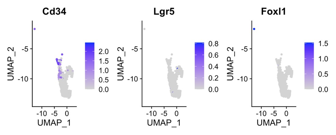
- I see the genetic validation of the role of telocytes or HGF signals are crucial to assure the significance of this manuscript. I recommend either of two experiments. a. testing the role of HGF signals by deleting the Hgf gene in telocytes. Using Wnt11-Cre; Hgf f/f mice, the authors could address the role of HGF signals in periodontitis. CX3CR1-Cre; cMet f/f mice will delete HGF signals in monocyte-derived macrophages. This will be another verification, but not sure if the PDL macrophages are derived from yolk sac or monocytes. b. measuring the contribution of telocytes in the homeostasis or disease progression. The mouse model could be challenging though, the system if achieved will be very informative. The authors could first check the expression of telocyte enriched genes, such as Lgr5 or Foxl1 reported previously in other tissue telocytes. Delete those genes under the Wnt1-Cre driver and check if telocyte lineage is removed. The system would be very useful for next-level study. DTA model could be an alternative, but Wnt1-Cre is vastly expressed in neural crest lineage.
These are good suggestions but unfortunately not feasible as we do not have all the mouse lines (e.g., Hgf f/f mice). Lgr5 and Foxl1 are used in intestine but is not suitable for PDL tissue. CD34;DTA show CD34+ cells, however, we encountered challenges associated with induced genetic heterogeneity when using this model, preventing us from making concrete conclusions from the experiments using the CD34;DTA model. Lgf5/Foxl1 are either not expressed or overlap with CD34 in and therefore do not seem suitable for us to pursue.
- This paper points out that the M1/M2 hybrid state of macrophages appears upon periodontitis. The authors could further characterize the hybrid macrophages by the expression of more markers, production of cytokines, and morphology. Need to clarify if this means some macrophages are in M1 state and others are in M2 state, or one macrophage possesses both M1 and M2 phenotype. Please conduct either FACS or immunofluorescence to demonstrate if one macrophage expresses both markers. Please introduce more information about the M1/M2 hybrid state of macrophage based on other present literature.
Unlike our single cell sequencing data, we were unsuccessful in determining if one macrophage possesses both M1 and M2 phenotype by immunolabelling.
- In the introduction part, the author lists several markers that can be used for telocyte identification, such as CD34+CD31-, CD34+c-Kit+, CD34+Vim+, CD34+PDGFRα+. Could authors explain why they chose CD34 CD31, but not other markers?
As shown in the cluster images below, the other markers do not overlap very well with CD34 cells or in the case of Vim, expressed more ubiquitously. We generated a new supplementary figure (Supp Fig2) and explained this in the text (page 12, lines 235-238).
- In figure 5g, I don't think the yellow color cell shows the reduction trend in the Tivantinib treatment group compared with a control group. Please validate the observation by gene expression analysis, WB, etc. In addition, please show c-Met+ cells level in the Tivantinib treatment group and control group.
New Supp Fig4 is included to show Met expression in homeostasis and periodontitis.
-
Evaluation Summary:
Drs Zhao and Sharpe have highlighted the role of a relatively understudied cell type, the telocyte, in periodontitis, using a mouse model. Periodontitis is a widely occurring inflammatory disease of the gums, that will eventually progress to bone resorption and teeth that are embedded less favorably and will eventually fall out. This disease is linked to many other illnesses, such as rheumatoid arthritis, cardiac disease and even Alzheimer's disease, so more in depth knowledge is needed on cell types that play a role in the progression of the disease.
(This preprint has been reviewed by eLife. We include the public reviews from the reviewers here; the authors also receive private feedback with suggested changes to the manuscript. Reviewer #3 agreed to share their name with the authors.)
-
Reviewer #1 (Public Review):
The authors have used many cleverly chosen mouse models (periodontitis models; various models that lead to an on-switch of genes) and methods (immune localizations of high quality; single cell RNA sequencing) for the quest of elucidating a role for telocytes. They describe that more telocytes are present around teeth in mice that had periodontitis. These cells proliferated, and they expressed a pattern of genes that allowed macrophages to differentiate into a different direction. In particular, they showed that telocytes in periodontitis express HGF, a molecule that steers macrophage differentiation towards a less inflammatory cell type, paving the way for recovery. As a weakness, one could state that an attempt to extrapolate to human cells is missing.
-
Reviewer #2 (Public Review):
Periodontitis is an inflammatory disease that results in bone and tooth loss. In this manuscript the authors analyzed the cell types and signaling pathways that constitute periodontitis compare to homeostasis situation by using single cell RNA sequencing of Molars in mouse periodontitis models. The Authors reveal Telocytes (interstitial cells), a cell type not previously identified in the periodontium, as a source for HGF signal which they claim to regulate macrophages behavior during periodontitis from M1(pro-inflammatory) to M2(anti-inflammatory) state. This discovery is important and may suggest new approaches to treat periodontitis. However, the conclusions are supported by inhibiting HGF signaling, systemically and not specifically from Telocytes, which may cause Telocyte-independent effects. In …
Reviewer #2 (Public Review):
Periodontitis is an inflammatory disease that results in bone and tooth loss. In this manuscript the authors analyzed the cell types and signaling pathways that constitute periodontitis compare to homeostasis situation by using single cell RNA sequencing of Molars in mouse periodontitis models. The Authors reveal Telocytes (interstitial cells), a cell type not previously identified in the periodontium, as a source for HGF signal which they claim to regulate macrophages behavior during periodontitis from M1(pro-inflammatory) to M2(anti-inflammatory) state. This discovery is important and may suggest new approaches to treat periodontitis. However, the conclusions are supported by inhibiting HGF signaling, systemically and not specifically from Telocytes, which may cause Telocyte-independent effects. In addition, following HGF signaling inhibition a moderate reduction in a M2 macrophages marker Arg1 was observed, which may suggest a change in macrophages behavior but would need to be carefully analyzed by in order to observe whether macrophages indeed changed to a M2 state. In summary, this study point on Telocytes as a source of HGF signal which regulate macrophages behavior in periodontitis however whether this has physiological relevance is still not clear.
-
Reviewer #3 (Public Review):
Zhao and Sharpe identified telocytes in the periodontium. To address their contribution to periodontal diseases, they conducted scRNA-seq analysis and lineage tracing in mice. They demonstrated that telocytes are activated in periodontitis. The activated telocytes send HGF signals to surrounding macrophages, converting M2 to M1/M2 hybrid status. The study implies that targeting telocytes and HGF signal for the potential treatment of periodontitis.
The significance of the study could be improved by authors testing if targeting telocytes or HGF signals could ameliorate periodontitis in the mouse model. The current form of the manuscript lacks the data that demonstrate the actual contribution of telocytes in the homeostasis of periodontium or progression of periodontitis.
Major comments:
I see the genetic …
Reviewer #3 (Public Review):
Zhao and Sharpe identified telocytes in the periodontium. To address their contribution to periodontal diseases, they conducted scRNA-seq analysis and lineage tracing in mice. They demonstrated that telocytes are activated in periodontitis. The activated telocytes send HGF signals to surrounding macrophages, converting M2 to M1/M2 hybrid status. The study implies that targeting telocytes and HGF signal for the potential treatment of periodontitis.
The significance of the study could be improved by authors testing if targeting telocytes or HGF signals could ameliorate periodontitis in the mouse model. The current form of the manuscript lacks the data that demonstrate the actual contribution of telocytes in the homeostasis of periodontium or progression of periodontitis.
Major comments:
I see the genetic validation of the role of telocytes or HGF signals are crucial to assure the significance of this manuscript. I recommend either of two experiments. a. testing the role of HGF signals by deleting the Hgf gene in telocytes. Using Wnt11-Cre; Hgf f/f mice, the authors could address the role of HGF signals in periodontitis. CX3CR1-Cre; cMet f/f mice will delete HGF signals in monocyte-derived macrophages. This will be another verification, but not sure if the PDL macrophages are derived from yolk sac or monocytes. b. measuring the contribution of telocytes in the homeostasis or disease progression. The mouse model could be challenging though, the system if achieved will be very informative. The authors could first check the expression of telocyte enriched genes, such as Lgr5 or Foxl1 reported previously in other tissue telocytes. Delete those genes under the Wnt1-Cre driver and check if telocyte lineage is removed. The system would be very useful for next-level study. DTA model could be an alternative, but Wnt1-Cre is vastly expressed in neural crest lienage.
This paper points out that the M1/M2 hybrid state of macrophages appears upon periodontitis. The authors could further characterize the hybrid macrophages by the expression of more markers, production of cytokines, and morphology. Need to clarify if this means some macrophages are in M1 state and others are in M2 state, or one macrophage possesses both M1 and M2 phenotype. Please conduct either FACS or immunofluorescence to demonstrate if one macrophage expresses both markers. Please introduce more information about the M1/M2 hybrid state of macrophage based on other present literature.
In the introduction part, the author lists several markers that can be used for telocyte identification, such as CD34+CD31-, CD34+c-Kit+, CD34+Vim+, CD34+PDGFRα+. Could authors explain why they chose CD34 CD31, but not other markers?
In figure 5g, I don't think the yellow color cell shows the reduction trend in the Tivantinib treatment group compared with a control group. Please validate the observation by gene expression analysis, WB, etc. In addition, please show c-Met+ cells level in the Tivantinib treatment group and control group.
-
