Infant brain regional cerebral blood flow increases supporting emergence of the default-mode network
Curation statements for this article:-
Curated by eLife

Evaluation Summary:
In this paper, the authors find a link between the emergence of functional connectivity (FC) and changes in regional Cerebral Blood Flow (rCBF) in human infancy from birth to 24 months of age, which will be of interest to the increasing field investigating how the establishment of the brain's functional organization is linked to neurodevelopmental and psychiatric conditions. The data quality and complementarity are impressive for infants over this developmental period (0-2 years). Most of the key claims of the manuscript are well supported by the data. However, the relatively sparse sample and cross-sectional nature does limit interpretation.
(This preprint has been reviewed by eLife. We include the public reviews from the reviewers here; the authors also receive private feedback with suggested changes to the manuscript. Reviewer #1 agreed to share their name with the authors.)
This article has been Reviewed by the following groups
Discuss this preprint
Start a discussion What are Sciety discussions?Listed in
- Evaluated articles (eLife)
Abstract
Human infancy is characterized by most rapid regional cerebral blood flow (rCBF) increases across lifespan and emergence of a fundamental brain system default-mode network (DMN). However, how infant rCBF changes spatiotemporally across the brain and how the rCBF increase supports emergence of functional networks such as DMN remains unknown. Here, by acquiring cutting-edge multi-modal MRI including pseudo-continuous arterial-spin-labeled perfusion MRI and resting-state functional MRI of 48 infants cross-sectionally, we elucidated unprecedented 4D spatiotemporal infant rCBF framework and region-specific physiology–function coupling across infancy. We found that faster rCBF increases in the DMN than visual and sensorimotor networks. We also found strongly coupled increases of rCBF and network strength specifically in the DMN, suggesting faster local blood flow increase to meet extraneuronal metabolic demands in the DMN maturation. These results offer insights into the physiological mechanism of brain functional network emergence and have important implications in altered network maturation in brain disorders.
Article activity feed
-

-

Author Response
Reviewer #1 (Public Review):
The actual description of the methods does not allow the reader to evaluate the precision of two important processing steps. First, rCBF measures are supposed to be restricted to the cortex, but given the pCASL image spatial resolution, partial volume effects with white matter probably exist, especially in younger infants. Furthermore, segmenting tissues on the basis of anatomical images (especially T1-weighted) is complicated in the first postnatal year. As rCBF measurements are very different between grey and white matter, the performed procedure might impact the measures at each age, or even lead to a systematic bias on age-dependent changes. Second, the methodology and accuracy of the brain registration across infants are little detailed whereas it is a challenging aspect given the …
Author Response
Reviewer #1 (Public Review):
The actual description of the methods does not allow the reader to evaluate the precision of two important processing steps. First, rCBF measures are supposed to be restricted to the cortex, but given the pCASL image spatial resolution, partial volume effects with white matter probably exist, especially in younger infants. Furthermore, segmenting tissues on the basis of anatomical images (especially T1-weighted) is complicated in the first postnatal year. As rCBF measurements are very different between grey and white matter, the performed procedure might impact the measures at each age, or even lead to a systematic bias on age-dependent changes. Second, the methodology and accuracy of the brain registration across infants are little detailed whereas it is a challenging aspect given the intense brain growth and folding, the changing contrast in T1w images at these ages, and the importance of this step to perform reliable voxelwise comparison across ages.
We thank the reviewer for this comment. We have added more descriptions in the methods to address this comment. Briefly, individual rCBF map was generated in the individual space and calibrated by phase contrast MRI to minimize the individual variations of processing parameters such as T1 of arterial blood (Aslan et al., 2010). Cortical segmentation was also conducted in individual space. Then different types of images including rCBF map and gray matter segmentation probability map in the individual space were normalized into the template space. An averaged gray matter probability map was generated after inter-subject normalization. After carefully testing multiple thresholds in the averaged gray matter probability maps, 40% probability minimizing the contamination of white matter and CSF while keeping the continuity of the cortical gray matter mask across the cerebral cortex was used to generate the binary gray matter mask shown on the left panel of Figure R1 below. Despite poor contrasts and poor cortical segmentation of T1-weighted images of younger infants rightfully pointed out by this reviewer, the poor cortical segmentation of younger infants was compensated by the averaged cortical mask and measurement of rCBF in the template space. As demonstrated in the right three panels in Figure R1, the rCBF measure in the cortical mask in the template space is consistent across ages for accurate and reliable voxelwise comparison across age.
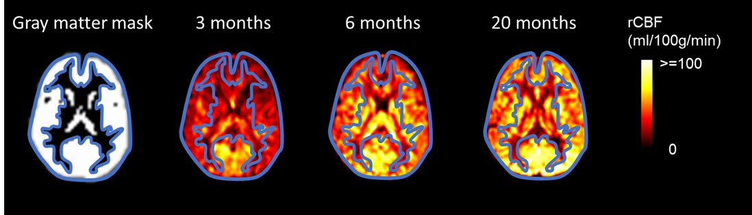
Figure R1. The gray matter mask and segmented cortical mask overlaid on rCBF map of three representative infants aged 3, 6, and 20 months in the template space. The gray matter mask on the left panel was created to minimize the contamination of white matter and CSF while keeping the continuity of the cortical gray matter mask across the cerebral cortex. The contour of the gray matter mask was highlighted with bule line.
The authors achieved their aim in showing that the rCBF increase differs across brain regions (the DMN showing intense changes compared to the visual and sensorimotor networks). Nevertheless, an analysis of covariance (instead of an ANOVA) including the infants' age as covariate (in addition to the brain region) would have allowed them to evaluate the interaction between age and region (i.e. different slopes of age-related changes across regions) in a more rigorous manner. Regarding the evaluation of the coupling between physiological (rCBF) and functional connectivity measures, the results only partly support the authors' conclusion. Actually, both measures strongly depend on the infants' age, as the authors highlight in the first parts of the study. Thus, considering this common age dependency would be required to show that the physiological and connectivity measurements are specifically related and that there is indeed a coupling.
We thank the reviewer for this comment. Following the reviewer’s suggestion, we conducted an analysis of covariance (ANCOVA) and found significant interaction between regions and age (F(6, 322) = 2.45, p < 0.05) with age as a covariate. This ANCOVA result is consistent with Figure 3c showing differential rCBF increase rates across brain regions. The ANCOVA result was added in the last paragraph in the Results section “Faster rCBF increases in the DMN hub regions during infant brain development”.
Regarding the evaluation of the coupling between physiological (rCBF) and functional connectivity measures (FC), the Figure 5, Figure 5–figure supplement 1 and 2 were generated exactly to test that the FC-rCBF coupling specifically localized in the DMN is not due to mutual age dependency. Briefly, Figure 5B demonstrated significant correlation only clustered in the DMN regions using the correlation method demonstrated in Figure 5-figure supplement 1. Furthermore, nonparametric permutation tests with 10,000 permutations were conducted. Such permutation tests are sensitive and effective with Figure 5c revealing significant coupling only in the DMN regions. If coupling is related to mutual age dependency, Figure 5c would demonstrate significant coupling in Vis and SM network regions too.
-
Evaluation Summary:
In this paper, the authors find a link between the emergence of functional connectivity (FC) and changes in regional Cerebral Blood Flow (rCBF) in human infancy from birth to 24 months of age, which will be of interest to the increasing field investigating how the establishment of the brain's functional organization is linked to neurodevelopmental and psychiatric conditions. The data quality and complementarity are impressive for infants over this developmental period (0-2 years). Most of the key claims of the manuscript are well supported by the data. However, the relatively sparse sample and cross-sectional nature does limit interpretation.
(This preprint has been reviewed by eLife. We include the public reviews from the reviewers here; the authors also receive private feedback with suggested changes to the …
Evaluation Summary:
In this paper, the authors find a link between the emergence of functional connectivity (FC) and changes in regional Cerebral Blood Flow (rCBF) in human infancy from birth to 24 months of age, which will be of interest to the increasing field investigating how the establishment of the brain's functional organization is linked to neurodevelopmental and psychiatric conditions. The data quality and complementarity are impressive for infants over this developmental period (0-2 years). Most of the key claims of the manuscript are well supported by the data. However, the relatively sparse sample and cross-sectional nature does limit interpretation.
(This preprint has been reviewed by eLife. We include the public reviews from the reviewers here; the authors also receive private feedback with suggested changes to the manuscript. Reviewer #1 agreed to share their name with the authors.)
-
Reviewer #1 (Public Review):
This study focuses on changes in regional cerebral blood flow (rCBF) during infancy, in relation to the emergence of the default mode network (DMN), a fundamental system associated with cognitive processes that are directed towards the self. The authors used cutting-edge complementary MRI modalities allowing them to measure rCBF (with pseudo-continuous arterial spin labelling -pCASL- and phase-contrast -PC- MRI) and functional connectivity (with resting-state -rs- functional MRI) in 48 sedated infants aged between 0 and 24 months. They first showed that rCBF dynamics differ across functional networks, with high changes in cortical regions supporting the DMN. Their analyses further aimed to reveal a coupling between the increase in rCBF in these regions and the increased functional connectivity strength …
Reviewer #1 (Public Review):
This study focuses on changes in regional cerebral blood flow (rCBF) during infancy, in relation to the emergence of the default mode network (DMN), a fundamental system associated with cognitive processes that are directed towards the self. The authors used cutting-edge complementary MRI modalities allowing them to measure rCBF (with pseudo-continuous arterial spin labelling -pCASL- and phase-contrast -PC- MRI) and functional connectivity (with resting-state -rs- functional MRI) in 48 sedated infants aged between 0 and 24 months. They first showed that rCBF dynamics differ across functional networks, with high changes in cortical regions supporting the DMN. Their analyses further aimed to reveal a coupling between the increase in rCBF in these regions and the increased functional connectivity strength within this network, to suggest relationships between physiological mechanisms and the emergence of functional networks.
* Major strengths of the study:
Relating physiological changes such as the increase in CBF across cortical regions and the development of functional connections supporting cognitive brain networks is a question that has been little asked until now because of the difficulty of acquiring such complementary data in infants. It is nevertheless highly relevant, especially in the context of the DMN development, which is known to be intense in early childhood. This study relies on reliable multimodal data, measured with robust and complementary methodologies, despite the challenge of studying such population. This allowed the authors to highlight spatiotemporal differences in rCBF during infancy, in particular across the DMN regions compared to the visual and sensorimotor networks. The provided analyses support the presented results.* Major weaknesses of the methods and results:
The actual description of the methods does not allow the reader to evaluate the precision of two important processing steps. First, rCBF measures are supposed to be restricted to the cortex, but given the pCASL image spatial resolution, partial volume effects with white matter probably exist, especially in younger infants. Furthermore, segmenting tissues on the basis of anatomical images (especially T1-weighted) is complicated in the first postnatal year. As rCBF measurements are very different between grey and white matter, the performed procedure might impact the measures at each age, or even lead to a systematic bias on age-dependent changes.
Second, the methodology and accuracy of the brain registration across infants are little detailed whereas it is a challenging aspect given the intense brain growth and folding, the changing contrast in T1w images at these ages, and the importance of this step to perform reliable voxelwise comparison across ages.* The authors achieved their aim in showing that the rCBF increase differs across brain regions (the DMN showing intense changes compared to the visual and sensorimotor networks). Nevertheless, an analysis of covariance (instead of an ANOVA) including the infants' age as covariate (in addition to the brain region) would have allowed them to evaluate the interaction between age and region (i.e. different slopes of age-related changes across regions) in a more rigorous manner.
Regarding the evaluation of the coupling between physiological (rCBF) and functional connectivity measures, the results only partly support the authors' conclusion. Actually, both measures strongly depend on the infants' age, as the authors highlight in the first parts of the study. Thus, considering this common age dependency would be required to show that the physiological and connectivity measurements are specifically related and that there is indeed a coupling.* As a whole, this work will have an important impact on the field, as this is a seminal study on the physiological bases of the emergence of functional networks during child development. In addition, both the data and described MRI methods are original and rare.
-
Reviewer #2 (Public Review):
In this work the authors sought to explore the relationship between changes in regional cerebral blood flow (rCBF) and functional connectivity (FC) in human infants across the first 24 months after birth. Understanding this is important, as it is increasingly appreciated that the brain's lifelong framework of functional connections is established across this period, with alterations seemingly associated with the development of conditions such as autism later in life. They show a relationship between developmental increases in rCBF with maturational changes in FC specific to the default mode network (DMN), a functional network which is known to rapidly mature during this period. Interestingly, this relationship was not significant when studying the primary visual and sensorimotor networks which are thought to …
Reviewer #2 (Public Review):
In this work the authors sought to explore the relationship between changes in regional cerebral blood flow (rCBF) and functional connectivity (FC) in human infants across the first 24 months after birth. Understanding this is important, as it is increasingly appreciated that the brain's lifelong framework of functional connections is established across this period, with alterations seemingly associated with the development of conditions such as autism later in life. They show a relationship between developmental increases in rCBF with maturational changes in FC specific to the default mode network (DMN), a functional network which is known to rapidly mature during this period. Interestingly, this relationship was not significant when studying the primary visual and sensorimotor networks which are thought to be already mature at birth.
The combination of measuring both rCBF and FC with pCASL and BOLD fMRI in this population is relatively novel, and would certainly have been challenging data to acquire which is a clear strength. That they validate the assumed (but largely not proven) link between rCBF and FC during development will be of use to the field, although this result is perhaps not surprising and given the cross sectional nature of the data, unfortunately, does not really provide further mechanistic insight into the relationship. The authors' suggestion that the results for example can tell us about brain metabolism is rather speculative and is not supported by the data (as they have no additional measure of oxygen extraction or glucose metabolism).
Due to the difficulties with infant cooperation (and movement), it is extremely rare to be able to measure rCBF in this population, so the results will be of interest in this context, particularly as they demonstrate that regional differences, with the strongest correlation with age appearing to be frontally and across the midline. Providing the CBF data and trajectories will be of great use to the field where it can be compared to other maturational brain changes identified in large studies such as the HBCD cohort. However, there is some caution about these results, as these were all infants who had imaging performed for clinical reasons (albeit with normal neurology) who received sedation for the scan. Furthermore, given the rapid development across this period (which is, of course, the major focus of the work), a further potential drawback is that the same acquisition sequence was used across all of the infants. This likely has implications, particularly for the rCBF estimation - as key factors such as partial voluming effects, the T1 of arterial blood, and arterial transit time are all rapidly developing across this time.
The FC analysis methods are largely appropriate and are in line with the broader literature which shows established connectivity in primary networks, but the apparent establishment of long distance organisation within the DMN (which is neatly shown in figure 3). The authors show that in a number of different ways, the identified relationships between rCBF and FC are network specific, which strengthens their conclusions that the two are linked in the developing brain. However, interpretation is somewhat limited by their approach of only considering three networks which have been empirically derived from the older infants.
-
Reviewer #3 (Public Review):
The authors evaluated the connection between regional blood flow changes and local cortical maturation in 48 human subjects aged 0-24 months, focusing on the default mode network, visual cortex, and somatomotor cortex. The major strength involves the use of sophisticated MR methods to non-invasively measure blood flow and cortical network maturation. The results are consistent with their conclusion that local cerebral flow increases to meet the demands of cortical maturation in regions undergoing rapid development, though other interpretations of the data are also possible. Their approach has the potential to improve our understanding of neurovascular coupling in the context of maturation.
-
