Single-cell and spatial transcriptomic analyses reveal the dynamic transcript profiles of myocardial lymphangiogenesis post-myocardial infarction
Curation statements for this article:-
Curated by eLife

eLife Assessment
This study presents useful albeit preliminary findings on transcriptome changes in cardiac lymphatic cells after myocardial infarction in mice. Despite revision, the conclusions of the authors remain uncertain as sample sizes in general are very low, and even sometimes too low to allow for valid statistical comparisons. Accordingly, there are concerns regarding statistical robustness, raised by both the editors and the reviewers. While the single-cell transcriptomic data were analyzed using solid advanced methodology, too few cells were included in the scRNA-seq data set and the spatial transcriptomics analyses. Thus, this study rather represents more a collection of preliminary transcriptomic data than a full scientific report that would definitively advance the field.
This article has been Reviewed by the following groups
Discuss this preprint
Start a discussion What are Sciety discussions?Listed in
- Evaluated articles (eLife)
Abstract
Background:
Cardiac lymphatics play an important role in myocardial edema and inflammation, however, the heterogeneity of cardiac lymphatic endothelial cells (LECs) and their biological functions have rarely been investigated.
Methods:
This study integrated single-cell sequencing data and spatial transcriptome data from mouse heart tissue at different time points post-myocardial infarction (MI), and then revealed LECs heterogeneity and biological functions by clustering, spatial localization, cell trajectory, and Cell-Chat analyses.
Results:
Four transcriptionally distinct subtypes of LECs were identified and localized in space. Cardiac LEC subgroups were found to be localized in different zones of infarcted heart related to different functions. LEC capillary III (LEC CaIII) may be involved in the direct regulation of myocardial injuries in an infarcted zone (IZ) from the perspective of metabolic stress, while LEC CaII may be related to the rapid immune inflammatory responses of the border zone (BZ) in the early stage of MI. LEC CaI, as well as LEC collection mainly participate in the regulation of myocardial tissue edema resolution in the middle and late stages post-MI. Cell trajectory and Cell-Chat analyses further identified that LECs may regulate myocardial edema through Aquaporin 1, and might affect the infiltration of macrophages through the galectin-9 (Gal-9)-CD44 pathway.
Conclusion:
This study revealed the dynamic transcriptional heterogeneity distribution of LECs in different regions of the infarcted heart, and these LECs formed different functional subgroups that might exhibit different bioeffects in myocardial tissue post-MI.
Article activity feed
-
-

-
-

eLife Assessment
This study presents useful albeit preliminary findings on transcriptome changes in cardiac lymphatic cells after myocardial infarction in mice. Despite revision, the conclusions of the authors remain uncertain as sample sizes in general are very low, and even sometimes too low to allow for valid statistical comparisons. Accordingly, there are concerns regarding statistical robustness, raised by both the editors and the reviewers. While the single-cell transcriptomic data were analyzed using solid advanced methodology, too few cells were included in the scRNA-seq data set and the spatial transcriptomics analyses. Thus, this study rather represents more a collection of preliminary transcriptomic data than a full scientific report that would definitively advance the field.
-
Reviewer #1 (Public review):
Summary:
Assessment of cardiac LEC transcriptomes post-MI may yield new targets to improve lymphatic function. scRNAseq is a valid approach as cardiac LECs are rare compared to blood vessel endothelial cells.
Strengths:
Extensive bioinformatics approaches employed by the group
Weaknesses:
Too few cells included in scRNAseq data set and the spatial transcriptomics data that was exploited has little relevance, or rather specificity, for cardiac lymphatics. This study seems more a collection of preliminary transcriptomic data than a true scientific report to help advance the field.
Comments on revisions:
Thank you for the revision that helps clarify some outstanding questions.
(1) I still have questions relating to the relevance of the spatial maps generated and shown in fig 3C. They are supposedly generated …
Reviewer #1 (Public review):
Summary:
Assessment of cardiac LEC transcriptomes post-MI may yield new targets to improve lymphatic function. scRNAseq is a valid approach as cardiac LECs are rare compared to blood vessel endothelial cells.
Strengths:
Extensive bioinformatics approaches employed by the group
Weaknesses:
Too few cells included in scRNAseq data set and the spatial transcriptomics data that was exploited has little relevance, or rather specificity, for cardiac lymphatics. This study seems more a collection of preliminary transcriptomic data than a true scientific report to help advance the field.
Comments on revisions:
Thank you for the revision that helps clarify some outstanding questions.
(1) I still have questions relating to the relevance of the spatial maps generated and shown in fig 3C. They are supposedly generated using a 'molecular finger print' specific to each sub-cluster of LECs. However, given that at early stages postMI most populations are exceedingly rare in your analyses, could you please explain or comment on the relevance of the spatial maps?
(2) Fig 3 s1 would indicate that the population CaII is the majoritarian one in healthy hearts, while quantifications in Fig 3A show that rather the LEC Co subpopulation is majoritarian. Further, in mouse hearts histological analyses have demonstrated that cardiac lymphatics are restricted to the outer layers of the heart. This is not seen in your spatial maps. This seems to be the case only for the LEc Co population in healthy hearts, but not for other subpopulation signatures. Please explain.
(3) Further, the population of CaI, with 1 cell analysed in d3, but appears very prevalent in the spatial maps at d3. Please explain.
(4) In your list of 12 genes used as matrix anchors to identify LEC subpopulations in your screens, it is not apparent how LEC CaI, II and III differ so much as to allow selective detection of subpopulations. This similitude of profiles is supported by Fig 2F, and further explanations are needed to explain how the spatial maps of LEC ca subpopulations appear as distinct as shown in fig 3 S1 and Fig 3C.
-
Reviewer #2 (Public review):
Summary:
This study integrated single-cell sequencing and spatial transcriptome data from mouse heart tissue at different time points post-MI. They identified four transcriptionally distinct subtypes of lymphatic endothelial cells and localized them in space. They observed that LECs subgroups are localized in different zones of infarcted heart with functions. Specifically, they demonstrated that LEC ca III may be involved in directly regulating myocardial injuries in the infarcted zone concerning metabolic stress, while LEC ca II may be related to the rapid immune inflammatory responses of the border zone in the early stage of MI. LEC ca I and LEC collection mainly participate in regulating myocardial tissue edema resolution in the middle and late stages post-MI. Finally, cell trajectory and Cell-Chat …
Reviewer #2 (Public review):
Summary:
This study integrated single-cell sequencing and spatial transcriptome data from mouse heart tissue at different time points post-MI. They identified four transcriptionally distinct subtypes of lymphatic endothelial cells and localized them in space. They observed that LECs subgroups are localized in different zones of infarcted heart with functions. Specifically, they demonstrated that LEC ca III may be involved in directly regulating myocardial injuries in the infarcted zone concerning metabolic stress, while LEC ca II may be related to the rapid immune inflammatory responses of the border zone in the early stage of MI. LEC ca I and LEC collection mainly participate in regulating myocardial tissue edema resolution in the middle and late stages post-MI. Finally, cell trajectory and Cell-Chat analyses further identified that LECs may regulate myocardial edema through Aqp1, and likely affect macrophage infiltration through the galectin9-CD44 pathway. The authors concluded that their study revealed the dynamic transcriptional heterogeneity distribution of LECs in different regions of the infarcted heart and that LECs formed different functional subgroups that may exert different bioeffects in myocardial tissue post-MI.
Strengths:
The study addresses a significant clinical challenge, and the results are of great translational value. All experiments were carefully performed, and their data support the conclusion.
-
Editors' comments (Public review):
Weaknesses:
(1) Figure 7C, 7E, 7I, and "Figure7-figure supplement 1 ": All data in these data panels are based on only n=3, which is insufficient. Sample sizes of n=3 are too low to correctly assess normality of distribution and, as a consequence, do not allow to select the appropriate parametric/non-parametric tests. Accordingly, no statistical comparison can be performed and all p values and symbols currently indicating statistically significant differences between groups must be removed.
(2) Figure 3A, 3B, or 3C: No information about n numbers per group. Should n numbers per group be n=4 or less, no statistical comparison can be performed and all p values and symbols indicating statistically significant differences between groups must be removed.
(3) Figure 4 E and 4F: No information about n numbers …
Editors' comments (Public review):
Weaknesses:
(1) Figure 7C, 7E, 7I, and "Figure7-figure supplement 1 ": All data in these data panels are based on only n=3, which is insufficient. Sample sizes of n=3 are too low to correctly assess normality of distribution and, as a consequence, do not allow to select the appropriate parametric/non-parametric tests. Accordingly, no statistical comparison can be performed and all p values and symbols currently indicating statistically significant differences between groups must be removed.
(2) Figure 3A, 3B, or 3C: No information about n numbers per group. Should n numbers per group be n=4 or less, no statistical comparison can be performed and all p values and symbols indicating statistically significant differences between groups must be removed.
(3) Figure 4 E and 4F: No information about n numbers per group. Should n numbers per group be n=4 or less, no statistical comparison can be performed and all p values and symbols indicating statistically significant differences between groups must be removed.
(4) Figure 5: No information about n numbers per group is provided. Should n numbers per group be n=4 or less, no statistical comparison can be performed and all p values and symbols indicating statistically significant differences between groups must be removed.
-
Author response:
The following is the authors’ response to the previous reviews.
Reviewer #1 (Recommendations for The Authors):
Q1: In response to reviewers you noted totally 292 sequenced LECs, however in reviewer figure 3 B the numbers seem to add up to 221. Please include mention of the total number of LEC sequences. Please mention line 119, page 4 the total number of explored LEC transcriptomes
Thank you for your carefully review. We have updated Fig 2A, 2C and 2E. It was 242 (not 292) LECs included in our initial analysis, which contains the sample of d5 post MI in raw data (E-MTAB-7895). We dropped d5 in our subsequent analysis because the change in d5 did not significant differ from d3. Therefore, we included 221 LECs in our final analysis as we updated in Fig2A, 2C and 2E.
Q2-1: Figure 3A supposedly shows % of LEC …
Author response:
The following is the authors’ response to the previous reviews.
Reviewer #1 (Recommendations for The Authors):
Q1: In response to reviewers you noted totally 292 sequenced LECs, however in reviewer figure 3 B the numbers seem to add up to 221. Please include mention of the total number of LEC sequences. Please mention line 119, page 4 the total number of explored LEC transcriptomes
Thank you for your carefully review. We have updated Fig 2A, 2C and 2E. It was 242 (not 292) LECs included in our initial analysis, which contains the sample of d5 post MI in raw data (E-MTAB-7895). We dropped d5 in our subsequent analysis because the change in d5 did not significant differ from d3. Therefore, we included 221 LECs in our final analysis as we updated in Fig2A, 2C and 2E.
Q2-1: Figure 3A supposedly shows % of LEC subpopulations relative to their numbers found in day 0 samples. However, there seem to be some errors, because for example the subpop LEC Cap I include 13 cells day 1 and 6 cells day 1, which corresponds to 46% of initial numbers. However, from your graph 3B the blue population seems to occupy 10%. Please revise or explain how these relative % were calculated.
Thank you for your question. In the Figure 3A, each column was calculated by dn/d0*100%, that is d0=57/57*100%=100%, and d1= 21/57*100%=36.84%, d3=9/57*100%=15.79%, d7, d14, d28...Therefor, Cap I in d0 (13 cells) is 13/57*100%=22.81%, and Cap I in d1(6 cells) is 6/57*100%= 10.53%.
Q2-2: Further, based on the relative % of LEC subpopulations, using the numbers mentioned in Fig 3B, it would appear that the relative frequency LEC cap II population is actually stable at around 20-30% of all LECs per time point throughout the study (except day 1 drop). This contrasts with line 136 p. 4 statement. I would also urge caution for interpreting too much into the variation of relative levels of LEC co, as these represent exceeding rare cells in your samples, and could reflect technical issues rather than true biological variation (total LEC co numbers analyzed ranging from 1-24 cells/ time point). The same could be said of LEC cap II and cap III.
We strongly agree with your comment on the proportion of LEC cell subtypes post MI. As you pointed out, we have revised the result description on Page 4, line 137-143 as followed.
“In the early stages of myocardial infarction (D1 and D3), the quantity of LECs decreased sharply. The number of LECs gradually increasing from day 7 and returning to normal levels by day 14 after MI. Moreover, from day 14 onwards, the number and proportion of Ca I type LECs significantly increased.”
Q3: Please list in supplement the gene features used to identify in spatial transcriptomics the different LEC subpopulations, as their profiles (notably for capillary LECs) don't appear to be very different based on data in Fig 2F.
We have supplied gene features in supplementary materials.
Q4: In section 2.7 you refer to Gal9 secretion. Please replace with expression as no measure of protein levels from LECs has been described in your study.
Thank you for your suggestion, we have replaced secretion with expression.
Q5: The updated method to exclude non-lymphatic cells from lymphatic vessel analyses by incorporating pdpn as an additional marker ('present costained areas wherever possible' line 350 p 10)
Thank you for your correction. We have updated the description as follows and lighted them in the manuscript: rabbit anti-Lyve1 (1:300, ab14917, Abcam, UK), [Syrian hamster anti-Podoplanin (1:100, 53-5381-82, Thermo, USA), rabbit anti-Prox1(1:300, ab199359, Abcam, UK), both anti-podoplain and anti-prox1 are additional markers co-stained with Lyve1 to exclude non-lymphatic cells from lymphatic vessel].
Q6: Fig 1B, it is highly surprising to see the lymphatic density in the BZ go from 25 um² at day 3 to more than 1000 um² only four days later (day 7). Is it possible that your day 3 measurements were in the infarct area, and not BZ area? The H&E image shown in Fig1a for d3 sample would seem to indicate the analysis was done in a dead area, rather than BZ. Please revise (perhaps select similar zone as shown for d1 in fig 6D, adjusted for subepicardial region and not mid-myocardial as seems to be the case currently), and also provide lymphatic area measures in healthy myocardium for day 0 samples. The unit used (um²) also would depend on the size of the area examined. Is this unit per image? If so please report total imaged area as a reference.
A6: Thank you for your reminding and advises. We have labeled each zone on H&E and IF images in Fig1-supplementary Fig2B, and updated a clearer histological photo taken at 3 days post MI in Fig1A. Furthermore, we recalculated the lymphatic vessel area ratio as you suggested by calculating the ratio of LEC co-stained area to total imaged area under 100-fold magnification.
Q7: The mention that CD68 antibody isn't compatible with lyve1 antibody could easily have been bridged by using other macrophage markers, such as F4/80, which is readily available and often used marker for macs in mice and comes notably as a rat anti-mouse F4-80. It would have added much more relevant information to exclude Lyve1-/F4/80+ cells as compared to the current analysis, which may indeed include in area measures Lyve1+ /Pdpn- single cells erroneously spotted as 'lymphatic vessels'
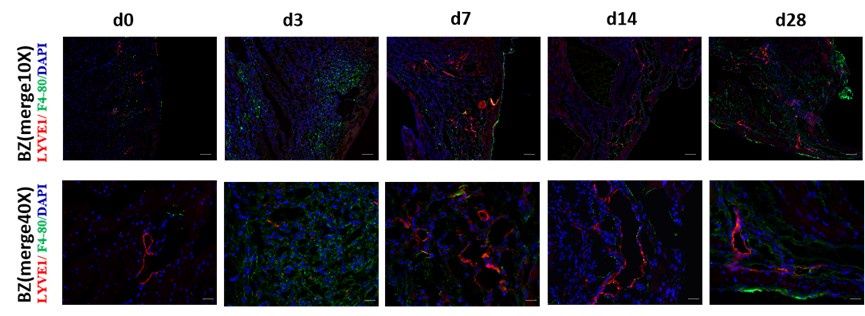
Thank you for your excellent suggestion. We co-stained the sample with F4/80 and LYVE1 and supplied in the Fig1-supplementary Figure 1E, as shown in Author response image 1.
Author response image 1.
Immunofluorescence (IF) co-staining of tissue section with F4/80 and LYVE1 in sham and MI mice model at d3, d7, d14, and d28 post-MI. LYVE1: lymphatic vessel endothelial hyaluronan receptor 1; DAPI: 4’6-diamidino-2-phenylindole; scale bar in 10×-100 μm, 40×-25μm.
Reviewer 2 (Recommendations for The Authors):
Q1: Language expression must be improved. Many incomplete sentences exist throughout the manuscript. A few examples: Line 70-71: In order to further elucidate the effects and regulatory mechanisms of the lymphatic vessels in the repair process of myocardial injury following MI. Line 71-73. This study, integrated single-cell sequencing and spatial transcriptome data from mouse heart tissue at different timepoints after MI from publicly available data (E-MTAB-7895, GSE214611) in the ArrayExpress and gene expression omnibus (GEO) databases. Line 88-89: Since the membrane protein LYVE1 can present lymphatic vessel morphology more clearly than PROX1.
Thank you for your correction. We have carefully inspected and corrected the whole manuscript.
Q2: The type of animal models (i.e., permanent MI or MI plus reperfusion) included in Array Express and gene expression omnibus (GEO) databases must be clearly defined as these two models may have completely different effects on lymphatic vessel development during post-MI remodeling.
Thank you for your excellent suggestion. The animal models used in both E-MTAB-7895 and GSE214611 are permanent MI. We have modified the model information in the methodology section (page 12, line 400-401).
Q3: Line 119-120: Caution must be taken regarding Cav1 as a lymphocyte marker because Cav1 is expressed in all endothelial cells, not limited to LEC.
Thanks for your reminding. Cav 1 used in our clustering is one of the marker gene for its different expression in sub-types of LECs, referred in article PMID: 31402260
Q4: Figure 1 legend needs to be improved. RZ, BZ, and IZ need to be labeled in all IF images. Day 0 images suggest that RZ is the tissue section from the right ventricle.
Thank you for your suggestion. We have labeled and updated the regions of RZ, BZ, and IZ in H&E and IF image in Figure1-Figure supplement 2B.
Q5: The discussion section needs to be improved and better focused on the findings from the current study.
Thank you for your good comment. Based on your suggestion, we have revised the first paragraph of the discussion from lines 250-256 (Page 7) as followed:
Cardiac lymphatics play an important role in myocardial edema and inflammation. This study, for the first time, integrated single-cell sequencing data and spatial transcriptome data from mouse heart tissue at different time points of post-MI, and identified four transcriptionally distinct subtypes of LECs and their dynamic transcriptional heterogeneity distribution in different regions of myocardial tissue post-MI. These subgroups of LECs were shown to form different function involved in the inflammation, apoptosis, ferroptosis, and water absorption related regulation of vasopressin during the process of myocardial repair after MI.
-
-
eLife Assessment
This study presents useful, yet preliminary, findings on the transcriptomic changes in cardiac lymphatic cells after myocardial infarction in mice. The conclusions of the authors remain uncertain as sample sizes for lymphatic endothelial cells are very low. The single-cell transcriptomic data were analyzed using solid advanced methodology and may be used as a starting point for future studies of the impact of lymphatic cells on heart disease.
-
Reviewer #1 (Public review):
Summary:
Assessment of cardiac LEC transcriptomes post-MI may yield new targets to improve lymphatic function. scRNAseq is a valid approach as cardiac LECs are rare compared to blood vessel endothelial cells.
Strengths:
Extensive bioinformatics approaches employed by the group
Weaknesses:
Too few cells included in scRNAseq data set and the spatial transcriptomics data that was exploited has little relevance, or rather specificity, for cardiac lymphatics. This study seems more a collection of preliminary transcriptomic data than a true scientific report to help advance the field.
-
Reviewer #2 (Public review):
Summary:
This study integrated single-cell sequencing and spatial transcriptome data from mouse heart tissue at different time points post-MI. They identified four transcriptionally distinct subtypes of lymphatic endothelial cells and localized them in space. They observed that LECs subgroups are localized in different zones of infarcted heart with functions. Specifically, they demonstrated that LEC ca III may be involved in directly regulating myocardial injuries in the infarcted zone concerning metabolic stress, while LEC ca II may be related to the rapid immune inflammatory responses of the border zone in the early stage of MI. LEC ca I and LEC collection mainly participate in regulating myocardial tissue edema resolution in the middle and late stages post-MI. Finally, cell trajectory and Cell-Chat …
Reviewer #2 (Public review):
Summary:
This study integrated single-cell sequencing and spatial transcriptome data from mouse heart tissue at different time points post-MI. They identified four transcriptionally distinct subtypes of lymphatic endothelial cells and localized them in space. They observed that LECs subgroups are localized in different zones of infarcted heart with functions. Specifically, they demonstrated that LEC ca III may be involved in directly regulating myocardial injuries in the infarcted zone concerning metabolic stress, while LEC ca II may be related to the rapid immune inflammatory responses of the border zone in the early stage of MI. LEC ca I and LEC collection mainly participate in regulating myocardial tissue edema resolution in the middle and late stages post-MI. Finally, cell trajectory and Cell-Chat analyses further identified that LECs may regulate myocardial edema through Aqp1, and likely affect macrophage infiltration through the galectin9-CD44 pathway. The authors concluded that their study revealed the dynamic transcriptional heterogeneity distribution of LECs in different regions of the infarcted heart and that LECs formed different functional subgroups that may exert different bioeffects in myocardial tissue post-MI.
Strengths:
The study addresses a significant clinical challenge, and the results are of great translational value. All experiments were carefully performed, and their data support the conclusion.
Weaknesses:
(1) Language expression must be improved. Many incomplete sentences exist throughout the manuscript. A few examples: Line 70-71: In order to further elucidate the effects and regulatory mechanisms of the lymphatic vessels in the repair process of myocardial injury following MI. Line 71-73. This study, integrated single-cell sequencing and spatial transcriptome data from mouse heart tissue at different time points after MI from publicly available data (E-MTAB-7895, GSE214611) in the ArrayExpress and gene expression omnibus (GEO) databases. Line 88-89: Since the membrane protein LYVE1 can present lymphatic vessel morphology more clearly than PROX1.
(2) The type of animal models (i.e., permeant MI or MI plus reperfusion) included in ArrayExpress and gene expression omnibus (GEO) databases must be clearly defined as these two models may have completely different effects on lymphatic vessel development during post-MI remodeling.
(3) Line 119-120: Caution must be taken regarding Cav1 as a lymphocyte marker because Cav1 is expressed in all endothelial cells, not limited to LEC.
(4) Figure 1 legend needs to be improved. RZ, BZ, and IZ need to be labeled in all IF images. Day 0 images suggest that RZ is the tissue section from the right ventricle. Was RZ for all other time points sampled from the right ventricular tissue section?
(5) The discussion section needs to be improved and better focused on the findings from the current study. -
Author response:
The following is the authors’ response to the original reviews.
Reviewer #1 (Recommendations for The Authors):
Q1: Please replace lymphocytes with lymphatic endothelial cells throughout the manuscript.
A1: Thank you for your conscientious review. Per your suggestion, we have replaced “lymphocytes” with “lymphatic endothelial cells (LECs)” throughout the manuscript.
Q2: Please re-analyse lymphatics using LYVE1 and CD68 or another macrophage marker, as Lyve1 is NOT specific for lymphatics.
A2: Thank you for your suggestion. We completely agree with your opinion. Because both the CD68 (CST,97778S) and LYVE1 antibodies (Abcam,ab14917) are rabbit multiclonal antibodies and to more accurately label cardiac lymphatics, we performed immunofluorescence co-staining using LYVE1 and PDPN antibodies (Thermo,53-5381-82) and …
Author response:
The following is the authors’ response to the original reviews.
Reviewer #1 (Recommendations for The Authors):
Q1: Please replace lymphocytes with lymphatic endothelial cells throughout the manuscript.
A1: Thank you for your conscientious review. Per your suggestion, we have replaced “lymphocytes” with “lymphatic endothelial cells (LECs)” throughout the manuscript.
Q2: Please re-analyse lymphatics using LYVE1 and CD68 or another macrophage marker, as Lyve1 is NOT specific for lymphatics.
A2: Thank you for your suggestion. We completely agree with your opinion. Because both the CD68 (CST,97778S) and LYVE1 antibodies (Abcam,ab14917) are rabbit multiclonal antibodies and to more accurately label cardiac lymphatics, we performed immunofluorescence co-staining using LYVE1 and PDPN antibodies (Thermo,53-5381-82) and re-measured the lymphatic vessel area using the Image J software (version 1.53). The result is shown in Figure 1A and 1B. Further, we performed co-staining with PDPN and CD68 to observe the relationship between macrophage and cardiac lymphatic vessel distributions at different time points post-myocardial infarction (MI) (Figure1-figure supplement 1F). Per your comment, some LYVE1 markers are positive, whereas PDPN markers may be negative for macrophages in the heart tissue. We have added notes on the catalog numbers of anti-PDPN and anti-CD68 in the methods (Page 10, Lines 351‒352) and updated them in the KRT template and MDAR checklist.
Q3: Rephrase title 2.6, 2.7 to fit the results in these sections that are purely descriptive and do not add any insight into the functional relevance of the findings.
A3: Thank you for your suggestion. We have rephrased titles 2.6 and 2.7 as follows:
2.6 AQP1 in LEC is correlated with myocardial edema occurrence and resolution post-MI.
2.7 Gal9 secreted by LEC can affect macrophage migration.
Q4: Please refrain from extensive discussion of non-significant findings, such as Figures 6D, and 7A, B, and M (ifng vs ifng + antiGal9 is n.s).
A4: Thank you for your suggestions. Lymphatic endothelial cells (LECs) are a type of cell that exists in the myocardial tissue in small quantities. Owing to the extremely small number of LECs, elucidating their biological functions and regulation may be challenging during MI. To gain a deeper understanding of the role of the lymphatic system post-MI, we attempted to analyze the transcriptomic changes of LEC subsets at different time points after MI by combining single-cell sequencing and spatial transcriptomics data. We have selected relevant molecules with significant differences in transcription levels and conducted the validation analysis in LECs at different time points after MI. Among them, AQP1 and GAL9 showed significant differences. CD44, as a receptor for GAL9, showed significant differences in its expression in macrophages at different time points after MI. Therefore, we have added the relevant information to the discussion section (marked with yellow) on Page 9, Lines 299‒312.
Q5: Please explain the method used to calculate lymphatic areas in Figure 1.
A5: Thank you for your observation. The method we used is consistent with that described in previous studies[1,2]. (PMID: 30582443 and PMID: 32404007). The detailed methods have been described in the Methods as follows (Page 10, Lines 358‒363):
For quantification of vessel area, vessels with visible co-staining were measured using Image J software. First, we selected an image, turned it into 8-bit, and then applied a suitable threshold adjustment (present co-stained areas wherever possible). Second, five equally sized squares were selected in the respective zones (remote, infarct, and border zones) of each slice. ROI manager tools were used to analyze the automatic signal intensity quantification by the software in the area inside this square. Finally, the GraphPad software was used to plot the results as a bar graph.
Q6: In Figure 1 supp C, the upper and lower panels don't seem to have the same zoom factor.
A6: Thank you for pointing this out. The upper and lower images in Figure S1C have the same magnification. To facilitate your review, we have added a 1× image and re-labeled the position and scale information of the image. The revised Figure S1C was added to the manuscript and is shown as follows:
Q7: In Figure 2d please include aqp1 among displayed genes.
A7: Thank you for your suggestion. The Aqp1 gene is already displayed in the 11th, and we have labeled it.
Q8: In Figure 2f include markers of LECs such as Prox1, Flt4, Itga9, and also show Aqp1 here.
A8: Thank you for your valuable comment. We have updated Figure 2f.
Q9: Please indicate in Figure 3a what the y axis means? % of total LECs? % of total LECs at a given time point? The data is really not clear.
A9: Thank you for your suggestion. The y-axis represents the percentage of the total number of LECs at d1, d3, d7, d14, and d28 post-MI, relative to the number of LECs at d0, which is used as the reference value set at 100%. Meanwhile, different colors were applied to represent the proportion of different cell subtypes at different time points. We have updated Figure 3a.
Q10:Add n of LECs per time points in Figures 3a and b.
A10: Thank you for your suggestion. We have updated Figure 3b.
Q11: For Figure 3c please explain what marker genes were used to identify LEC enriched areas. What was the spatial resolution of the transcriptomic screens? How do these images relate to the localization of lymphatics in the heart?
A11: We appreciate your observation. We have added the required information to the Methods on Page 13, Lines 442‒448, as follows:
“We conducted spatial transcriptome data analysis using the deconvolution algorithm. The deconvolution algorithm refers to the application of feature genes to infer the full matrix information of single-cell transcriptome of cell subclusters. We then compared and anchored the matrix information of the single-cell transcriptome with the information of each SPOT in the spatial transcriptome, predicting cell types based on the similarity between the two sets of information.”
Q12:Figure 6 explains the y-axis in panel A, the timepoint in panel G, and absence of aqp1 staining in blood vessels in images d1 and d3 in panel D.
A12: Thank you for your suggestion. The y-axis in Figure 6A (Figure to reviewer 7A) shows Aqp1 expression in LECs at different time points from the sc-RNA sequence data. We have also added the timepoint in Figure 6G, which is for 24 hours. To clarify the expression trend of APQ1 more clearly, we performed immunofluorescence staining of APQ1 and LYVE1 at different time points after MI (d0, d1, d3, d7, and d14). The results are shown in Figure to reviewer 7C. APQ1 expression was found to be increased in the border zone of infarction at d3 post-MI adjacent to LYVE1 staining positive area.
Q13: Explain the y-axis unit in Figure 7a.
A13: Thank you for your comment. The y-axis in Figure 7A shows Lgals9 gene expression in LECs at different time points from the Sc-RNA sequence data.
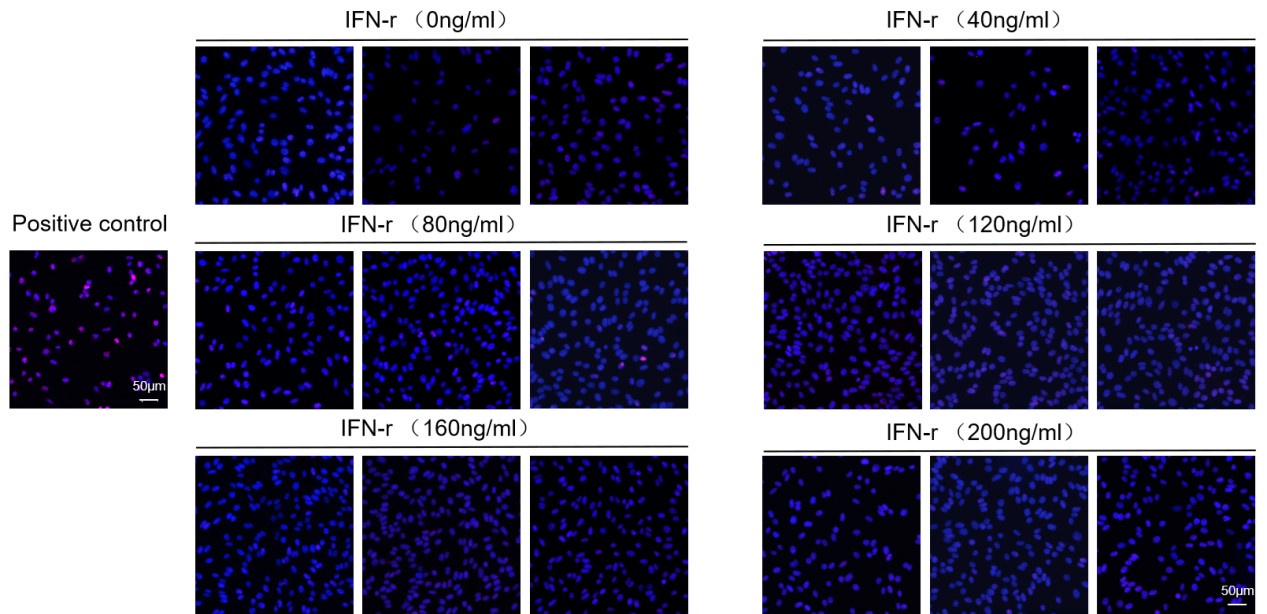
Q14: In Figure 7c, d how was the induction of cell death excluded as a cause of IFNg-mediated effects in LECs?
A14: Thank you for your suggestion. To remove the interference of apoptosis on the results, we performed TUNEL staining of LECs after stimulation with different concentrations of IFN-r for 24 h. As shown in the Figure to reviewer 9, little apoptosis of LECs was observed in this concentration gradient range. Therefore, we can exclude the potential impact of IFN-r-induced cell apoptosis.
Author response image 1.
TUNEL staining of LECs after stimulation with different concentrations of IFN-r for 24 h.
Q15: Results with hypoxia in Figure 7 are mentioned but not shown.
A15: Thank you for your observation. In the revised article, we supplemented the detection of Gal9 expression after hypoxic stimulation. We conducted hypoxia intervention experiments using two methods. First, we applied 1% oxygen concentration stimulation to detect the expression of Gal9 at 0 h, 2 h, 4 h, 8 h, 12 h, and 24 hours. Second, we applied CoCl2 intervention to activate HIF1α expression and simulated cell hypoxia stimulation to detect Gal9 expression. Both results confirmed that hypoxia could not stimulate LECs to secrete galectin 9. The results are presented in Figure 7-figure Supplement 1 (A-D).
Reviewer #3 (Recommendations For The Authors):
Q1: In Figure 1, the so-called "LYVE1-labeled lymphatic capillaries with discontinuous walls" might be macrophages. The authors measured lymphatic area by measuring "vessels with visible lumens", which is unclear. This may underestimate the number of capillaries that expand after MI in the border zone of the infarct area. The authors need to use CD68 and Pdpn markers, as Lyve1 is not specific for lymphatics and also stains macrophages, and Pdpn is more reliable for assessing lymphatic identity.
A1: Thank you for your good suggestion. We totally agree with your opinion. Because both the CD68 (CST,97778S) and LYVE1 antibodies (Abcam,ab14917) are rabbit multiclonal antibodies and to more accurately label cardiac lymphatics, we performed immunofluorescence co-staining using LYVE1 and PDPN antibodies(Thermo,53-5381-82) and re-measured the lymphatic vessel area using the Image J software (version 1.53). The result is shown in Figure to reviewer 1 (Figure 1A and 1B in manuscript). Further, we performed co-staining with PDPN and CD68 to observe the relationship between macrophage and cardiac lymphatic vessel distributions at different time points post-myocardial infarction (Figure to reviewer 2,and Figure1-figure supplement 1F in manuscript). Per your comment, some LYVE1 markers are positive, whereas PDPN markers may be negative for macrophages in the heart tissue. We have added notes on the catalog numbers of anti-PDPN and anti-CD68 in the methods (Page 10, Lines 351‒352) and updated them in the KRT template and MDAR checklist.
Q2: It is not clear how they analyse the lymphatic area in Figure 1, please explain.
A2: Thank you for your observation. The method we used is consistent with that described in previous studies[1,2]. (PMID: 30582443 and PMID: 32404007). The detailed methods have been described in the Methods as follows (Page 10, Lines 347‒352):
For quantification of vessel area, vessels with visible co-staining were measured using Image J software. First, we selected an image, turned it into 8-bit, and then applied a suitable threshold adjustment (present co-stained areas wherever possible). Second, five equally sized squares were selected in the respective zones (remote, infarct, and border zones) of each slice. ROI manager tools were used to analyze the automatic signal intensity quantification by the software in the area inside this square. Finally, the GraphPad software was used to plot the results as a bar graph.
Q3: Figure 1-supplement 1D: The authors claim that the observed structure is a lymphatic valve, however in 2D sections, this shape might result from membrane destruction due to the cutting and staining process. To accurately identify valves, the authors should employ 3D imaging of the lymphatic network, such as using a clearing protocol followed by lightsheet microscopy.
A3: Thank you for your good suggestion. We performed a 3D scan using a confocal microscope on another slice. The results are shown in Figure 1-supplement 1D. We believe it is more like the lymphatic valve than chips from membrane destruction.
Q4: In Figure 2, the number of LECs is too little. Indeed, 242 LECs were identified over 44860 total cell numbers and 5688 endothelial cells cannot be representative and cannot afford to distinguish 4 different clusters.
A4: We further analyzed the percentage of LEC in the adult mouse heart in the physiological state on day d0 based on the results of single-cell nuclear sequencing from public databases (GSE214611). A total of 292 LEC cells were obtained from 26,779 cells captured on board in three samples, meaning that the percentage of LEC cells in the normal adult mouse heart is 1.09%. Cardiac LECs are really rare, and enrichment methods such as flow cytometry and magnetic beads separation for cardiac LECs are under marked probing, which might exhibit more irrefutable evidence in future studies.
Q5: The authors claimed that there is transcriptional heterogeneity in regenerated cardiac LECs post-MI, based on their over-clusterization. However, to substantiate this claim, they need to include a control comparison. Currently, the observed differences in cardiac LEC profiles lack a direct connection to the disease condition.
A5: Thank you for pointing this out. Because we could not download spatial transcriptome data for day d0 in the public database (GSE214611) or from the authors, we have used data of 1 h after IR as a reference for approximating the physiological state in Figure 3 and in Supplemental Figure 1.
Q6: Line 131, what is the regeneration ratio the authors cite here?
A6: Thank you for the comment. Regeneration ratio is an inappropriate use of the word, and we apologize for this confusion. We were actually referring to the regenerative potential of LECs.
Q7: Line 132, it is not clear what is the "normal myocardial tissue" in the graphs presented Figures 3A and B. Is it d0 time point?
A7: Thank you for your suggestion. The d0 time point means LECs in the normal adult mouse heart.
Q8: In Figure 2D, please add more lymphatic markers as Ccl21, Flt4, Itga9, FoxC2 and Aqp1.
A8: Thank you for your suggestion. We have added these markers (Except Ccl21, whose gene expression is too low to mark) in Figure 2D in the revised manuscript.
Q9: The authors must replace "lymphocyte" with "lymphatic" from 2.5, where they start to present interactions between lymphatic and immune cells.
A9: Thank you for your good comments. We have corrected these words.
Q10: In Figure 3, please indicate what the color scale means.
A10: Thank you for your suggestion. We have supplied a color scale label.
Q11: In Figures 3C and D, the authors distinguished the same LECs clusters in the spatial transcriptomic as in the scRNAseq analysis. This is not clear whether they used the same markers.
A11: We appreciate your observation. We have added the required information to the Methods on Page 12, Lines 429‒434, as follows:
“We conducted spatial transcriptome data analysis using the deconvolution algorithm. The deconvolution algorithm refers to the application of feature genes to infer the full matrix information of single-cell transcriptome of cell subclusters. We then compared and anchored the matrix information of the single-cell transcriptome with the information of each SPOT in the spatial transcriptome, predicting cell types based on the similarity between the two sets of information.”
Q12: In 2.5, it is not clear whether the main message is about macrophage interactions with lymphocytes or with lymphatics(LEC interact with others)
A12: Thank you for your suggestion. We have revised the title 2.5 as “Assessment of Cell-Cell Communication between LECs and immune cells,” which is clearer for the reader.
Q13: In 2.6, the authors claim that they reveal "that fluid retention occurs in LEC ca I and LEC co. They don't show any data supporting this.
A13: Thank you for your comment. “…that fluid retention occurs in LEC ca I and LEC co” is mainly supported by Figure 3D KEGG enrichment. LEC Ca I is related to vasopressin-regulated water reabsorption, and LEC co is related to renin secretion.
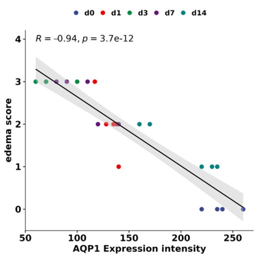
Q14: In Figure 6A, please add statistical values, as the authors claim a significant correlation. Please also add a figure to support the correlation between Aqp1 and edema score, as mentioned in 2.6.
A14: Thank you for pointing this out. We have presented the information on statistical values in Figure 6A. Moreover, we calculated the correlation between Aqp1 and edema score in Figure 6D (shown in Author response image 2).
Author response image 2.
Correlation between Aqp1 expression intensity and edema score.
Q15: In Figure 6B, myocardial edema assessment using H&E staining is not accurate. If the authors wish to analyse cardiac edema, they must use gravimetry or MRI techniques.
A15: Thank you for your comment. We totally agree with your opinion. However, owing to limitations in experimental conditions, we could not perform MRI detection of mouse myocardial injury. To evaluate whether edema occurred in the mouse heart tissue, we used classic pathological evaluation methods described in the literature (PMID: 30582443). This method has been described in detail as follows (Page 11, Lines 365‒370):
Four high-power (40×) representative images were chosen per animal under the H&E stained section; each image must have a clear border of the section visible. Images were blinded, and five visual fields per sample were evaluated. Subsequently, an edema score was determined for each sample (Score 1=no edema, 2=mild edema, 3=severe edema). Graphs represent the average score value per animal.
Q16: Line 227, please correct "LVEC" with "LEC".
A16: Thank you for your careful review. We have revised this in the manuscript.
Q17: In Figure 6D, IF co-staining of Aqp1 and lymphatic vessels is mentioned as "significantly reduced". However, we don't see any quantification data supporting this.
A17: Thank you for your comment. To clarify the expression trend of APQ1 more clearly, we performed immunofluorescence staining of APQ1 and LYVE1 at different time points post-MI (d0, d1, d3, d7, and d14). The results are shown in the corrected Figure 6-figure supplement 1A. The result showed that APQ1 expression increased in the border zone of infarction in d3 post-MI adjacent to LYVE1 staining positive area.
Q18: As Gal9 was not significantly impaired in LECs post. MI, Figure 7A does not support any real finding concerning the role of this molecule in monocytes/macrophages interaction with cardiac lymphatics.
A18: Thank you for your comment. The Lgals9 gene is significantly impaired in LEC post-MI, as well as the Cd44 gene in macrophage. We have updated them in Figures 7A and 7B.
Q19: In Figure 7, please correct INF by IFN.
A19: Thank you for your careful review. We have revised this in the manuscript.
-
-
eLife assessment
This study presents useful, yet preliminary findings on the transcriptomic changes in cardiac lymphatic cells after myocardial infarction in mice. The conclusions of the authors remain uncertain as sample sizes for lymphatic endothelial cells are very low. The single-cell transcriptomic data were analyzed using solid advanced methodology and may be used as a starting point for future studies of the impact of lymphatic cells on heart disease.
-
Reviewer #1 (Public Review):
Summary:
Assessment of cardiac LEC transcriptomes post-MI may yield new targets to improve lymphatic function. scRNAseq is a valid approach as cardiac LECs are rare compared to blood vessel endothelial cells.
Strengths:
Extensive bioinformatics approaches employed by the group.
Weaknesses:
Too few cells are included in scRNAseq data set and the spatial transcriptomics data that was exploited has little relevance, or rather specificity, for cardiac lymphatics. This study seems more like a collection of preliminary transcriptomic data than a conclusive scientific report to help advance the field.
-
Reviewer #2 (Public Review):
Summary:
This study integrated single-cell sequencing and spatial transcriptome data from mouse heart tissue at different time points post-MI. They identified four transcriptionally distinct subtypes of lymphatic endothelial cells and localized them in space. They observed that LECs subgroups are localized in different zones of infarcted heart with functions. Specifically, they demonstrated that LEC ca III may be involved in directly regulating myocardial injuries in the infarcted zone concerning metabolic stress, while LEC ca II may be related to the rapid immune inflammatory responses of the border zone in the early stage of MI. LEC ca I and LEC collection mainly participate in regulating myocardial tissue edema resolution in the middle and late stages post-MI. Finally, cell trajectory and Cell-Chat …
Reviewer #2 (Public Review):
Summary:
This study integrated single-cell sequencing and spatial transcriptome data from mouse heart tissue at different time points post-MI. They identified four transcriptionally distinct subtypes of lymphatic endothelial cells and localized them in space. They observed that LECs subgroups are localized in different zones of infarcted heart with functions. Specifically, they demonstrated that LEC ca III may be involved in directly regulating myocardial injuries in the infarcted zone concerning metabolic stress, while LEC ca II may be related to the rapid immune inflammatory responses of the border zone in the early stage of MI. LEC ca I and LEC collection mainly participate in regulating myocardial tissue edema resolution in the middle and late stages post-MI. Finally, cell trajectory and Cell-Chat analyses further identified that LECs may regulate myocardial edema through Aqp1, and likely affect macrophage infiltration through the galectin9-CD44 pathway. The authors concluded that their study revealed the dynamic transcriptional heterogeneity distribution of LECs in different regions of the infarcted heart and that LECs formed different functional subgroups that may exert different bioeffects in myocardial tissue post-MI.
Strengths:
The study addresses a significant clinical challenge, and the results are of great translational value. All experiments were carefully performed, and their data support the conclusion.
Weaknesses:
(1) Language expression must be improved. Many incomplete sentences exist throughout the manuscript. A few examples: Lines 70-71: In order to further elucidate the effects and regulatory mechanisms of the lymphatic vessels in the repair process of myocardial injury following MI. Lines 71-73: This study, integrated single-cell sequencing and spatial transcriptome data from mouse heart tissue at different time points after MI from publicly available data (E-MTAB-7895, GSE214611) in the ArrayExpress and gene expression omnibus (GEO) databases. Line 88-89: Since the membrane protein LYVE1 can present lymphatic vessel morphology more clearly than PROX1.
(2) The type of animal models (i.e., permeant MI or MI plus reperfusion) included in ArrayExpress and gene expression omnibus (GEO) databases must be clearly defined as these two models may have completely different effects on lymphatic vessel development during post-MI remodeling.
(3) Lines 119-120: Caution must be taken regarding Cav1 as a lymphocyte marker because Cav1 is expressed in all endothelial cells, not limited to LEC.
(4) Figure 1 legend needs to be improved. RZ, BZ, and IZ need to be labeled in all IF images. Day 0 images suggest that RZ is the tissue section from the right ventricle. Was RZ for all other time points sampled from the right ventricular tissue section?
(5) The discussion section needs to be improved and better focused on the findings from the current study.
-
Reviewer #3 (Public Review):
Summary:
It has been demonstrated that cardiac lymphatics are essential for cardiac health and function. Moreover, post-myocardial infarction, targeting lymphatics by stimulating lymphangiogenesis has been shown to improve cardiac inflammation, fibrosis, and function. Then, the aim of this study was to evaluate the transcriptomic changes of cardiac lymphatic endothelial cells (LECs) after a myocardial infarction, which could reveal new therapeutic targets targeting lymphatic function. Moreover, investigating the cell-cell communication between lymphatic and immune cells would give critical information for a better understanding of the disease.
Strengths:
The use of scRNAseq data to evaluate LECs is an effective strategy considering the small proportion of LECs compared to blood endothelial cells. The …
Reviewer #3 (Public Review):
Summary:
It has been demonstrated that cardiac lymphatics are essential for cardiac health and function. Moreover, post-myocardial infarction, targeting lymphatics by stimulating lymphangiogenesis has been shown to improve cardiac inflammation, fibrosis, and function. Then, the aim of this study was to evaluate the transcriptomic changes of cardiac lymphatic endothelial cells (LECs) after a myocardial infarction, which could reveal new therapeutic targets targeting lymphatic function. Moreover, investigating the cell-cell communication between lymphatic and immune cells would give critical information for a better understanding of the disease.
Strengths:
The use of scRNAseq data to evaluate LECs is an effective strategy considering the small proportion of LECs compared to blood endothelial cells. The extensive bioinformatic analysis used by the authors for three different data sets.
Weaknesses:
Among a total of 44,860 cells, only 242 LECs and 5,688 endothelial cells were identified. This small number of LECs is not representative and is insufficient to reliably distinguish four different clusters. The bioinformatic analysis is not supported by significant results in their in vivo and in vitro experiments.
-
